Review
. 2022 Dec;298(12):102628.
doi: 10.1016/j.jbc.2022.102628. Epub 2022 Oct 20.

Scaffold proteins as dynamic integrators of biological processes

Affiliations
Review

Scaffold proteins as dynamic integrators of biological processes

Christopher J DiRusso et al. J Biol Chem. 2022 Dec.

Abstract

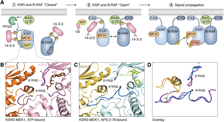
Scaffold proteins act as molecular hubs for the docking of multiple proteins to organize efficient functional units for signaling cascades. Over 300 human proteins have been characterized as scaffolds, acting in a variety of signaling pathways. While the term scaffold implies a static, supportive platform, it is now clear that scaffolds are not simply inert docking stations but can undergo conformational changes that affect their dependent signaling pathways. In this review, we catalog scaffold proteins that have been shown to undergo actionable conformational changes, with a focus on the role that conformational change plays in the activity of the classic yeast scaffold STE5, as well as three human scaffold proteins (KSR, NEMO, SHANK3) that are integral to well-known signaling pathways (RAS, NF-κB, postsynaptic density). We also discuss scaffold protein conformational changes vis-à-vis liquid-liquid phase separation. Changes in scaffold structure have also been implicated in human disease, and we discuss how aberrant conformational changes may be involved in disease-related dysregulation of scaffold and signaling functions. Finally, we discuss how understanding these conformational dynamics will provide insight into the flexibility of signaling cascades and may enhance our ability to treat scaffold-associated diseases.

Keywords: KSR; NEMO; SHANK3; STE5; conformational change; liquid–liquid phase separation; scaffold protein; signaling.


Conflict of interest statement

Conflict of interest The authors declare that they have no conflicts of interest with the contents of this article.

Figures

Figure 1
Figure 1
Scaffold STE5 has pathway regulatory properties mediated via conformational changes. On the left is the traditional stable platform depiction of the three-tiered MAPK cascade modulated by STE5 in yeast, where α-factor binds to STE2 and a phosphorylation cascade involving STE11, STE7, and FUS3 is initiated. This leads to the eventual phosphorylation of the STE12 transcription factor to initiate transcription of mating response genes. On right is a more detailed depiction of the complexity of STE5’s role in the pathway. In unstimulated cells, STE5 is in a closed state where FUS3 binding is blocked. After α-factor binds to its receptor STE2, STE5 is recruited to PIP2 at the plasma membrane. This causes a conformational change in STE5 to transition it to an open state such that FUS3 can bind to STE5. Upon binding to STE5, FUS3 then undergoes a conformational change to be properly presented to STE7 for phosphorylation.
Figure 2
Figure 2
Conformational change in KSR during RAS signaling.A, in the human MAPK pathway, the KSR–MEK complex exists in a closed conformation in the cytosol. Upon activation of the pathway, PP2A is recruited by RAS to dephosphorylate KSR in order to release its inhibition by 14-3-3 and IMP (1). The release of 14-3-3 and IMP causes a conformational change in KSR such that its CA3 domain can be tethered to the plasma membrane and KSR can dimerize with active B-RAF (2). Upon binding active B-RAF, KSR undergoes a second conformational change to present MEK for phosphorylation in trans via a separate KSR/B-RAF complex for signal propagation (3). B, view of the crystal structure of KSR2-MEK1 bound to ATP (brown) (PDB 2Y4I). Activation loop and p-loop (dark purple) are “closed” surrounding MEK1 218/222 (gray), reflecting the inactive RAF-inaccessible state of the complex. C, view of the crystal structure of KSR2-MEK1 bound to APS-2-79 (light green) (PDB 5KKR). Activation loop and p-loop (dark blue) are “closed” in a different state, reflecting another inactive, RAF-inaccessible conformation of the resting state. D, simplified superposition of the difference in the KSR active site bound to APS-2-79 (dark blue) and ATP (dark purple), centered around MEK 218/222 (gray), demonstrating the conformational flexibility of these segments in inactive complexes. Crystal structure images adapted from Dhawan et al. (29) and Brennan et al. (23). Images compiled in PyMOL 2.5. A-loop, activation loop; PDB, Protein Data Bank.
Figure 3
Figure 3
Conformational changes in NEMO during NF-κB signaling.A, dimerized NEMO exists as an inactive extended coiled coil in the cytosol (1). Once it binds ubiquitin at its UBAN region and IκB at its zinc finger, NEMO undergoes a series of conformational changes to present a bound IκB to IKKβ for phosphorylation (2). This conformational shift in NEMO, induced by ubiquitination, promotes liquid–liquid phase separation of NEMO and its associated client proteins for efficient signaling (3). (p50 and p65 represent NF-κB, which is bound to IκB.) B, schematic representation of the inactive IKK complex, IKKβ (PBD 4KIK) is in green, NEMO KBD and IKKβ NEMO binding domain (PDB 3BRV); NEMO CoZi domain (PDB 2ZVO); NEMO ZF (2JVX) are in orange, and IκB (PDB 1IKN, p50/p65 omitted for simplicity) is in dark cyan. Dotted orange lines represent regions lacking structural data within NEMO. C, schematic representation of active IKK complex with conformational rearrangement centered around the central structurally uncharacterized region to bring IκB in proximity to IKKβ. Blue dotted lines with arrowheads represent the stabilization of the active conformation when NEMO residues 384 to 389 are bound to IKKβ, which is induced by the binding of linear ubiquitin (pink). Images compiled in PyMOL 2.5. PDB, Protein Data Bank; ZF, zinc finger.
Figure 4
Figure 4
Conformational change of SHANK3 during synaptic signaling.A, in the postsynaptic density (PSD), oligomerized SHANK3 is bound by RAP1 via its SPN and ANK domains, promoting its closed conformation. SHANK3 sequence C-terminal to the SAM domain omitted for simplicity (1). A signal, possibly an increase in zinc concentration at the PSD, causes a conformational change in SHANK3 to the open conformation. This causes RAP1 to be released from SHANK3, reduces the oligomeric state of SHANK3, promotes actin binding to SHANK3, and leads to the recruitment of actin regulators such as AbiI, CaMKKIIα, and SHARPIN to promote actin polymerization in the PSD (2). B, crystal structure of the SPN-ANK domain of SHANK3 (PDB 5G4X). The ANK domain is in light green and the SPN domain is in pink. Actin-binding sites are buried to prevent actin binding. C, schematic representation of the N52R mutant of SHANK3, which is constitutively in the open state, wherein RAP1 is unbound and actin-binding sites are exposed. The ANK domain is in blue and the SPN domain is in purple. Crystal structure images adapted from Salomaa et al. (48). Images compiled in PyMOL 2.5. PDB, Protein Data Bank.

References

    1. Good M.C., Zalatan J.G., Lim W.A. Scaffold proteins: hubs for controlling the flow of cellular information. Science. 2011;332:680–686. - PMC - PubMed
    1. Shaw A.S., Filbert E.L. Scaffold proteins and immune-cell signalling. Nat. Rev. Immunol. 2009;9:47–56. - PubMed
    1. Buday L., Tompa P. Functional classification of scaffold proteins and related molecules. FEBS J. 2010;277:4348–4355. - PubMed
    1. Hu J., Neiswinger J., Zhang J., Zhu H., Qian J. Systematic prediction of scaffold proteins reveals new design principles in scaffold-mediated signal transduction. PLoS Comput. Biol. 2015;11 - PMC - PubMed
    1. Alexa A., Varga J., Reményi A. Scaffolds are 'active' regulators of signaling modules. FEBS J. 2010;277:4376–4382. - PubMed

Publication types

MeSH terms

LinkOut - more resources