Review
. 2022 Oct 6:13:979719.
doi: 10.3389/fmicb.2022.979719. eCollection 2022.

Glutathione deficiency in the pathogenesis of SARS-CoV-2 infection and its effects upon the host immune response in severe COVID-19 disease

Affiliations
Review

Glutathione deficiency in the pathogenesis of SARS-CoV-2 infection and its effects upon the host immune response in severe COVID-19 disease

Carlos A Labarrere et al. Front Microbiol. .

Abstract

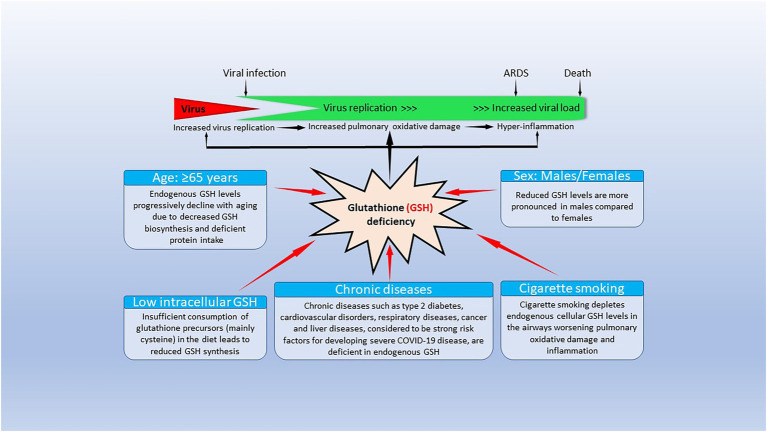
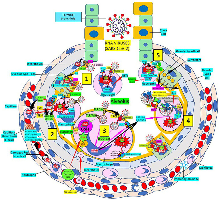
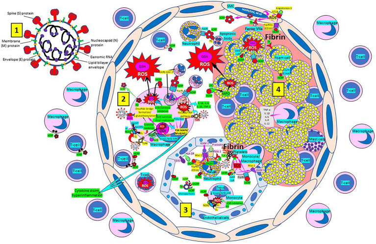
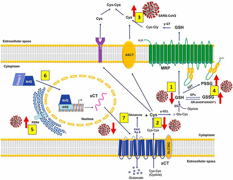
Severe acute respiratory syndrome coronavirus 2 (SARS-CoV-2) that causes coronavirus disease 19 (COVID-19) has numerous risk factors leading to severe disease with high mortality rate. Oxidative stress with excessive production of reactive oxygen species (ROS) that lower glutathione (GSH) levels seems to be a common pathway associated with the high COVID-19 mortality. GSH is a unique small but powerful molecule paramount for life. It sustains adequate redox cell signaling since a physiologic level of oxidative stress is fundamental for controlling life processes via redox signaling, but excessive oxidation causes cell and tissue damage. The water-soluble GSH tripeptide (γ-L-glutamyl-L-cysteinyl-glycine) is present in the cytoplasm of all cells. GSH is at 1-10 mM concentrations in all mammalian tissues (highest concentration in liver) as the most abundant non-protein thiol that protects against excessive oxidative stress. Oxidative stress also activates the Kelch-like ECH-associated protein 1 (Keap1)-Nuclear factor erythroid 2-related factor 2 (Nrf2)-antioxidant response element (ARE) redox regulator pathway, releasing Nrf2 to regulate the expression of genes that control antioxidant, inflammatory and immune system responses, facilitating GSH activity. GSH exists in the thiol-reduced and disulfide-oxidized (GSSG) forms. Reduced GSH is the prevailing form accounting for >98% of total GSH. The concentrations of GSH and GSSG and their molar ratio are indicators of the functionality of the cell and its alteration is related to various human pathological processes including COVID-19. Oxidative stress plays a prominent role in SARS-CoV-2 infection following recognition of the viral S-protein by angiotensin converting enzyme-2 receptor and pattern recognition receptors like toll-like receptors 2 and 4, and activation of transcription factors like nuclear factor kappa B, that subsequently activate nicotinamide adenine dinucleotide phosphate (NADPH) oxidase (NOX) expression succeeded by ROS production. GSH depletion may have a fundamental role in COVID-19 pathophysiology, host immune response and disease severity and mortality. Therapies enhancing GSH could become a cornerstone to reduce severity and fatal outcomes of COVID-19 disease and increasing GSH levels may prevent and subdue the disease. The life value of GSH makes for a paramount research field in biology and medicine and may be key against SARS-CoV-2 infection and COVID-19 disease.

Keywords: COVID-19; Glutathione; SARS-CoV-2; acute respiratory distress syndrome; atherosclerosis; atherothrombosis; oxidative stress; reactive oxygen species.


Conflict of interest statement

The authors declare that the research was conducted in the absence of any commercial or financial relationships that could be construed as a potential conflict of interest.

Figures

Figure 1
Figure 1
Factors causing endogenous glutathione (GSH) deficiency and GSH deficiency-mediated mechanisms contributing to coronavirus disease 19 (COVID-19) pathogenesis and outcomes. The bottom part of the figure shows that risk factors for severe COVID-19 infection lead to decrease/depletion of intracellular GSH. The top part of the figure shows potential GSH deficiency-mediated mechanisms that could influence clinical manifestations and outcomes in COVID-19 disease. Modified from Polonikov (2020).
Figure 2
Figure 2
Severe acute respiratory syndrome coronavirus 2 (SARS-CoV-2) pulmonary infection, oxidative stress and antioxidant defenses. [1] After entry of SARS-CoV-2 into the alveolus, viruses invade type II alveolar cells through angiotensin-converting enzyme 2 receptors (ACE2) and glycosaminoglycans (GAGs), and infected cells increase reactive oxygen species (ROS) production, reduce Kelch-like ECH-associated protein 1 (Keap1)-Nuclear factor erythroid 2-related factor 2 (Nrf2)-antioxidant response element (ARE) redox regulator pathway and become defective for surfactant production. Infected cells activate nuclear factor (NF)-κB and release cytokines like interleukin (IL)-8. Alveolar type I cells augment ROS production via toll-like receptors (TLRs) 2 and 4. SARS-CoV-2 enhances neutrophil extracellular trap (NET) release and increases ROS production [2] SARS-CoV-2 augments macrophage’s ROS production, inhibiting Nrf2 activation and enhancing NF-κB upregulation. ROS are counterbalanced by enzymes like superoxide dismutase (SOD), catalase (Cat), glutathione S-transferase (GST) and glutathione peroxidase (GPx) to protect cells from oxidative damage caused by nicotinamide adenine-dinucleotide phosphate (NADPH) oxidase 2 (NOX2), superoxide (O2), hydrogen peroxide (H2O2), and myeloperoxidase (MPO). Capillary neutrophils migrate to and from alveoli by trans-endothelial (TEM) and reverse transmigration (rTEM), respectively. SARS-CoV-2 infection can cause excessive ROS production in capillaries, red blood cell (RBC) dysfunction, thrombosis and alveolar damage. [3] SARS-CoV-2-infected macrophages (via ACE2 and TLRs) reduce enzymes like SOD and Cat, among others, and activate NF-κB. NOX2 activation increases ROS production that enhance NF-κB activation. Activated alveolar macrophages release increased levels of IL-1β, IL-6, IL-8 and tumor necrosis factor (TNF)-α. Glutathione (GSH) precursors (Cystine, cysteine, N-acetyl cysteine, NAC), and selenium (Se) restore GSH and GPx, respectively, to counteract the effects of ROS. [4] Alveolar macrophages engulf SARS-CoV-2-infected apoptotic cells via Fc (γ/α/μ) and scavenger receptors and/or pattern recognition protein receptors (PRPRs) leading to increased ROS production, NFκB activation and cytokine release; and infected alveolar type II cells enhance inflammation. [5] Neutrophils contribute to O2 production, lipid peroxidation and increased oxidative stress, Keap-1-Nrf2-ARE signaling pathway reduction and NFκB activation promoting cytokine storm. Abbreviations: TMPRSS2: Transmembrane protease Serine 2; PRPs: pattern recognition proteins.
Figure 3
Figure 3
Severe acute respiratory syndrome coronavirus-2 (SARS-CoV-2) enhances oxidative stress and atherosclerosis progression. [1] SARS-CoV-2 structure. [2] SARS-CoV-2 viruses facilitate oxidative stress and inflammation in the arterial intima. Native C-reactive protein (nCRP), a marker of severe SARS-CoV-2 produced in liver, macrophages, lymphocytes, smooth muscle cells (SMC) and other cells, promotes inflammation through monomeric CRP (mCRP) enhancing intimal oxidative stress. SARS-CoV-2 binds macrophage toll-like receptor (TLR) 4 and facilitates nicotinamide adenine dinucleotide phosphate (NADP) H oxidase 2 (Nox2) activity and superoxide (O2) production causing cysteine oxidation, disulfide bridge formation and S-glutathionylation. Xanthine oxidase (XO) and inhibition of superoxide dismutase (SOD)/catalase further facilitate O2 cellular activity and ROS generation. SARS-CoV-2 can bind TLRs 2 and 4 and activate transcription factors like nuclear factor (NF)-κB facilitating cytokine storm and hyperinflammation. Excessive mitochondrial reactive oxygen species (ROS) generation further enhances cytokine production. CRP (nCRP, mCRP) can facilitate macrophage and neutrophil uptake of SARS-CoV-2-infected apoptotic cells through Fcγ and Fcα receptors, respectively (FcRs). Oxidative stress also activates the Kelch-like ECH-associated protein 1 (Keap1)-Nuclear factor erythroid 2-related factor2 (Nrf2)-antioxidant response element (ARE) redox regulator pathway in monocytes (see [3] and macrophages, releasing Nrf2 to regulate the expression of genes that control antioxidant enzymes like glutathione S-transferase (GST)), facilitating glutathione (GSH) activity. Macrophages, Tlymphocytes, neutrophils and SMCs can generate mCRP increasing inflammation. [3] Monocytes, macrophages, neutrophils, endothelial cells and microparticles can generate mCRP, increase O2 and ROS formation and reactive nitrogen species like peroxinitrite (ONOO), and tissue factor (TF) expression enhancing oxidation, inflammation and thrombosis. TLR 4-mediated SARS-CoV-2-binding to platelets promotes thrombosis, mCRP binding to lipid rafts and FcγRs enhances inflammation and endothelial activation allows intimal cell migration. [4] Foam cells and smooth muscle cells associated with atherosclerotic plaques enhance ROS formation, cytokine release and tissue factor (TF)-mediated fibrin deposition. MAPK/ERK, mitogen-activated protein kinase/extracellular signal-regulated kinase; AT1R, Angiotensin II type 1 receptor; PC, phosphorylcholine; LPC, lysophosphatidylcholine; MPO, myeloperoxidase; nnCRP, non-native CRP; TNF, tumor necrosis factor; IL, interleukin; ACE, angiotensin converting enzyme; MyD88/TRIF, myeloid differentiation primary response88/TIR-domain-containing adapter-inducing interferon-β; PI3K/Akt, phosphatidylinositol-3-kinase/protein kinase B; AP-1, activator protein 1; CD31, cluster of differentiation 31; ICAM-1, intercellular adhesion molecule-1; Mac-1, macrophage-1 antigen; PSGL-1, P-selectin glycoprotein ligand-1; HLA-DR, Human Leukocyte Antigen – DR isotype.
Figure 4
Figure 4
Severe acute respiratory syndrome coronavirus 2 (SARS-CoV-2) infection alters metabolism and redox function of cellular glutathione (GSH). SARS-CoV-2 markedly decreases GSH levels [1], that could be explained by lower intake of the GSH precursor cysteine (Cys) [2] and increased efflux of cellular thiols [3]. Increased levels of oxidized glutathione (GSSG) and protein glutathionylation [4] along with upregulation of endoplasmic reticulum stress marker protein kinase R (PKR)-like endoplasmic reticulum kinase (PERK) [5] are also observed. Antivirals activate the Kelch-like ECH-associated protein 1 (Keap1)-Nuclear factor erythroid 2-related factor2 (Nrf2)-antioxidant response element (ARE) redox regulator pathway, releasing Nrf2 [6] to regulate the expression of genes that control antioxidant, inflammatory and immune system responses (including the cystine (cys-cys)/glutamate transporter xCT and the membrane transporter multidrug resistance protein [MRP], which are decreased and markedly upregulated, respectively, during infection); restoring GSH levels in the infected cells and facilitating GSH synthesis [7] and activity. Abbreviations: γ-GT: γ-glutamyl transferase; ASCT: alanine-serine-cysteine transporter; LAT: L-type amino acid transporter; PSSG: S-glutathionylated Proteins; GST: glutathione-S-transferase; γ-GCL: γ-glutamate cysteine ligase; γ-glutamyl cysteine; GST: glutathione-S-transferase; GPx: glutathione peroxidase; GR: glutathione reductase; NADPH: reduced NADP+; NADP+: Nicotinamide adenine dinucleotide phosphate; GS: glutathione synthetase; xCT (SLC7A11)/SLC3A2: cystine/glutamate transporter light (xCT [SLC7A11]) and heavy (SLC3A2) chains. Modified from Bartolini et al. (2021).
Figure 5
Figure 5
Severe acute respiratory syndrome coronavirus 2 (SARS-CoV-2)-related glutathione (GSH) cellular depletion, repletion treatment options and a multiweapon defense approach. [1] SARS-CoV-2 exacerbates oxidative stress, inflammation and coagulation, reducing GSH levels mainly in hosts with added risk factors. [2] SARS-CoV-2 invades host cells through virus protein ligands, like trimeric spike glycoprotein interacting with cellular receptors like angiotensin converting enzyme 2 (ACE2), and host proteases, like transmembrane protease serine 2 (TMPRSS2), that proteolytically activates virus ligands, promoting virus entry, replication and ACE2 down-regulation; N-acetylcysteine (NAC) can block this interaction. SARS-CoV-2-related ACE2 downregulation facilitates angiotensin II/angiotensin types 1 and 2 receptor (AT1/2R)-mediated nicotinamide adenine dinucleotide phosphate (NADPH) oxidase activation and reactive oxygen species (ROS) production; and inhibits interaction of angiotensin 1–7 and MAS1 oncogene/G protein-coupled (MAS/G) receptor. [3] A multiweapon approach includes enhancing host response to viral particles and peptides by monocytes/macrophages and T-cells, as well as innate and adaptive B-cell/plasma cells producing antibodies to reduce cytokine production and the subsequent cytokine storm. Reactive oxygen species (ROS) cell production enhances proinflammatory cytokine release while reducing anti-inflammatory cytokines. [4] Modulation of CD4+ and CD8+ T cells will facilitate SARS-CoV-2 removal. [5] SARS-CoV-2 binding and cleavage of ACE2 receptor leads to shedding of host ACE2 receptor contributing to the loss of ACE2 function and systemic release of S1/ACE2 complex. SARS-CoV-2 reduces Nrf2 and GSH allowing ROS and RNS to damage the cell. [6] Liposomal GSH (see [2], vitamin D3 by increasing glutamate cysteine ligase and glutathione reductase activity), and N acetyl cysteine (NAC), see [2] participate in GSH synthesis. Increased intracellular GSH reduces ROS and reactive nitrogen species (RNS), as well as NF-κB activation. Sulforaphane and resveratrol enhance Nrf2 production and Nrf2 negatively regulates the endoplasmic-reticulum-resident protein stimulator of interferon genes (STING) reducing interferon secretion. Increased antioxidant defense (cystine, cysteine, NAC, liposomal GSH, vitamin D3, sulforaphane, resveratrol, and others) reestablishes cell homeostasis [7]. Increased nuclear factor-κB (NF-κB) activity enhances interleukin (IL)-6 secretion and cytokine storm, while decreased nuclear NF-κB allows activation of nuclear factor erythroid 2-related factor2 (Nrf2)-dependent antioxidant genes and enzyme transcription (HO-1, NQO-1, and others); Nrf2 inhibition of M1 and upregulation of M2 induced genes; decreased pro-inflammatory and increased anti-inflammatory cytokine expression; and decreased cytokine storm. C-Src, proto-oncogene tyrosine-protein kinase sarcoma; PKC, protein kinase C; Rac-1, Ras-related C3 botulinum toxin substrate 1; IL1RA, IL1 receptor antagonist; MCP1, monocyte chemoattractant protein-1; MIP, macrophage inflammatory protein; PDGFB, Platelet Derived Growth Factor B; VEGF-A, vascular endothelial growth factor A; iNOS, inducible nitric oxide synthase; TCR, T-cell receptor; MHCI/II, major histocompatibility complex class I/II; GCSF, granulocyte colony-stimulating factor; GMCSF, granulocyte-macrophage colony-stimulating factor; FGF, fibroblast growth factor; IP10, interferon gamma-induced protein 10; NAb, natural antibody; SOD, superoxide dismutase; HO-1, heme oxygenase-1; NQO1, NAD(P) H quinone dehydrogenase 1; β2M, β2 microglobulin; GSSG, glutathione disulfide; Keap 1, Kelch-like ECH-associated protein 1; ERK, extracellular signal-regulated protein kinase; FcμR, Fcμ receptor; IgM, immunoglobulin M.

Similar articles

Cited by

References

    1. Abbas M., Verma S., Verma S., Siddiqui S., Khan F. H., Raza S. T., et al. . (2021). Association of GSTM1 and GSTT1 gene polymorphisms with COVID-19 susceptibility and its outcome. J. Med. Virol. 93, 5446–5451. doi: 10.1002/jmv.27076, PMID: - DOI - PMC - PubMed
    1. Abd El-Aziz T. M., Al-Sabi A., Stockand J. D. (2020). Human recombinant soluble ACE2 (hrs ACE2) shows promise for treating severe COVID19. Signal Transduct. Target. Ther. 5:258. doi: 10.1038/s41392-020-00374-6, PMID: - DOI - PMC - PubMed
    1. Abdool Karim S. S., Abdool K. Q. (2021). Omicron SARS-CoV-2 variant: a new chapter in the COVID-19 pandemic. Lancet 398, 2126–2128. doi: 10.1016/S0140-6736(21)02758-6, PMID: - DOI - PMC - PubMed
    1. Aboudounya M. M., Heads R. J. (2021). COVID-19 and toll-like receptor 4 (TLR4): SARS-CoV-2 may bind and activate TLR4 to increase ACE2 expression, facilitating entry and causing hyperinflammation. Mediat. Inflamm. 2021:8874339. doi: 10.1155/2021/8874339, PMID: - DOI - PMC - PubMed
    1. Ader F., Bouscambert-Duchamp M., Hites M., Peiffer-Smadja N., Poissy J., Belhadi D., et al. . (2022). Remdesivir plus standard of care versus standard of care alone for the treatment of patients admitted to hospital with COVID-19 (dis CoVeRy): a phase 3, randomised, controlled, open-label trial. Lancet Infect. Dis. 22, 209–221. doi: 10.1016/S1473-3099(21)00485-0, PMID: - DOI - PMC - PubMed