

Comparative Evaluation of Implants with Different Surface Treatments Placed in Human Edentulous Mandibles: A 1-Year Prospective Study
- PMID: 36274893
- PMCID: PMC9474755
- DOI: 10.1007/s12663-021-01600-6
Comparative Evaluation of Implants with Different Surface Treatments Placed in Human Edentulous Mandibles: A 1-Year Prospective Study
Abstract
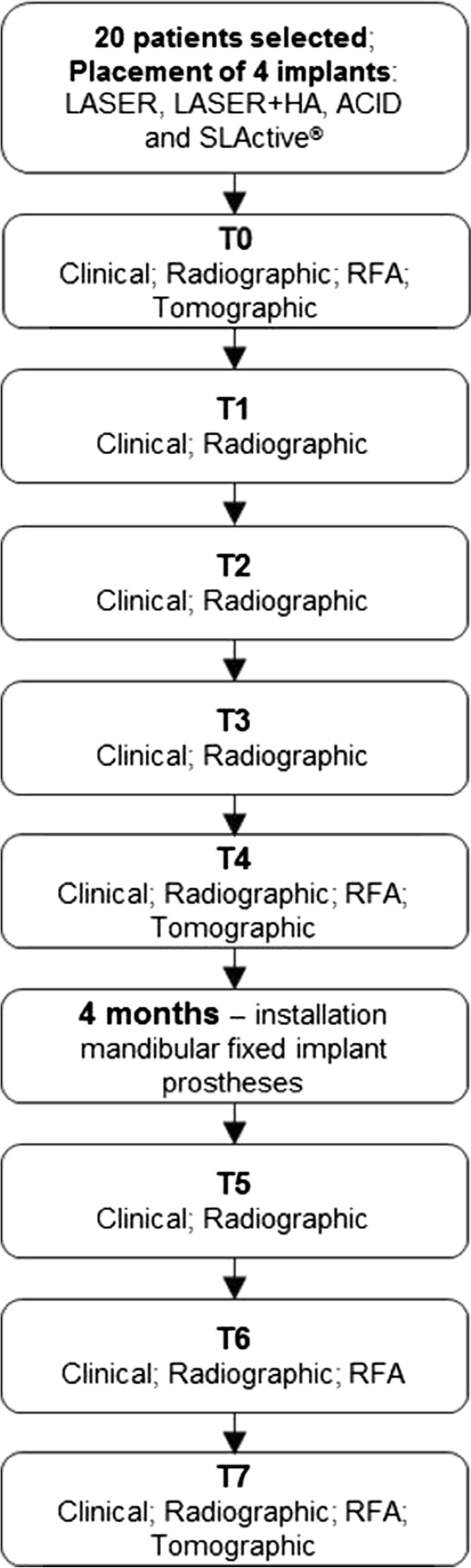
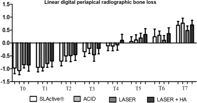
The aims of this study were to analyze prospectively and comparatively the peri-implant bone crest levels, bone density, stability and success rate of implants with different surface treatments in human edentulous mandibles. Twenty edentulous patients were selected. Four different implants were placed between the mental foramen. Four groups were evaluated: (1) laser-modified surface (LASER), (2) surface modified by laser with deposition of apatites (LASER + HA), (3) surface modified by double acid etching (ACID, Implacil De Bortoli) and (4) surface modified by sandblasting and acid etching (SLActive®, Straumann). Clinical, radiographic, resonance frequency and tomographic analyses were used. After 4 months, mandibular fixed implant prostheses were installed. Clinical and radiographic analyses were performed at times T0 (immediately after implant placement), T1 (15 days), T2 (30 days), T3 (60 days), T4 (90 days), T5 (120 days), T6 (180 days) and T7 (360 days), post-implant placement. The resonance frequency analysis (RFA) was measured at T0, T4, T6 and T7. The tomographic analysis was performed at T0, T4 and T7. In the radiographic bone density analysis, a statistical difference was found between the SLActive® and LASER + HA groups at T4 (p < 0.05). Statistical differences were observed in RFA at T4 (90 days), between the SLActive® and LASER groups (p < 0.05) and between the SLActive® and LASER + HA groups (p < 0.05). At T6 and T7, statistical differences were found between the SLActive® group and all other implant surfaces (p < 0.01). The experimental surfaces analyzed showed encouraging positive outcomes compared to those of the SLActive® surface. Long-term follow-up should be performed to confirm these results.
Keywords: Dental implants; Imaging; Lasers; Resonance frequency analysis; Surface.
© The Association of Oral and Maxillofacial Surgeons of India 2021.
Conflict of interest statement
Conflict of interestThe authors declare that they have no conflict of interest.
Figures


References
-
- Al-Hamdan SH, Al-Hamdan K, Junker R, et al. Effect of implant surface properties on peri-implant bone healing: implant stability and microcomputed tomographic analysis. Int J Oral Maxillofac Implants. 2012;27:77–83. - PubMed
LinkOut - more resources
Full Text Sources