. 2022 Oct 5:13:981884.
doi: 10.3389/fpls.2022.981884. eCollection 2022.

The anatomy of the seed-coat includes diagnostic characters in the subtribe Eugeniinae (Myrteae, Myrtaceae)

Affiliations

The anatomy of the seed-coat includes diagnostic characters in the subtribe Eugeniinae (Myrteae, Myrtaceae)

Patricia Gonçalves Sbais et al. Front Plant Sci. .

Abstract

The subtribe Eugeniinae comprises of two genera, Eugenia (ca. 1,100 species) and Myrcianthes (ca. 40 species). Eugenia is the largest genus of neotropical Myrtaceae and its latest classification proposes 11 sections. This study describes the seed anatomy of forty-one species of Eugeniinae in order to provide possible diagnostic characteristics. Following standard anatomical techniques, flower buds, flowers, and fruits were processed and analyzed using microtome sections and light microscopy. The phylogeny used the regions ITS, rpl16, psbA-trnH, trnL-rpl32, and trnQ-rps16, following recent studies in the group. Ancestral character reconstruction uncovered that: (1) the ancestral ovule in Eugeniinae was campylotropous (98.9% probability), bitegmic (98.5% probability), and unitegmic ovules arose on more than one lineage independently within Eugenia; (2) the pachychalazal seed-coat appeared with a 92% probability of being the ancestral type; (3) non-lignified seed-coat (24,5% probability) and aerenchymatous mesotesta (45.8% probability) are diagnostic characters in Myrcianthes pungens (aerenchymatous mesotesta present in the developing seed-coat) and in the species of E. sect. Pseudeugenia until the species of E. sect. Schizocalomyrtus and it is the type of seed-coat that predominates in most basal sections on the tree; (4) the partial sclerification (only in the exotesta-exotestal seed-coat) is mainly observed in species of E. sect. Excelsae, E. sect. Jossinia (group X), and E. sect. Racemosae (22.2% probability); (5) and in the species of the recent lineages of Eugenia, with a probability of 27.2%, predominate the exomesotestal or testal construction of the seed-coat [character observed in almost all species analyzed of E. sect. Jossinia (group Y) and E. sect. Umbellatae]. A dehiscent fruit is considered as a plesiomorphic state in Myrtaceae; the ancestor of this family had seeds with a completely sclerified testa, and the other testa types described for the current species with dehiscent and indehiscent fruits are simplified versions of this ancestral type. Perhaps, this means that the sclerified layers in the seed-coat have remained in whole or in part as a plesiomorphic condition for taxa with a capsule and bacca. Maintaining the plesiomorphic condition may have represented a selective advantage at some point in the evolutionary history of the family and its groups.

Keywords: Myrcianthes; Pseudeugenia; Racemosae; Umbellatae; pachychalaza; perichalaza; testa; trait evolution.


Conflict of interest statement

The authors declare that the research was conducted in the absence of any commercial or financial relationships that could be construed as a potential conflict of interest.

Figures

FIGURE 1
FIGURE 1
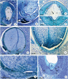
Longitudinal (A–F,H–I,K) and transversal (G,J) sections of the ovule of flower buds (B,C), flowers at anthesis (A,E–K) and post-anthesis (D) of Myrcianthes pungens (A,H); Eugenia neoverrucosa (B); E. uniflora (C–E); E. florida (F); Eugenia arenosa (G); E. myrciantes (I); E. hiemalis (J) and E. longipedunculata (K). (A) Bitegmic and anatrapous tending to campylotropus ovule and perichalaza (∙) beginning its differentiation. (B) Campylotropus ovule evidencing the integuments development. (C–E) Development of the campylotropous and unitegmic ovule. Note in photo (C) the development of the single integument; in photo (D) an obturator in contact to the mycropyle; and in photo (E) the pachychalaza (*). (F) Campylotropous and unitegmic ovule. Note the extensive pachychalaza and the single integument restricted to the vicinity of the micropyle. (G) Ovule showing the ramification of the rapheal bundle in the pachychalazal integument. (H–K) Details evidencing the integument layers. Ca, chalaza; Es, embryo sac; Fu, funicle; Ii, inner integument; Mc, micropylar channel; Nu, nucellus; Ob, obturator; Oi, outer integument; Pb, pachychalazal vascular bundle; Pc, pachychalaza; Ra, raphe; Rb, rapheal bundle; Si, single integument; ▶, micropyle. Scale bars: 25 μm (C,D,H,J,K), 50 μm (A,E), 100 μm (B,I), 200 μm (F,G).
FIGURE 2
FIGURE 2
Longitudinal (A,B,E–H) and transversal (C,D) sections of developing (A,B,H) and mature seeds (C–G) of Eugenia longipedunculata (A,E,G), E. paracatuana (B), E. gracillima (C,D), E. involucrata (F) and E. pyriformis (H). (A) General aspect. Note the non-linear micropylar channel, the nuclear endosperm, the remains of the nucellus, the anticlinal divisions (▶) in the anti-raphe and the limit of the pachychalaza (*). (B) Detail evidencing the hypostase, proliferating nucellus, radially elongated exotestal and endotestal cells and mesotesta with divisions in various planes. (C,D) Detail showing the perichalazal integument. (E,F) General aspect of the markedly campylotropous seed. Note the eugenioid embryo occupying the seed cavity and the limit of the pachychalaza (*). (G) Detail of the micropylar region. (H) Detail evidencing the multiplicative tegmen and exotesta constituted by radially elongated cells in vicinity of the micropyle. Eb, embryo; Et, endostome; En, nuclear endosperm; Es, endotesta; Ex, exotesta; Hy, hypostase; Hr, hypocotyl-radicle axis; Mc, micropylar channel; Ms, mesotesta; Np, nucellus proliferation; Nu, nucellus; Pr, pre-raphe; Pt, pachychalazal integument; Pz, perichalaza; Tg, tegmen; Tp, multiplicative tegmen; Ts, testa; Xt, exostome. Scale bars = 200 μm (A,B,E–H), 500 μm (C,D).
FIGURE 3
FIGURE 3
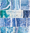
Details of transversal (B–D,J) and longitudinal (A,E–I) sections showing the seed-coat in the immature (E–J) and mature seed (A–D) of Myrcianthes pungens (A–D), Eugenia myrcianthes (E), E. longipedunculata (F), E. uniflora (G), E. pyriformis (H), E. involucrata (I) and E. gracillima (J). (A) Hilar region evidencing hypostase with cells of phenolic content and exotesta constituted by macroesclereids (note the aerenchymatic mesotesta). (B–D) Seed-coat in perichalaza [pre-raphe – (B) and anti-raphe – (C)], and in the testa (D), respectively. Note the thick-walled tabular obliquely elongated exotestal cells, the crushed aerenchymatous mesotesta and the cells of phenolic content in the perichalaza (*). (E) Seed-coat in pachychalaza. Note the thin-walled tabular obliquely elongated exotestal cells, the multiplicative mesotesta (outer aerenchymatic), inner and endotesta cells with phenolic content. (F,G) Seed-coat in testa and pachychalaza. Note the thin-walled obliquely elongated tabular exotestal cells, the aerenchymatic mesotesta and endotesta crushing. (H,I) Seed-coat in testa. Note the obliquely elongated exotestal cells and the aerenchymatic outer mesotesta. (J) Seed-coat in rapheal region. Note the remains of the nucellus, the hypostase and the rapheal vascular bundle. Ct, cotyledonous; Eb, embryo; Es, endotesta; Ex, exotesta; Hl, hilum; Hy, hypostase; Im, inner mesotesta; Ms, mesotesta; Np, nucellus proliferation; Om, outer mesotesta; Pc, pericarp; Ph, perichalaza; Pt, pachychalazal integument; Rb, rapheal vascular bundle; Sc, secretory cavity; Ts, testa; Vp, pachychalazal vascular bundle; ▶, intercellular space. Scale bars = 50 μm (D,F–H), 100 μm (B,C), 200 μm (A,I,J), 500 μm (E).
FIGURE 4
FIGURE 4
Details of longitudinal sections showing the pachychalazal (A,B,D–F,H) and testal (C,G) seed-coat in the immature (A–C) and mature seed (D–H) Eugenia subterminalis (A), E. modesta (B), E. goiapabana (C), E. arenosa (D,E), E. myrcianthes (F), E. brasiliensis (G), and E. acutata (H). (A,B) Note in photo (A) the inner mesotesta with cells stretched longitudinally following the vascular tissue and in photo (B) sclereid groups. (C) Note that the exotesta cells extend radially and are arranged in a palisade. (D–F) Note the sclerified inner mesotesta (inner mesotestal seed). (G,H) Note in photo (G) the thin-walled exotestal cells and the most cells in the mesotesta and endotesta with phenolic content (non-lignified seed-coat) and in photo (H) groups of sclereids in outer mesotesta (outer mesotestal seed). Eb, embryo; Es, endotesta; Ex, exotesta; Im, inner mesotesta; Ms, mesotesta; Om, outer mesotesta; Pc, pericarp; Sd, sclereids; Vp, pachychalazal vascular bundle; ▶, intercellular space. Scale bars = 25 μm (A), 100 μm (B,C), 200 μm (D–G).
FIGURE 5
FIGURE 5
Details of longitudinal sections of mature seed showing the pachychalazal (A–C,F–J) and testal (D,K–M) seed-coat of Eugenia subterminalis (A), E. excelsa (B), E. florida (C), E. modesta (D), E. paracauana (E), E. bahiensis (F), E. hirta (G), E. leptoclada (H), E. neoverrucosa (I), E. flavescens (J), E. batingabranca (K), E. egensis (L), and E. hiemalis (M). Note the exotesta constituted by a palisade of radially elongated lignified macroesclereids (A–C,E–L). In photos (D,M) these cells are cuboidal in shape. Note the exotestal seed in photos (A,D,E,H,I,K,L), exomesotestal in photos (B,C) and testal in photos (F,G). Eb, embryo; Es, endotesta; Ex, exotesta; Im, inner mesotesta; Ms, mesotesta; Om, outer mesotesta; Pc, pericarp; Vp, pachychalazal vascular bundle; ▶, intercellular space. Scale bars = 25 μm (M), 50 μm (G,L), 100 μm (B–D,F,H,J,K), 200 μm (I), 400 μm (A).
FIGURE 6
FIGURE 6
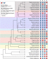
Eugeniinae phylogeny with species character states of ovule: curvature (A), integuments number (B), outer integument layers (C), and inner integument layers (D) (formula image Eugenia sect. Pseudeugenia;formula image E. sect. Hexachlamys;formula image E. sect. Eugenia;formula image E. sect. Pilothecium;formula image E. sect. Phyllocalyx;formula image E. sect. Schizocalomyrtus;formula image E. sect. Jossinia;formula image E. sect. Racemosae;formula image E. sect. Speciosae;formula image E. sect. Umbellatae).
FIGURE 7
FIGURE 7
Eugeniinae phylogeny with species character states of mature seed: exotestal cell shape (A), exotesta cell wall (B), exotesta surface (C), mesotesta (D), outer and/or inner mesotesta (E), endotesta lignification (F), mature seed coat (G), and chalaza (H) (formula image Eugenia sect. Pseudeugenia;formula image E. sect. Hexachlamys;formula image E. sect. Eugenia;formula image E. sect. Pilothecium;formula image E. sect. Excelsae;formula image E. sect. Phyllocalyx;formula image E. sect. Schizocalomyrtus;formula image E. sect. Jossinia;formula image E. sect. Racemosae;formula image E. sect. Speciosae;formula image E. sect. Umbellatae).

References

    1. Anderson L. C. (1963). Studies on Petradoria (Compositae): Anatomy, cytology, taxonomy. Trans. Kansas Acad. Sci. 66 632–684. 10.2307/3626813 - DOI
    1. Andrade R. N. B., Ferreira A. G. (2000). Germinação e armazenamento de sementes de uvaia (Eugenia pyriformis Camb.) Myrtaceae. Rev. Bras. Sem. 22 118–125. 10.17801/0101-3122/rbs.v22n2p118-125 - DOI
    1. Beech E., Rivers M., Oldfield S., Smith P. P. (2017). Global tree search: The first complete global database of tree species and country distributions. J. Sustain. For. 36 454–489. 10.1080/10549811.2017.1310049 - DOI
    1. Berg O. (1855-1856). Revisio Myrtacearum Americae. Linnaea 27 1–472.
    1. Berg O. (1857). “Myrtaceae,” in Flora brasiliensis, Vol. 14 ed. von Martius C. F. P. (Munich: Monachii et Lipsiae; ), 1–468. 10.5962/bhl.title.454 - DOI

LinkOut - more resources