. 2022 Oct 5:9:989809.
doi: 10.3389/fmolb.2022.989809. eCollection 2022.

Smooth muscle Acid-sensing ion channel 1a as a therapeutic target to reverse hypoxic pulmonary hypertension

Affiliations

Smooth muscle Acid-sensing ion channel 1a as a therapeutic target to reverse hypoxic pulmonary hypertension

Selina M Garcia et al. Front Mol Biosci. .

Abstract

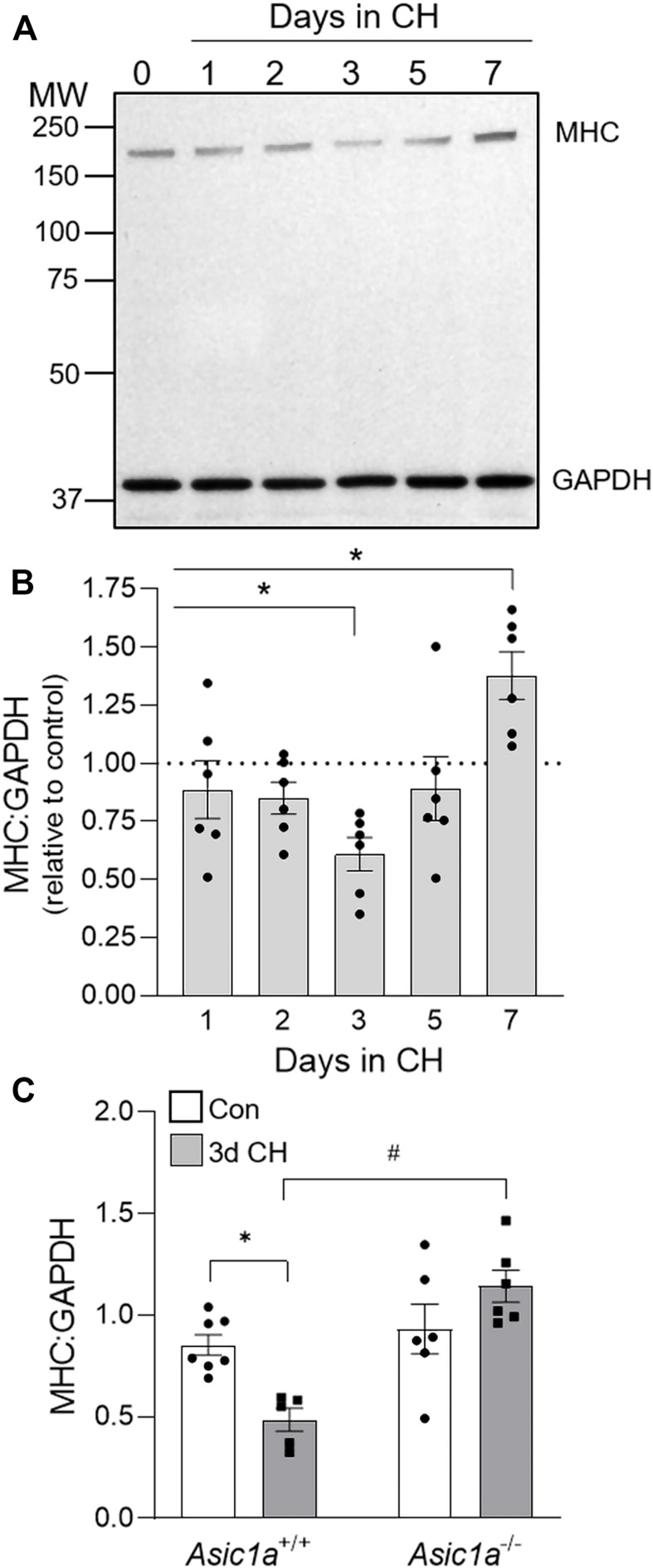
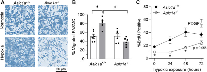
Acid-sensing ion channel 1a (ASIC1a) is a voltage-independent, non-selective cation channel that conducts both Na+ and Ca2+. Activation of ASIC1a elicits plasma membrane depolarization and stimulates intracellular Ca2+-dependent signaling pathways in multiple cell types, including vascular smooth muscle (SM) and endothelial cells (ECs). Previous studies have shown that increases in pulmonary vascular resistance accompanying chronic hypoxia (CH)-induced pulmonary hypertension requires ASIC1a to elicit enhanced pulmonary vasoconstriction and vascular remodeling. Both SM and EC dysfunction drive these processes; however, the involvement of ASIC1a within these different cell types is unknown. Using the Cre-LoxP system to generate cell-type-specific Asic1a knockout mice, we tested the hypothesis that SM-Asic1a contributes to CH-induced pulmonary hypertension and vascular remodeling, whereas EC-Asic1a opposes the development of CH-induced pulmonary hypertension. The severity of pulmonary hypertension was not altered in mice with specific deletion of EC-Asic1a (TekCre-Asic1a fl/fl). However, similar to global Asic1a knockout (Asic1a -/-) mice, mice with specific deletion of SM-Asic1a (MHCCreER-Asic1a fl/fl) were protected from the development of CH-induced pulmonary hypertension and right heart hypertrophy. Furthermore, pulmonary hypertension was reversed when deletion of SM-Asic1a was initiated in conditional MHCCreER-Asic1a fl/fl mice with established pulmonary hypertension. CH-induced vascular remodeling was also significantly attenuated in pulmonary arteries from MHCCreER-Asic1a fl/fl mice. These findings were additionally supported by decreased CH-induced proliferation and migration of pulmonary arterial smooth muscle cells (PASMCs) from Asic1a -/- mice. Together these data demonstrate that SM-, but not EC-Asic1a contributes to CH-induced pulmonary hypertension and vascular remodeling. Furthermore, these studies provide evidence for the therapeutic potential of ASIC1a inhibition to reverse pulmonary hypertension.

Keywords: endothelium; migration; proliferation; right heart hypertrophy; vascular remodeling.


Conflict of interest statement

The authors declare that the research was conducted in the absence of any commercial or financial relationships that could be construed as a potential conflict of interest.

Figures

FIGURE 1
FIGURE 1
CH-induced proliferation of vascular cells is ASIC1a dependent. (A) Representative immunofluorescence images showing Ki-67 (red), α-SMA (green, top row), CD31 (green, bottom row), and To-Pro-3 (blue) in small pulmonary arteries (<100 µm) from Asic1a +/+ mice and (B) summary data showing the percent Ki-67 positive pulmonary arterial endothelial cells (PAECs, blue circles) and pulmonary arterial smooth muscle cells (PASMCs, orange squares) under control conditions or following exposure to CH (3-, 7-, or 28-days). n = 3 animals per group (∼20 vessels were averaged for each animal); analyzed as one-way ANOVA for each cell type and individual groups compared with Šídák’s multiple comparisons tests. Summary data showing percent Ki-67 positive (C) PAECs and (D) PASMCs in small pulmonary arteries (<100 µm) from Asic1a +/+ and Asic1a −/- mice under control conditions or following 3-days CH exposure. n = five to eight animals (∼15 vessels were averaged for each animal); analyzed by two-way ANOVA. Significant interactions between the individual groups (p < 0.0001 for both PAECs and PASMCs) were compared with Šídák’s multiple comparisons tests; *p < 0.05 vs. control; #p < 0.05 vs. corresponding 3-days CH; ns = not significant.
FIGURE 2
FIGURE 2
CH-induced loss of contractile protein, MHC, is ASIC1a dependent. (A) Representative western blot and (B) the effect of CH exposure (1–7 days) on MHC to GAPDH protein expression in whole lung homogenates in Asic1a +/+ animals. The scanned image of the film was converted to greyscale and adjusted for brightness/contrast. n = 6/group; analyzed by one-way ANOVA and individual groups compared with Šídák’s multiple comparisons tests. (C) Summary data for MHC to GAPDH in whole lungs from Asic1a +/+ and Asic1a −/- mice under control conditions or following 3-days CH exposure. n = 5-7 animals/group; analyzed by two-way ANOVA. Significant interactions between the individual groups (p = 0.0022) were compared with the Šídák’s multiple comparisons tests; *p < 0.05 vs. control; #p < 0.05 vs. Asic1a +/+ mice.
FIGURE 3
FIGURE 3
Characterization of transgenic mice. (A) Representative immunofluorescence for ASIC1 (red), CD31 (green), and SMA (blue) in pulmonary arteries from lung sections of Asic1a fl/fl (TAM), TekCre-Asic1a fl/fl, and MHCCreER-Asic1a fl/fl mice treated with vehicle or tamoxifen (TAM). White arrowheads show punctate immunofluorescence of ASIC1 in EC or SM. The dotted line in each image indicates the line profile shown in (B) of relative fluorescence units across the artery wall from lumen to adventitia. Summary analysis of ASIC1 expression in either (C) EC or (D) SM of pulmonary arteries from lung sections. n = 5 animals per group; analyzed by one-way ANOVA and individual groups compared with Šídák’s multiple comparisons tests. (E) PCR of tail DNA showing Cre-mediated LoxP recombination and excision of targeted Asic1a in the same MHCCreER-Asic1a fl/fl mouse before (-) and after (+) tamoxifen (TAM) treatment. (F) Asic1a mRNA expression in brain tissue and intrapulmonary arteries (PA) in MHCCreER-Asic1a fl/fl mice with vehicle (-) or TAM (+). Scanned images of the blots were inverted and adjusted for brightness/contrast.
FIGURE 4
FIGURE 4
SM-specific knockout of Asic1a protects against development of pulmonary hypertension and reverses established hypoxic pulmonary hypertension. (A) Experimental design showing treatments of no TAM, preventative (pTAM, administered before CH), and therapeutic (tTAM, administered following established pulmonary hypertension). (B) Right ventricular systolic pressure (RVSP, mmHg) and (C) Fulton’s Index (RV/LV + S) in Asic1a +/+, Asic1a −/-, Asic1a fl/fl (pTAM), TekCre-Asic1a fl/fl, MHCCreER-Asic1a fl/fl, MHCCreER-Asic1a fl/fl (pTAM), or MHCCreER-Asic1a fl/fl (tTAM) mice under control conditions (white bars, circles) or following 6 weeks CH (grey bars, squares). SM-Asic1a knockout was induced by treatment with tamoxifen as a preventive (pTAM; before exposure to CH) or therapeutic approach (tTAM; following establishment of CH-induced pulmonary hypertension). n = 5-8/group; analyzed by two-way ANOVA. Significant interactions between the individual groups (p < 0.0001 for both RVSP and RV/LV + S) were compared with Šídák’s multiple comparisons tests; *p < 0.05 vs. control; #p < 0.05 vs. respective genetic control.
FIGURE 5
FIGURE 5
SM ASIC1a contributes to vascular remodeling following CH. (A) Representative SMA immunofluorescence (black) images of small pulmonary arteries in lung sections from TekCre-Asic1a fl/fl, MHCCreER-Asic1a fl/fl, MHCCreER-Asic1a fl/fl (pTAM), or MHCCreER-Asic1a fl/fl (tTAM) mice under control conditions (white bars) or following 6 weeks CH (filled bars). Fluorescence images were digitally inverted to provide better contrast and visibility of immunofluorescence. (B) Percent muscularization calculated as percent thresholded SMA area divided by total arterial area based on arterial diameter: <25 μm (n = ∼30 vessels from four animals/group), 25–50 μm (n = ∼100 vessels from four animals/group), or 50–100 μm (n = ∼50 vessels from four animals/group); analyzed by two-way ANOVA. Significant interactions between the individual groups (p < 0.0001 for all vessel diameter ranges) compared with Šídák’s multiple comparisons tests. *p < 0.05 vs. control; #p < 0.05 vs. (-) TAM; and τ p < 0.05 pTAM vs. tTAM.
FIGURE 6
FIGURE 6
SM ASIC1a contributes to CH-induced PASMC proliferation. (A) Representative immunofluorescence images showing Ki-67 (red), α-SMA (green), and Sytox (blue) in small pulmonary arteries (<100 µm) from control and 3 days CH MHCCreER-Asic1a fl/fl mice following treatment with vehicle or tamoxifen (TAM). (B) Summary data for percent Ki-67 positive cells in endothelial cells (EC, blue) and smooth muscle cells (SMC, orange). Summary data for control mice are not shown since there were no detectable Ki-67 positive cells. n = 75 vessels from five animals (15 vessels each) per group; analyzed by unpaired t-test; #p < 0.05 vs. (-) TAM 3-days CH; ns = not significant.
FIGURE 7
FIGURE 7
ASIC1a contributes to mPASMC migration and proliferation. (A) Representative brightfield images of coomassie-stained mPASMC and (B) summary data for % of migrated mPASMC from Asic1a +/+ and Asic1a −/- mice exposed to normoxia (5% CO2, 95% air; orange circles) or hypoxia (5% CO2, 2% O2; blue squares) for 24 h n = 6 animals/group; *p < 0.05 vs. hypoxia and #p < 0.05 Asic1a +/+; analyzed by two-way ANOVA. Significant interactions between the individual groups (p = 0.0003) compared using Šídák’s multiple comparisons tests. (C) Flow cytometry analysis of percent of mPASMC from Asic1a +/+ and Asic1a −/- mice with BrdU incorporation under normoxia (0 h; 5% CO2, 95% air) or hypoxia (5% CO2, 2% O2) for 24, 48, or 72 h. Normoxic mPASMC from Asic1a +/+ were incubated with PDGF-BB (20 ng/ml) for 72 h as a positive control. n = five to eight animals/group, analyzed by unpaired t-tests at each time point, *p < 0.05.
FIGURE 8
FIGURE 8
ASIC1 contributes to human PASMC migration and proliferation. (A) Representative Western blots of ASIC1 (predicted ∼65 kDa) and corresponding coomassie-stained blot and (B) summary data showing ASIC1 expression (normalized to entire lane on coomassie-stained blot) in human PASMC (hPASMC) exposed to normoxia (norm, 5% CO2, 95% air; white bars/circles) or 12 h hypoxia (hyp, 5% CO2, 2% O2; grey bars/squares). Scanned images of the film were converted to greyscale and adjusted for brightness/contrast. (C) Summary data showing Western blot analysis for biotinylated (plasma membrane) and cytosolic ASIC1 protein expression in hPASMC exposed to normoxia or hypoxia. (D) Brightfield images of hPASMC immediately following the scratch (baseline time 0; top row of images) or 12 h post-exposure (bottom row of images) to normoxia (5% CO2, 95% air; orange circles) or hypoxia (5% CO2, 2% O2; blue squares) and (E) summary data showing percent reinvasion of hPASMC into the wounded area in the presence of vehicle, amiloride or PcTX1. n = 8-11/group; analyzed by two-way ANOVA. Significant interactions between the individual groups (p = 0.0032) were compared using Šídák’s multiple comparisons tests. (F) increase in the number of cells after 24 h of normoxia or hypoxia in the presence of vehicle, amiloride, or PcTX1. hPASMC were initially plated at a density of 15,000 cells/well. n = 6/group; analyzed by one-way ANOVA and individual groups compared using Šídák’s multiple comparisons tests. *p < 0.05 vs. normoxic group; #p < 0.05 vs. corresponding vehicle group.

References

    1. Akanji O., Weinzierl N., Schubert R., Schilling L. (2019). Acid sensing ion channels in rat cerebral arteries: Probing the expression pattern and vasomotor activity. Life Sci. 227, 193–200. 10.1016/j.lfs.2019.04.054 - DOI - PubMed
    1. Bierer R., Nitta C. H., Friedman J., Codianni S., de Frutos S., Dominguez-Bautista J. A., et al. (2011). NFATc3 is required for chronic hypoxia-induced pulmonary hypertension in adult and neonatal mice. Am. J. Physiol. Lung Cell. Mol. Physiol. 301 (6), L872–L880. 10.1152/ajplung.00405.2010 - DOI - PMC - PubMed
    1. Bonnet S., Michelakis E. D., Porter C. J., Andrade-Navarro M. A., Thébaud B., Bonnet S., et al. (2006). An abnormal mitochondrial-hypoxia inducible factor-1alpha-kv channel pathway disrupts oxygen sensing and triggers pulmonary arterial hypertension in fawn hooded rats: Similarities to human pulmonary arterial hypertension. Circulation 113 (22), 2630–2641. 10.1161/circulationaha.105.609008 - DOI - PubMed
    1. Broughton B. R., Jernigan N. L., Norton C. E., Walker B. R., Resta T. C. (2010). Chronic hypoxia augments depolarization-induced Ca2+ sensitization in pulmonary vascular smooth muscle through superoxide-dependent stimulation of RhoA. Am. J. Physiol. Lung Cell. Mol. Physiol. 298 (2), L232–L242. 10.1152/ajplung.00276.2009 - DOI - PMC - PubMed
    1. Budhiraja R., Tuder R. M., Hassoun P. M. (2004). Endothelial dysfunction in pulmonary hypertension. Circulation 109 (2), 159–165. 10.1161/01.CIR.0000102381.57477.50 - DOI - PubMed