. 2022 Oct 7:13:1033367.
doi: 10.3389/fimmu.2022.1033367. eCollection 2022.

GLDC mitigated by miR-30e regulates cell proliferation and tumor immune infiltration in TNBC

Affiliations

GLDC mitigated by miR-30e regulates cell proliferation and tumor immune infiltration in TNBC

Huaying Xie et al. Front Immunol. .

Abstract

Background: TNBC, whose clinical prognosis is poorer than other subgroups of breast cancer, is a malignant tumor characterized by lack of estrogen receptors, progesterone hormone receptors, and HER2 overexpression. Due to the lack of specific targeted drugs, it is crucial to identify critical factors involved in regulating the progression of TNBC.

Methods: We analyzed the expression profiles of TNBC in TCGA and the prognoses values of GLDC. Correlations of GLDC and tumor immune infiltration were also identified. CCK8 and BrdU incorporation assays were utilized to determine cell proliferation. The mRNA and protein levels were examined by using Real-time PCR and Western blot analysis.

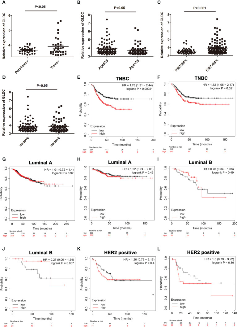
Results: In the present study, we analyzed the mRNA expression profiles of TNBC in TCGA and found that GLDC, a key enzyme in glycine cleavage system, was significantly up-regulated in TNBC tissues and higher expression of GLDC was correlated with a worse prognosis in TNBC. Moreover, the expression of GLDC was negatively correlated with macrophage and monocyte and positively correlated with activated CD4 T cell and type 2 T helper cell in TNBC. Overexpression of GLDC facilitated the proliferation of TNBC cells, whereas GLDC knockdown had the opposite effects. Additionally, miR-30e acts as a functional upstream regulator of GLDC and the inhibitory effects of miR-30e on cell proliferation were mitigated by the reintroduction of GLDC.

Conclusions: These results imply that miR-30e-depressed GLDC acts as a tumor suppressive pathway in TNBC and provides potential targets for the treatment of TNBC.

Keywords: GLDC; TNBC; miR-30e; proliferation; tumor immune.


Conflict of interest statement

The authors declare that the research was conducted in the absence of any commercial or financial relationships that could be construed as a potential conflict of interest. The reviewer GL declared a shared parent affiliation with the authors HX, TY, YD, SX, YK, LY, LZ to the handling editor at the time of the review.

Figures

Figure 1
Figure 1
Identification of differentially expressed genes in TNBC tissues. (A) The overall distribution of differentially expressed genes by volcano plots. (B) The heat map of 100 differential genes. (C): The biological processes, molecular function, and cellular component enriched by GO analysis. (D) The enriched KEGG pathways. (E, F) The expression of GLDC was significantly up-regulated in TNBC tissues compared with normal breast tissues by analyzing TCGA (E) and the UALCAN database (F).
Figure 2
Figure 2
The expression profiles of GLDC and its prognoses value in TNBC tissues. (A) The expression of GLDC was significantly higher in the TNBC tissues compared with their adjacent normal tissues (n=33). (B) The expression of GLDC is higher in the patients younger than 55 years (age ≤ 55) than the patients older than 55 years (age>55). (C) GLDC expression in the patients with ki67 ≤ 30% was less than that with ki67>30%. (D) No significant differences were detected on the expression of GLDC between positive lymph nodes and no positive lymph nodes. (E, F) TNBC patients with high levels of GLDC were associated with the shorter RFS (E) and the worse DMFS (F). (G, H) There were no significant differences on the RFS (G) and DMFS (H) between the low levels of GLDC and the high levels of GLDC in Luminal A breast cancer. (I, J) No significant differences were detected on the RFS (I) and DMFS (J) between the two groups in Luminal B breast cancer. (K, L) There were no significant differences on the RFS (K) and DMFS (L) between the two groups in HER2 positive breast cancer.
Figure 3
Figure 3
The relationship between GLDC and tumor immune infiltration. (A) The infiltration abundance of immune cells based on the levels of GLDC in TNBC patients. (B) High levels of macrophage, neutrophil, CD56 bright natural killer cell and plasmacytoid dendritic cell and low levels of activated CD4 T cell, central memory CD4 T cell, and type 2 T helper cell were acquired in the group with low expression of GLDC in TNBC. (C) The expression of GLDC was negatively correlated with macrophage, plasmacytoid dendritic cell, and monocyte and positively correlated with activated CD4 T cell and type 2 T helper cell. (D) No significant correlations were observed between GLDC and the four immune checkpoint molecules in TNBC. *P < 0.05, **P < 0.01, ***P < 0.001, and ns indicates P>0.05.
Figure 4
Figure 4
Cell proliferation is promoted by GLDC in TNBC cells. (A) Real-time PCR was utilized to determine the overexpression efficiency. (B, C) Overexpression of GLDC increased cell viability (B) and facilitated BrdU incorporation (C). (D, E) GLDC expression was positively correlated with the expression of PCNA (D) and MKI67 (E) in TNBC tissues. (F) Knockdown efficiency was determined by Real-time PCR. (G, H) Knockdown of GLDC mitigated cell viability (G) and repressed BrdU incorporation (H). *P < 0.05.
Figure 5
Figure 5
GLDC is negatively regulated by miR-30e in TNBC. (A) There was no significant difference on the promoter methylation level of GLDC between TNBC and normal breast tissues. (B) The schematic diagram of the protocol utilized for identifying the potential functional regulator of GLDC. (C) The expression of GLDC was negatively correlated with miR-30e expression in the same TNBC tissues. (D) The potential binding site of miR-30e conserved in the 3′UTR of GLDC. (E) Treatment with miR-30e led to the decreased luciferase activity of the WT (wild type) group. (F, G) MiR-30e significantly depressed the mRNA (F) and protein (G) expression of GLDC in TNBC cells. (H, I) Treatment with anti-miR-30e led to the increased expression of GLDC at both mRNA (H) and protein (I) levels. *P < 0.05.
Figure 6
Figure 6
The inhibitory effects of miR-30e on cell proliferation are mitigated by GLDC. (A) The expression of miR-30e was negatively correlated with PCNA expression in the same TNBC tissues. (B) High levels of miR-30e were associated with better overall survival. (C, D) Treatment with anti-miR-30e increased cell viability (C) * indicates p < 0.05 compared with anti-NC+siControl group, # indicates p < 0.05 compared with anti-miR-30e+siControl group and promoted BrdU incorporation. (D), which was attenuated by the knockdown of GLDC. (E, F): Restoration of GLDC depressed the regulatory effects of miR-30e on cell viability (E) * indicates p < 0.05 compared with NC+Vector group, # indicates p < 0.05 compared with miR-30e+Vector group and BrdU incorporation (F).

References

    1. Siegel RL, Miller KD, Fuchs HE, Jemal A. Cancer statistics, 2021. CA Cancer J Clin (2021) 71(1):7–33. doi: 10.3322/caac.21654 - DOI - PubMed
    1. Advani P, Cornell L, Chumsri S, Moreno-Aspitia A. Dual HER2 blockade in the neoadjuvant and adjuvant treatment of HER2-positive breast cancer. Breast Cancer (Dove Med Press) (2015) 7:321–35. doi: 10.2147/BCTT.S90627 - DOI - PMC - PubMed
    1. Maddocks ODK, Athineos D, Cheung EC, Lee P, Zhang T, van den Broek NJF, et al. . Modulating the therapeutic response of tumours to dietary serine and glycine starvation. Nature (2017) 544(7650):372–6. doi: 10.1038/nature22056 - DOI - PubMed
    1. Liu R, Zeng LW, Gong R, Yuan F, Shu HB, Li S. mTORC1 activity regulates post-translational modifications of glycine decarboxylase to modulate glycine metabolism and tumorigenesis. Nat Commun (2021) 12(1):4227. doi: 10.1038/s41467-021-24321-3 - DOI - PMC - PubMed
    1. Alptekin A, Ye B, Yu Y, Poole CJ, van Riggelen J, Zha Y, et al. . Glycine decarboxylase is a transcriptional target of MYCN required for neuroblastoma cell proliferation and tumorigenicity. Oncogene (2019) 38(50):7504–20. doi: 10.1038/s41388-019-0967-3 - DOI - PMC - PubMed

Publication types