

"Rogue" neutrophil-subset [DEspR+CD11b+/CD66b+] immunotype is an actionable therapeutic target for neutrophilic inflammation-mediated tissue injury - studies in human, macaque and rat LPS-inflammation models
- PMID: 36275710
- PMCID: PMC9581391
- DOI: 10.3389/fimmu.2022.1008390
"Rogue" neutrophil-subset [DEspR+CD11b+/CD66b+] immunotype is an actionable therapeutic target for neutrophilic inflammation-mediated tissue injury - studies in human, macaque and rat LPS-inflammation models
Abstract
Background and objective: The correlation (Rs > 0.7) of neutrophils expressing the dual endothelin1/signal peptide receptor (DEspR+CD11b+/CD66b+) with severity of hypoxemia (SF-ratio) and multi-organ failure (SOFA-score) in patients with acute respiratory distress syndrome (ARDS) suggest the hypothesis that the DEspR+ neutrophil-subset is an actionable therapeutic target in ARDS. To test this hypothesis, we conducted in vivo studies to validate DEspR+ neutrophil-subset as therapeutic target and test efficacy of DEspR-inhibition in acute neutrophilic hyperinflammation models.
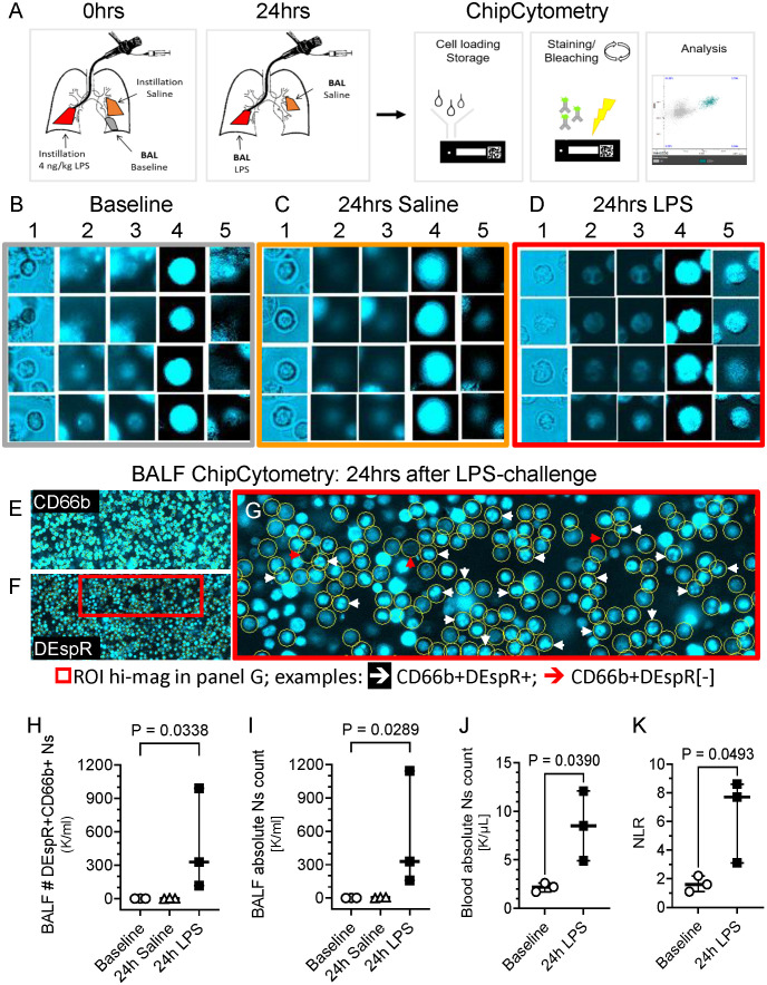
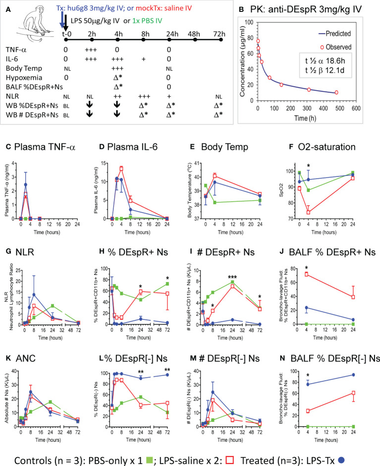
Methods: We performed tests in lipopolysaccharide (LPS)-induced acute neutrophilic inflammation in three species - human, rhesus macaque, rat - with increasing dose-dependent severity. We measured DEspR+CD66b+ neutrophils in bronchoalveolar lavage fluid (BALF) in healthy volunteers (HVs) 24-hours after segmental LPS-challenge by ChipCytometry, and DEspR+CD11b+ neutrophils in whole blood and BALF in an LPS-induced transient acute lung injury (ALI) model in macaques. We determined anti-DEspR antibody efficacy in vivo in LPS-ALI macaque model and in high-mortality LPS-induced encephalopathy in hypertensive rats.
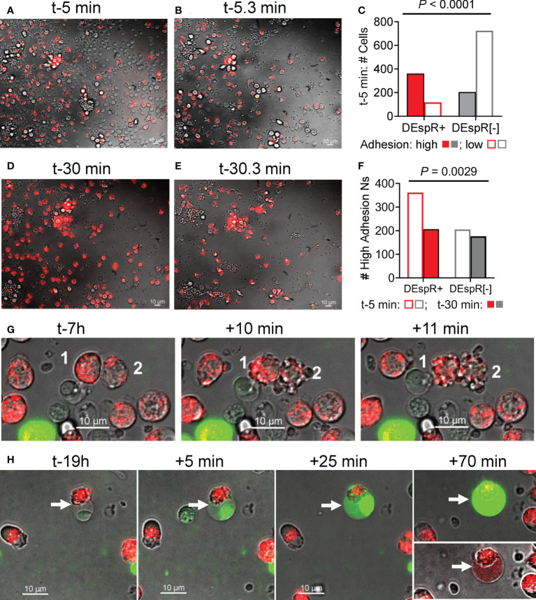
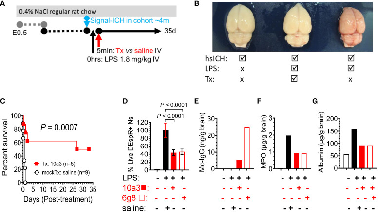
Results: ChipCytometry detected increased BALF total neutrophil and DEspR+CD66b+ neutrophil counts after segmental LPS-challenge compared to baseline (P =0.034), as well as increased peripheral neutrophil counts and neutrophil-lymphocyte ratio (NLR) compared to pre-LPS level (P <0.05). In the LPS-ALI macaque model, flow cytometry detected increased DEspR+ and DEspR[-] neutrophils in BALF, which was associated with moderate-severe hypoxemia. After determining pharmacokinetics of single-dose anti-DEspR[hu6g8] antibody, one-time pre-LPS anti-DEspR treatment reduced hypoxemia (P =0.03) and neutrophil influx into BALF (P =0.0001) in LPS-ALI vs vehicle mock-treated LPS-ALI macaques. Ex vivo live cell imaging of macaque neutrophils detected greater "intrinsic adhesion to hard-surface" in DEspR+ vs DEspR[-] neutrophils (P <0.001). Anti-DEspR[hu6g8] antibody abrogated intrinsic high adhesion in DEspR+ neutrophils, but not in DEspR[-] neutrophils (P <0.001). In the LPS-encephalopathy rat model, anti-DEspR[10a3] antibody treatment increased median survival (P =0.0007) and exhibited brain target engagement and bioeffects.
Conclusion: Detection of increased DEspR+ neutrophil-subset in human BALF after segmental LPS-challenge supports the correlation of circulating DEspR+ neutrophil counts with severity measure (SOFA-score) in ARDS. Efficacy and safety of targeted inhibition of DEspR+CD11b+ neutrophil-subset in LPS-induced transient-ALI and high-mortality encephalopathy models identify a potential therapeutic target for neutrophil-mediated secondary tissue injury.
Keywords: DEspR; LPS-acute inflammation tissue injury models; LPS-brain encephalopathy; acute lung injury; neutrophil subset; segmental LPS challenge.
Copyright © 2022 Carstensen, Müller, Tan, Pasion, Hohlfeld, Herrera and Ruiz-Opazo.
Conflict of interest statement
Boston University holds awarded and pending patents on DEspR. VH and NR-O are co-inventors filed by Boston University. These patents comprise the option granted for exclusive license to NControl Therapeutics, Inc. VH, NR-O are scientific co-founders of NControl Therapeutics, and paid consultants with equity in NControl Therapeutics, Inc. NControl Therapeutics was not involved in the design, conception, data interpretation, or manuscript preparation. The remaining authors declare that the research was conducted in the absence of any commercial or financial relationships that could be construed as a potential conflict of interest.
Figures
Similar articles
-
A targetable 'rogue' neutrophil-subset, [CD11b+DEspR+] immunotype, is associated with severity and mortality in acute respiratory distress syndrome (ARDS) and COVID-19-ARDS.Sci Rep. 2022 Apr 4;12(1):5583. doi: 10.1038/s41598-022-09343-1. Sci Rep. 2022. PMID: 35379853 Free PMC article.
-
Increased Neutrophil-Subset Associated With Severity/Mortality In ARDS And COVID19-ARDS Expresses The Dual Endothelin-1/VEGFsignal-Peptide Receptor (DEspR): An Actionable Therapeutic Target.Res Sq [Preprint]. 2021 Sep 13:rs.3.rs-846250. doi: 10.21203/rs.3.rs-846250/v1. Res Sq. 2021. PMID: 34545358 Free PMC article. Preprint.
-
Anti-DEspR antibody treatment improves survival and reduces neurologic deficits in a hypertensive, spontaneous intracerebral hemorrhage (hsICH) rat model.Sci Rep. 2023 Feb 15;13(1):2703. doi: 10.1038/s41598-023-28149-3. Sci Rep. 2023. PMID: 36792616 Free PMC article.
-
Drug repurposing of cyclin-dependent kinase inhibitors for neutrophilic acute respiratory distress syndrome and psoriasis.J Adv Res. 2025 Jun;72:485-500. doi: 10.1016/j.jare.2024.07.026. Epub 2024 Jul 31. J Adv Res. 2025. PMID: 39089617 Free PMC article. Review.
-
Review: Toll-like receptors and NOD-like receptors in pulmonary antibacterial immunity.Innate Immun. 2010 Jun;16(3):201-10. doi: 10.1177/1753425910366058. Epub 2010 Apr 23. Innate Immun. 2010. PMID: 20418253 Free PMC article. Review.
Cited by
-
Circulating neutrophil extracellular trap (NET)-forming 'rogue' neutrophil subset, immunotype [DEspR+CD11b+], mediate multi-organ failure in COVID-19 - an observational study.Res Sq [Preprint]. 2023 Feb 1:rs.3.rs-2479844. doi: 10.21203/rs.3.rs-2479844/v1. Res Sq. 2023. Update in: Transl Med Commun. 2023;8(1):12. doi: 10.1186/s41231-023-00143-x. PMID: 36778407 Free PMC article. Updated. Preprint.
-
Circulating neutrophil extracellular trap (NET)-forming 'rogue' neutrophil subset, immunotype [DEspR + CD11b +], mediate multi-organ failure in COVID-19-an observational study.Transl Med Commun. 2023;8(1):12. doi: 10.1186/s41231-023-00143-x. Epub 2023 Apr 18. Transl Med Commun. 2023. PMID: 37096233 Free PMC article.
-
The double-edged role of neutrophil heterogeneity in inflammatory diseases and cancers.MedComm (2020). 2023 Jul 23;4(4):e325. doi: 10.1002/mco2.325. eCollection 2023 Aug. MedComm (2020). 2023. PMID: 37492784 Free PMC article. Review.
-
Circulating neutrophil extracellular trap-forming neutrophils in rheumatoid arthritis exacerbation are majority dual endothelin-1/signal peptide receptor+ subtype.Clin Exp Immunol. 2024 Oct 16;218(2):163-168. doi: 10.1093/cei/uxae072. Clin Exp Immunol. 2024. PMID: 39110036 Free PMC article.
-
Neutrophils in the Spotlight-An Analysis of Neutrophil Function and Phenotype in ARDS.Int J Mol Sci. 2024 Nov 22;25(23):12547. doi: 10.3390/ijms252312547. Int J Mol Sci. 2024. PMID: 39684262 Free PMC article.
References
Publication types
MeSH terms
Substances
Grants and funding
LinkOut - more resources
Full Text Sources
Medical
Research Materials