

Perforated Gallbladder into the Abdominal Wall
- PMID: 36275925
- PMCID: PMC9584729
- DOI: 10.1155/2022/4782539
Perforated Gallbladder into the Abdominal Wall
Abstract
Objective: Perforation of the gallbladder (PG) is a dreaded complication of an acute cholecystitis and is associated with increased morbidity and mortality. Cholecystocutaneous abscess (CCA) is an extremely rare complication. There is usually a history of cholecystolithiasis or neglected chronic gallbladder disease. We report a case of perforated gallbladder into the abdominal wall.
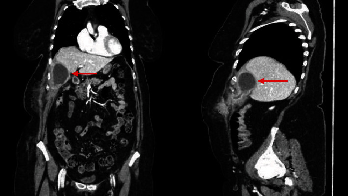
Methods: A 65-year-old female, obese, was admitted to our department complaining of right upper quadrant abdominal pain. The diagnosis of acute cholecystitis was based on the clinical picture, laboratory test, and ultrasound findings. She was treated with oral antibiotics for 10 days and readmitted due to a painful, erythematous mass on the right subcostal region. An abdominal computed tomography showed the presence of a subparietal formation in communication with the gallbladder, and a gallbladder perforation was postulated. The treatment consisted of percutaneous drainage of the abdominal wall abscess followed by laparoscopic cholecystectomy in a two-stage protocol. Anatomical pathology analysis found chronic inflammation and excluded malignancy. The postoperative follow-up was uneventful. Discussion. This case demonstrates a very rare presentation of PG that created an abscess into the muscles of the abdominal wall. This kind of PG is rarely seen due to medicine improvements. When the conditions of the patient are good, rather than perform immediate surgery that could lead to serious complications, we propose a two-stage approach.
Conclusion: CCA is a possible complication of gallbladder's pathology that all surgeons have to know. There is no standard baseline management for this pathology, due to the few numbers of cases and to the differences in the quality of the patients' illness. We suggest a two-stage approach with drainage of the abscess followed by laparoscopic cholecystectomy with abscess debridement.
Copyright © 2022 M. Puglisi et al.
Conflict of interest statement
The authors declare that they have no conflicts of interest.
Figures


References
Publication types
LinkOut - more resources
Full Text Sources
