Review
. 2022 Oct 5:12:1001318.
doi: 10.3389/fonc.2022.1001318. eCollection 2022.

Amino acid metabolism in primary bone sarcomas

Affiliations
Review

Amino acid metabolism in primary bone sarcomas

Jennifer A Jiménez et al. Front Oncol. .

Abstract

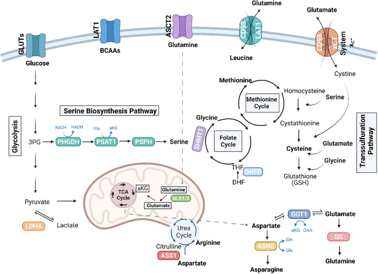
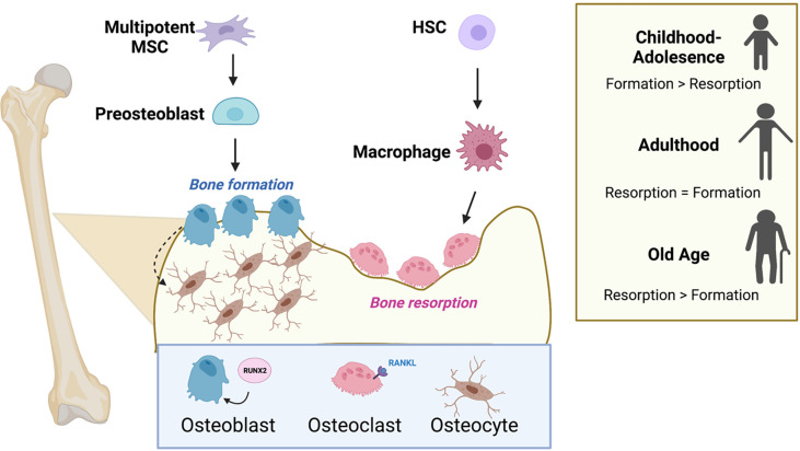
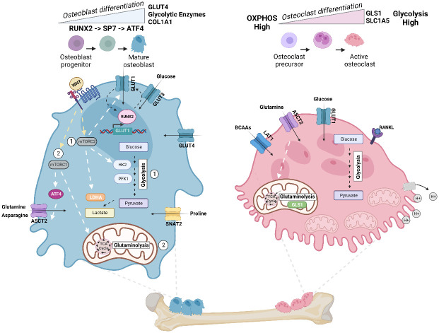
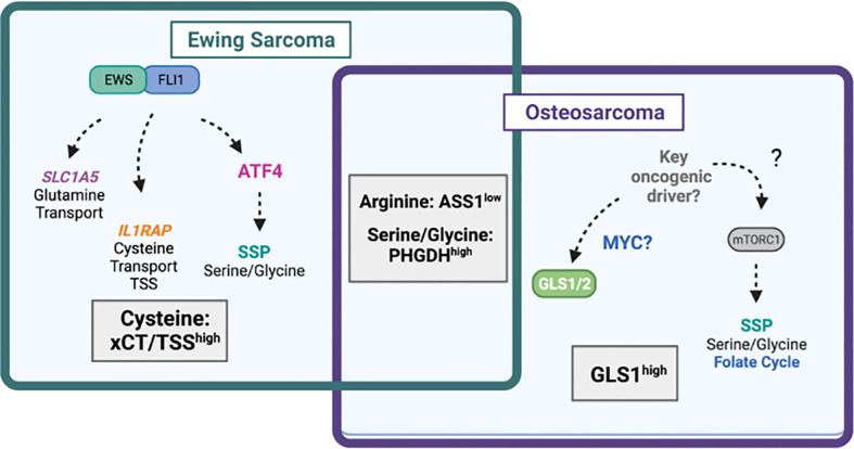
Primary bone sarcomas, including osteosarcoma (OS) and Ewing sarcoma (ES), are aggressive tumors with peak incidence in childhood and adolescence. The intense standard treatment for these patients consists of combined surgery and/or radiation and maximal doses of chemotherapy; a regimen that has not seen improvement in decades. Like other tumor types, ES and OS are characterized by dysregulated cellular metabolism and a rewiring of metabolic pathways to support the biosynthetic demands of malignant growth. Not only are cancer cells characterized by Warburg metabolism, or aerobic glycolysis, but emerging work has revealed a dependence on amino acid metabolism. Aside from incorporation into proteins, amino acids serve critical functions in redox balance, energy homeostasis, and epigenetic maintenance. In this review, we summarize current studies describing the amino acid metabolic requirements of primary bone sarcomas, focusing on OS and ES, and compare these dependencies in the normal bone and malignant tumor contexts. We also examine insights that can be gleaned from other cancers to better understand differential metabolic susceptibilities between primary and metastatic tumor microenvironments. Lastly, we discuss potential metabolic vulnerabilities that may be exploited therapeutically and provide better-targeted treatments to improve the current standard of care.

Keywords: Ewing sarcoma; amino acid metabolism; osteoblast; osteoclast; sarcoma; tumor metabolism.


Conflict of interest statement

CL has received consulting fees from Astellas Pharmaceuticals, Odyssey Therapeutics, and T-Knife Therapeutics, and is an inventor on patents pertaining to Kras regulated metabolic pathways, redox control pathways in pancreatic cancer, and targeting the GOT1-pathway as a therapeutic approach (US Patent No: 2015126580-A1, 05/07/2015; US Patent No: 20190136238, 05/09/2019; International Patent No: WO2013177426-A2, 04/23/2015). The remaining authors declare that the research was conducted in the absence of any commercial or financial relationships that could be construed as a potential conflict of interest.

Figures

Figure 1
Figure 1
Amino acid pathways rewired in cancer. Illustration depicting amino acid pathways linked in an intricate metabolic network. Essential amino acids, including the branched chain amino acids (BCAAs), leucine, isoleucine, and valine, are imported from the extracellular environment, in part via the LAT1 (SLC7A5) transporter. Glutamine (Gln) can be conditionally essential and is imported through ASCT2 (SLC1A5). Glutaminase enzymes (GLS1/2) convert glutamine to glutamate (Glu), which can further be converted to alpha-ketoglutarate (αKG) to enter the TCA cycle. The TCA cycle also generates intermediates, which give rise to non-essential amino acids, including aspartate and arginine. The de novo synthesis of arginine involves the conversion of citrulline to aspartate by argininosuccinate synthetase 1 (ASS1). TCA-cycle derived aspartate is converted to asparagine via ASNS, and GOT1 can convert aspartate to glutamate, which is further converted to glutamine by glutamine synthetase (GS). Serine, another non-essential amino acid, can be imported or synthesized de novo in the serine biosynthesis pathway (SBP), which diverts the 3P-glycerate (3PG) intermediate of glycolysis to generate serine via the enzymes, phosphoglycerate dehydrogenase (PHGDH), phosphoserine aminotransferase (PSAT1), and phosphoserine phosphatase (PSPH). Serine can further be converted to glycine by serine hydroxymethyltransferase (SHMT) (cytosolic SHMT1 isozyme shown above) to generate one-carbon units to fuel the folate cycle and the methionine cycles. Dihydrofolate reductase (DHFR) in the folate cycle catalyzes the reduction of dihydrofolate to tetrahydrofolate (THF). Cysteine is another non-essential amino acid that can be synthesized in the transsulfuration pathway or imported as cystine via the cystine/glutamate antiporter, system xC-. The SBP, transsulfuration pathway, along with ASS1 and glutamine metabolism have been implicated in sarcoma biology. Figure was created with BioRender.com.
Figure 2
Figure 2
Osteoblasts, osteoclasts, and the cycle of bone remodeling. Bone remodeling balances the competing actions of the bone-building osteoblasts and the osteoclasts that promote bone resorption. Osteoblasts and osteocytes originate from common precursor bone marrow mesenchymal stem cells (MSCs), while osteoclasts are multinucleated and originate from hematopoietic stem cells (HSCs) in the bone marrow. Successful differentiation of progenitor cells into osteoblasts and osteoclasts requires the transcription factor, RUNX2 and RANK-ligand (RANKL), respectively. The level of bone formation versus bone resorption is dynamic and changes with age. Formation exceeds resorption during adolescence, and the scale tips the opposite direction favoring resorption in old age. Figure was created with BioRender.com.
Figure 3
Figure 3
Energy metabolism in osteoblast and osteoclast. Metabolic pathways rewired in osteoblast (Left) and osteoclast (Right) during cellular differentiation. Left: In mature osteoblasts, glycolysis accounts for 80% of ATP production. Osteoblast precursors sequentially express the transcription factor RUNX2, followed by Sp7 (osterix (Osx)), and collagen-promoting ATF4 at late stages of differentiation. Illustration depicts the feed-forward mechanism where high intercellular glucose inhibits the proteasomal degradation of RUNX2, and RUNX2 increases the levels of GLUT1 transcription. WNT signaling regulates osteoblast activity and has been shown to (1) activate glycolytic enzymes (HK2 and PFK1), GLUT1, and LDHA via mTORC2 and (2) promote glutaminolysis and TCA cycle activation via mTORC1 and downstream regulation of ATF4 and glutamine transporter ASCT2 (SLC1A5). Import of asparagine via ASCT2 and proline via SNAT2 (SLC38A2) also facilitates osteoblast differentiation. Right: To dissolve bone and degrade collagen, osteoclasts require the generation of protons (H+) by hydrolyzing ATP. Thus, osteoclasts exhibit a significant demand for ATP and contain a high number of mitochondria. ATP demand is met by upregulating both glycolysis and glutaminolysis, and differentiation increases glycolysis and expression of glumaminase 1 (GLS1) and ASCT2. Mid-late stage osteoclast differentiation is also dependent on increased branched chain amino acid (BCAA) pools. Figure was created with BioRender.com.
Figure 4
Figure 4
Amino acid dependencies in Ewing sarcoma and osteosarcoma. Venn-diagram depicting shared and unique amino acid vulnerabilities in Ewing sarcoma and osteosarcoma. The EWS::FLI1 fusion protein has been shown to directly regulate expression of glutamine transporter SLC1A5 (ASCT2) leading to high glutamine import. EWS::FLI1 also regulates expression of IL1RAP and downstream cysteine availability via the transsulfuration pathway (TSS) and xCT (SLC7A11). EWS::FLI1 indirectly regulates the serine biosynthesis pathway (SSP) via ATF4. In osteosarcoma, the SSP also drives tumorgenicity through undefined mechanisms likely involving mTORC1. The mitochondrial enzyme GLS1, which may be under the control of MYC is highly expressed in osteosarcoma and predicts worse survival. Diagram was created with BioRender.com.

Similar articles

Cited by

References

    1. Palmerini E, Righi A, Staals EL. Rare primary malignant bone sarcomas. Cancers (Basel) (2020) 12(11):3092. doi: 10.3390/cancers12113092 - DOI - PMC - PubMed
    1. Molina ER, Chim LK, Barrios S, Ludwig JA, Mikos AG. Modeling the tumor microenvironment and pathogenic signaling in bone sarcoma. Tissue Eng Part B Rev (2020) 26(3):249–71. doi: 10.1089/ten.teb.2019.0302 - DOI - PMC - PubMed
    1. Hadjidakis DJ, Androulakis II. Bone remodeling. Ann N Y Acad Sci (2006) 1092:385–96. doi: 10.1196/annals.1365.035 - DOI - PubMed
    1. Borle AB, Nichols N, Nichols G, Jr. Metabolic studies of bone in vitro. i. normal bone. J Biol Chem (1960) 235:1206–10. doi: 10.1016/S0021-9258(18)69506-7 - DOI - PubMed
    1. Cohn DV, Forscher BK. Aerobic metabolism of glucose by bone. J Biol Chem (1962) 237:615–8. doi: 10.1016/S0021-9258(18)60342-4 - DOI - PubMed

LinkOut - more resources