

Infiltration of the spinal cord and peripheral nerves in multiple myeloma
- PMID: 36276061
- PMCID: PMC9584647
- DOI: 10.3389/fonc.2022.991246
Infiltration of the spinal cord and peripheral nerves in multiple myeloma
Abstract
Background: Multiple myeloma (MM) is a hematological malignancy, and intramedullary spinal cord metastasis is extremely rare.
Methods: Clinical and radiological data were collected from electronic medical records as well as a literature review of reported cases.
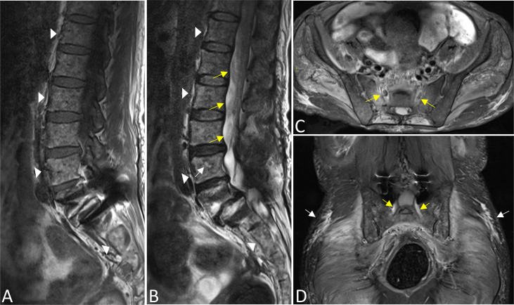
Results: We report a rare case of IgA-LAM stage IIB MM with involvement of the spinal cord and peripheral nervous system. Laboratory studies showed elevated levels of serum β2-macroglobulin and cerebrospinal fluid protein. Electromyography revealed a demyelinating process with motor conduction blocks. On MRI, the lesions of MM bone marrow are characterized as a type of diffuse infiltration. MR neurography demonstrated an enhanced nodule in the thoracic segment with swelling of the cervicothoracic segments of the spinal cord. Moreover, swelling and hypertrophy of the entire nerve branchial, lumbosacral plexus, and cauda equina were detected, accompanied by myofascitis and denervated muscles. Ultimately, the condition of the patient deteriorated quickly and she died with a diagnosis of refractory MM.
Conclusion: MRI not only has the advantage of displaying the primary involved site of the bone marrow but also facilitates detecting extramedullary hematopoietic MM, such as infiltrating sites of the central and/or peripheral nervous system.
Keywords: infiltration; magnetic resonance neurography; multiple myeloma; peripheral nerves; spinal cord.
Copyright © 2022 Su, Kong, Kong, Lu and Zheng.
Conflict of interest statement
The authors declare that the research was conducted in the absence of any commercial or financial relationships that could be construed as a potential conflict of interest.
Figures

Similar articles
-
Seropositive Neuromyelitis Optica in a Case of Undiagnosed Ankylosing Spondylitis: A Neuro-Rheumatological Conundrum.Qatar Med J. 2022 Jul 7;2022(3):29. doi: 10.5339/qmj.2022.29. eCollection 2022. Qatar Med J. 2022. PMID: 35864917 Free PMC article.
-
A case report of secondary neurolymphomatosis showing selective nerve infiltration and massive lumbar plexus enlargement.BMC Neurol. 2021 Jul 27;21(1):296. doi: 10.1186/s12883-021-02330-5. BMC Neurol. 2021. PMID: 34311723 Free PMC article.
-
Multiple myeloma presenting as an intramedullary spinal cord tumor: a case report and review of the literature.J Med Case Rep. 2020 Oct 16;14(1):189. doi: 10.1186/s13256-020-02496-5. J Med Case Rep. 2020. PMID: 33059729 Free PMC article. Review.
-
Multiple myeloma with intracranial and spinal intradural metastasis: A case report.Biomedicine (Taipei). 2020 Sep 1;10(3):45-49. doi: 10.37796/2211-8039.1079. eCollection 2020. Biomedicine (Taipei). 2020. PMID: 33854927 Free PMC article.
-
Intramedullary-Extramedullary Breast Metastasis to the Caudal Neuraxis Two Decades after Primary Diagnosis: Case Report and Review of the Literature.World Neurosurg. 2020 Aug;140:26-31. doi: 10.1016/j.wneu.2020.05.002. Epub 2020 May 11. World Neurosurg. 2020. PMID: 32437992 Review.
Cited by
-
Visual impairment as the initial presentation in multiple myeloma: a case report and literature review.Transl Cancer Res. 2024 Sep 30;13(9):5149-5156. doi: 10.21037/tcr-24-511. Epub 2024 Sep 18. Transl Cancer Res. 2024. PMID: 39430820 Free PMC article.
References
LinkOut - more resources
Full Text Sources
Miscellaneous