. 2022 Oct 13:2022:3888798.
doi: 10.1155/2022/3888798. eCollection 2022.

The lncRNA KIF9-AS1 Accelerates Hepatocellular Carcinoma Growth by Recruiting DNMT1 to Promote RAI2 DNA Methylation

Affiliations

The lncRNA KIF9-AS1 Accelerates Hepatocellular Carcinoma Growth by Recruiting DNMT1 to Promote RAI2 DNA Methylation

Yong Yu et al. J Oncol. .

Abstract

Background: Hepatocellular carcinoma (HCC) is a very common malignant tumor. Long noncoding RNAs (lncRNAs) enable discoveries of new therapeutic tumor targets. We aimed to study the role and potential regulatory mechanisms of the lncRNA KIF9-AS1 in HCC.

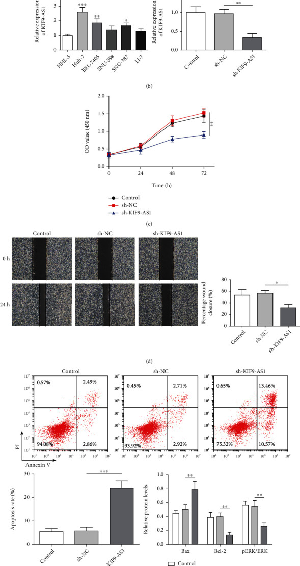
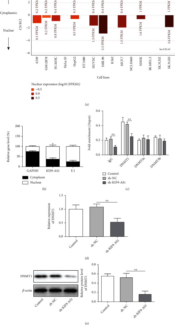
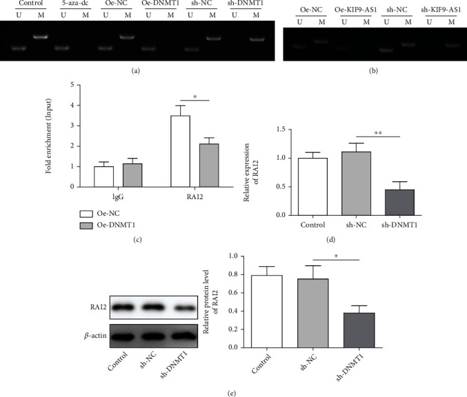
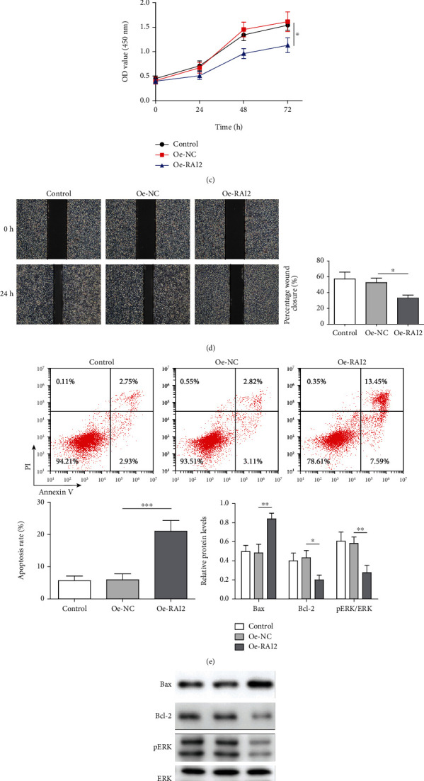
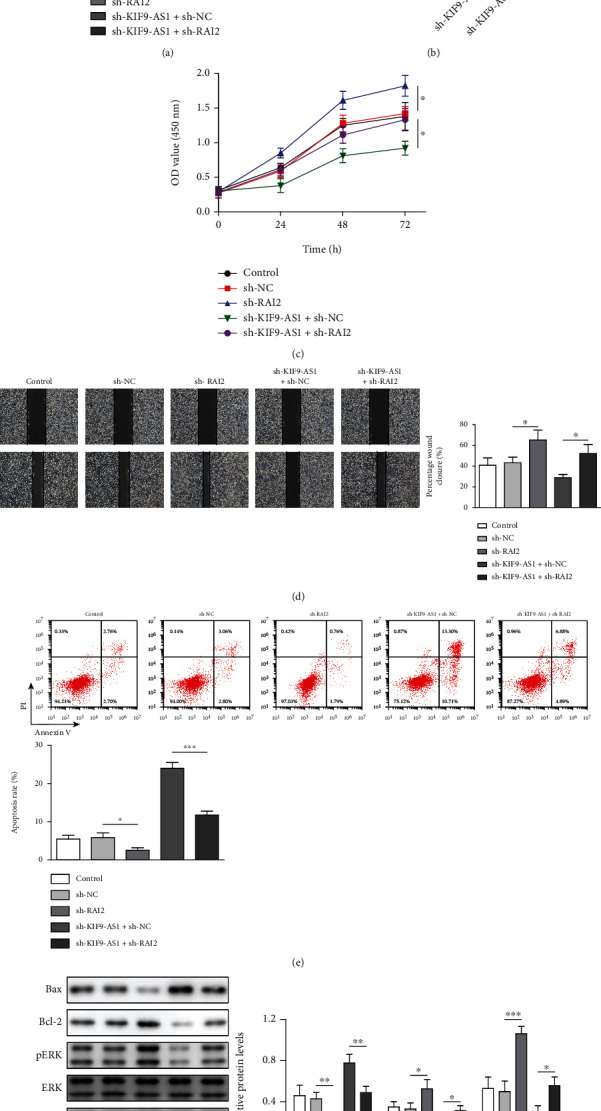
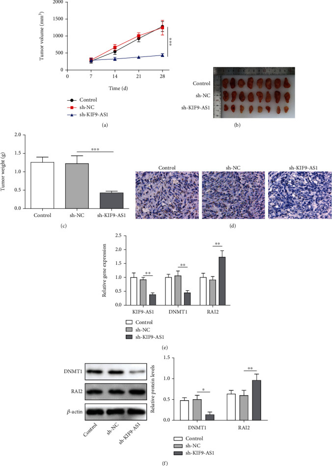
Methods: CCK-8, scratch assay, and flow cytometry were used to detect cell proliferation, migration, and apoptosis, respectively. Bax, Bcl-2, ERK, and pERK expression were measured by western blotting. StarBase predicted KIF9-AS1 expression in HCC and paracancerous tissues. RPISeq predicted the interaction score of KIF9-AS1 and DNMT1, and MethyPrimer revealed the CpG island distribution in the RAI2 promoter. MSP was performed to measure RAI2 methylation. RIP and ChIP were performed to examine lncRNA KIF9-AS1, DNMT1, and RAI2 interactions. Finally, the effect of KIF9-AS1 knockdown on HCC was verified with nude mice.

Results: We found that KIF9-AS1 expression was increased in HCC tissues. KIF9-AS1 knockdown inhibited the proliferation and migration, and facilitated the apoptosis of HCC cells. lncRNA KIF9-AS1-mediated RAI2 expression led to DNMT1 recruitment and regulated RAI2 DNA methylation. RAI2 overexpression inhibited the proliferation and migration and promoted the apoptosis of HCC cells. KIF9-AS1 knockdown inhibited subcutaneous tumor formation in vivo.

Conclusion: This study shows that KIF9-AS1 accelerates HCC growth by inducing DNMT1 promotion of RAI2 DNA methylation.


Conflict of interest statement

The authors declare that there is no conflict of interest.

Figures

Figure 1
Figure 1
Knocking down lncRNA KIF9-AS1 expression inhibited the proliferation and migration and promoted the apoptosis of HCC cells. Huh-7 cells were transfected with short hairpin (sh)-NC and sh-KIF9-AS1. (a) StarBase was used to predict lncRNA KIF9-AS1 expression in HCC and paracancerous tissues. (b) lncRNA KIF9-AS1 expression in normal liver cells (HHL-5 cells) and HCC cells (Huh-7, BEL-7405, SNU-398, SNU-387, and Li-7 cells) was detected by qRT–PCR. (c) The proliferative capacity of Huh-7 cells was determined by Cell Counting Kit-8 (CCK-8) assay. (d) The migratory ability of Huh-7 cells was analyzed with a scratch assay. (e) The Huh-7 cell apoptosis rate was measured by flow cytometry. (f) Western blotting was performed to detect Bax, Bcl-2, ERK, and pERK expression. The data are expressed as the means ± SD. P < 0.05, ∗∗P < 0.01, and ∗∗∗P < 0.001.
Figure 2
Figure 2
The lncRNA KIF9-AS1 interacted with DNMT1 in HCC. (a) Prediction of the subcellular localization of the lncRNA KIF9-AS1. (b) Expression of lncRNA KIF9-AS1 in the nucleus and cytoplasm. (c) The interaction between lncRNA KIF9-AS1 and DNMT1 and DNMT3A and DNMT3b was detected by RNA immunoprecipitation (RIP). (d and e) qRT–PCR and western blotting were performed to detect DNMT1 expression. The data are expressed as the means ± SD. P < 0.05, ∗∗P < 0.01, and ∗∗∗P < 0.001.
Figure 3
Figure 3
DNMT1 regulated RAI2 expression by regulating RAI2 DNA methylation. (a) After DNMT1 knockdown or overexpression, RAI2 promoter methylation was detected by methylation-specific PCR (MSP). (b) After the knockdown or overexpression of the lncRNA KIF9-AS1, MSP revealed RAI2 promoter methylation. (c) Chromatin immunoprecipitation (ChIP) was performed to measure the extent of the interaction between DNMT1 and the RAI2 promoter. (d and e) qRT–PCR and western blot analyses of RAI2 mRNA and protein expression, respectively. The data are expressed as the means ± SD. P < 0.05, ∗∗P < 0.01, and ∗∗∗P < 0.001.
Figure 4
Figure 4
Overexpression of RAI2 inhibited the proliferation and migration and promoted the apoptosis of HCC cells. Huh-7 cells were transfected with overexpressing (oe)-NC and oe-RAI2 vectors. (a) qRT–PCR was performed to detect RAI2 expression in normal liver cells (HHL-5 cells) and HCC cells (Huh-7, BEL-7405, SNU-398, SNU-387, and Li-7 cells). (b) The expression of RAI2 was detected by western blotting (WB). (c) A Cell Counting Kit-8 (CCK-8) assay was performed to assess Huh-7-cellcell proliferation. (d) A scratch assay was performed to determine the migratory ability of Huh-7 cells. (e) The Huh-7 cell apoptosis rate was determined by flow cytometry. (f) Bax, Bcl-2, ERK, and pERK expression were measured by WB. The data are expressed as the mean ± SD. P < 0.05, ∗∗P < 0.01, and ∗∗∗P < 0.001.
Figure 5
Figure 5
Knocking down RAI2 expression reversed lncRNA KIF9-AS1-knockdown effects on the proliferation, migration, and apoptosis of HCC cells. Huh-7 cells were transfected with short hairpin (sh)-NC, sh − RAI2, sh − KIF9 − AS1 + sh − NC, and sh − KIF9 − AS1 + sh − RAI2. (a) The expression of KIF9-AS1 and RAI2 was detected by qRT–PCR. (b) The expression of RAI2 was measured by western blotting (WB). (c) A Cell Counting Kit-8 (CCK-8) assay revealed the Huh-7-cell proliferative ability. (d) The migratory ability of Huh-7 cells was determined with a scratch assay. (e) The Huh-7 cell apoptosis rate was analyzed by flow cytometry. (f) WB was performed to detect Bax, Bcl-2, ERK, and pERK expression. The data are expressed as the means ± SD. P < 0.05, ∗∗P < 0.01, and ∗∗∗P < 0.001.
Figure 6
Figure 6
Knocking down lncRNA KIF9-AS1 expression inhibited subcutaneous HCC tumor formation in nude mice. A subcutaneous tumor-forming model in nude mice was established by injecting Huh-7 cells transfected with sh-NC or sh-KIF9-AS1. (a–c) Tumor volume, size, and weight in the nude mice. (d) Ki67 expression was determined by immunohistochemistry (IHC). (e) The expression of KIF9-AS1, DNMT1, and RAI2 was measured by qRT–PCR. (f) Western blot analysis was performed to detect DNMT1 and RAI2 expression. The data are expressed as the means ± SD, n = 8. P < 0.05, ∗∗P < 0.01, and ∗∗∗P < 0.001.

References

    1. Craig A. J., von Felden J., Garcia-Lezana T., Sarcognato S., Villanueva A. Tumour evolution in hepatocellular carcinoma. Nature Reviews. Gastroenterology & Hepatology . 2020;17(3):139–152. doi: 10.1038/s41575-019-0229-4. - DOI - PubMed
    1. Hartke J., Johnson M., Ghabril M. The diagnosis and treatment of hepatocellular carcinoma. Seminars in Diagnostic Pathology . 2017;34(2):153–159. doi: 10.1053/j.semdp.2016.12.011. - DOI - PubMed
    1. Yang C., Huang X., Liu Z., Qin W., Wang C. Metabolism-associated molecular classification of hepatocellular carcinoma. Molecular Oncology . 2020;14(4):896–913. doi: 10.1002/1878-0261.12639. - DOI - PMC - PubMed
    1. Xu L. X., He M. H., Dai Z. H., et al. Genomic and transcriptional heterogeneity of multifocal hepatocellular carcinoma. Annals of Oncology . 2019;30(6):990–997. doi: 10.1093/annonc/mdz103. - DOI - PMC - PubMed
    1. Sung H., Ferlay J., Siegel R. L., et al. Global cancer statistics 2020: GLOBOCAN estimates of incidence and mortality worldwide for 36 cancers in 185 countries. CA: a Cancer Journal for Clinicians . 2021;71(3):209–249. doi: 10.3322/caac.21660. - DOI - PubMed