

Spontaneously Recanalised Coronary Thrombus in the Left Anterior Descending Artery Presenting as Ventricular Tachycardia
- PMID: 36276506
- PMCID: PMC9524615
- DOI: 10.17925/HI.2020.14.2.123
Spontaneously Recanalised Coronary Thrombus in the Left Anterior Descending Artery Presenting as Ventricular Tachycardia
Abstract
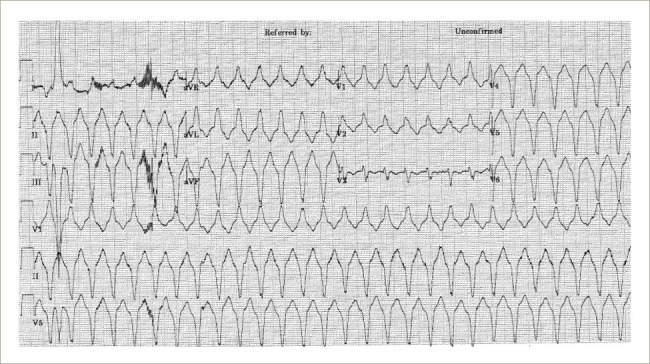
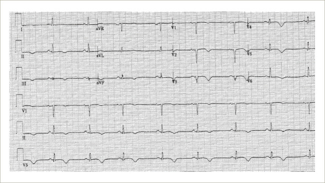
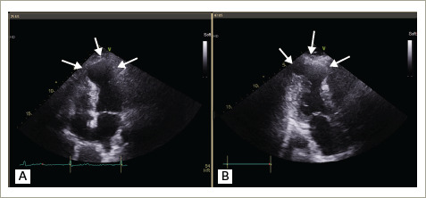
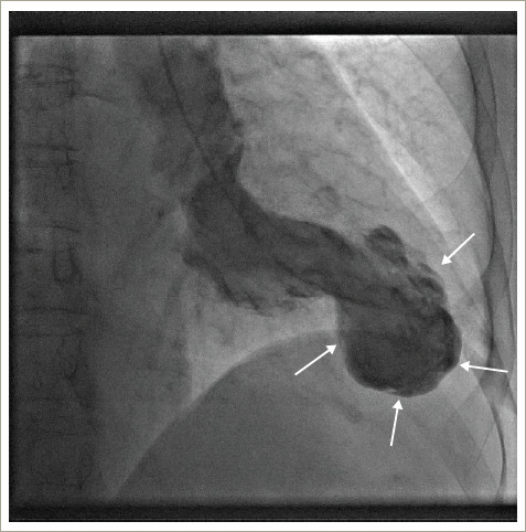
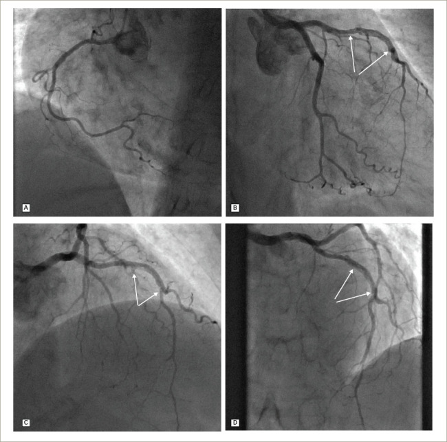
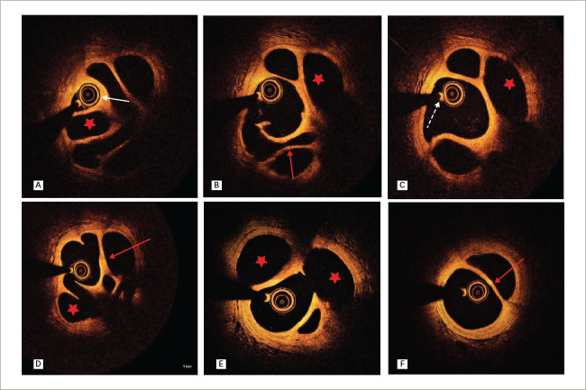
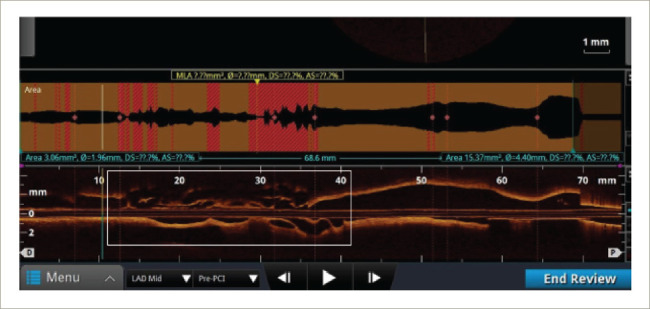
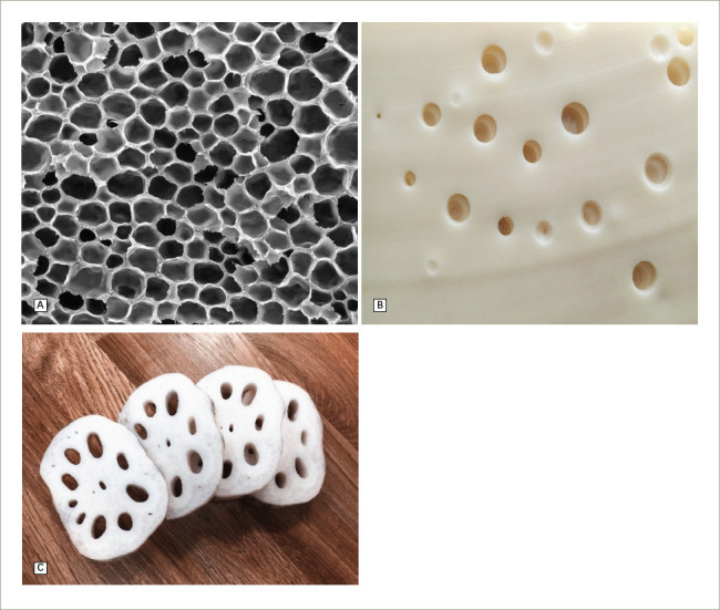
Spontaneously recanalized coronary thrombus (SRCT), also known as honeycomb, lotus root or Swiss-cheese lesion, is an increasingly recognised finding in patients undergoing coronary angiography. It is thought to arise from partial resorption of an initially occlusive thrombus. Most patients present with angina or exertional breathlessness. We describe a case of a 69-year-old patient who presented with ventricular tachycardia and was found to have SRCT in the left anterior descending artery on coronary angiography. Echocardiography and left ventricular (LV) angiography showed an akinetic, aneurysmal, thin-walled LV apex, diagnostic of an old anterior infarct. We highlight the role of optical coherence tomography in making the diagnosis and discuss the available management options of this condition.
Keywords: Spontaneously recanalized coronary thrombus; honeycomb lesion; lotus root lesion; optical coherence tomography; ventricular tachycardia.
© Touch Medical Media 2020.
Conflict of interest statement
Disclosure: Jie Man Low, Noah Kimit, Rizwan Rashid and Magdi El-Omar have no financial or non-financial relationships or activities to declare in relation to this article.
Figures
References
-
- Kang SJ, Nakano M, Virmani R. et al. OCT findings in patients with recanalization of organized thrombi in coronary arteries. JACC Cardiovasc Imaging. 2012;5:725–32. - PubMed
-
- Souteyrand G, Valladier M, Amabile N. et al. Diagnosis and management of spontaneously recanalized coronary thrombus guided by optical coherence tomography – lessons from the French “Lotus Root” Registry. Circ J. 2018;82:783–90. - PubMed
-
- Kadowaki H, Taguchi E, Kotono Y. et al. A lotus root-like appearance in both the left anterior descending and right coronary arteries. Heart Vessels. 2016;31:124–8. - PubMed
-
- Toutouzas K, Karanasos A, Stathogiannis K. et al. A honeycomb-like structure in the left anterior descending coronary artery: Demonstration of recanalized thrombus by optical coherence tomography. JACC Cardiovasc Interv. 2012;5:688–9. - PubMed
Publication types
LinkOut - more resources
Full Text Sources
Miscellaneous