

Spontaneous regression of an orbital Langerhans cell histiocytosis after biopsy: A case report. Spontaneous regression of an orbital Langerhans cell histiocytosis
- PMID: 36277239
- PMCID: PMC9583201
- DOI: 10.1177/20363613221135987
Spontaneous regression of an orbital Langerhans cell histiocytosis after biopsy: A case report. Spontaneous regression of an orbital Langerhans cell histiocytosis
Abstract
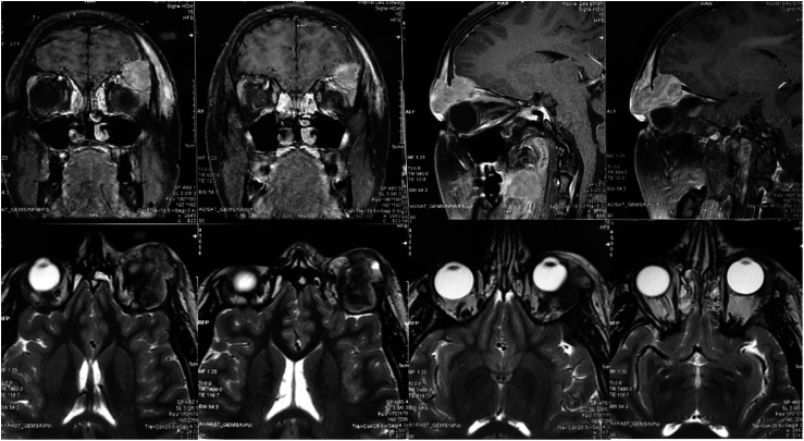
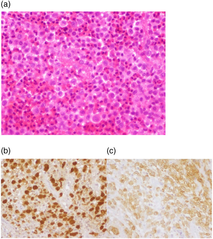
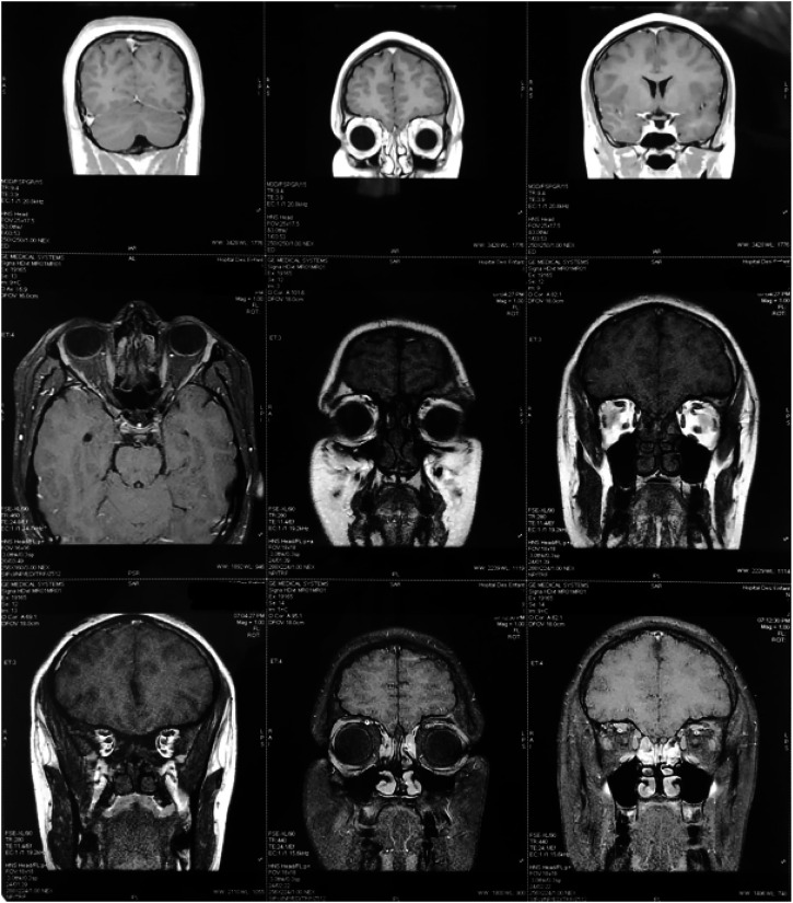
Langerhans histiocytosis or Langerhans cell histiocytosis (LCH) is a rare benign pathology representing less than 1% of orbital tumors. It can cause either localized or generalized lesions, leading to the destruction of hard and soft tissues. Eosinophilic granuloma is the most benign form and the predominant clinical presentation of LCH. We report a case of eosinophilic granuloma with orbital involvement in an 18-year-old male patient. Orbital radiotherapy was initially planned, but finally it was not performed due to a spontaneous regression of the lesion after the incisional biopsy. The presented case supports an expectant attitude given the possibility of a spontaneous regression after the biopsy, especially in small lesions. However, long-term follow-up is essential given the risk of recurrence.
Keywords: Langerhans cell histiocytosis; biopsy; orbital tumors; spontaneous regression.
© The Author(s) 2022.
Conflict of interest statement
The author(s) declared no potential conflicts of interest with respect to the research, authorship, and/or publication of this article.
Figures
References
-
- Desbarats C, Adnot J, Bastien AV, et al. Histiocytose langerhansienne révélée par un désordre de l’appareil manducateur : rapport d’un cas et revue de la littérature des atteintes crâniofaciales. Rev Médecine Interne Janv 2020; 41(1): 50–53. - PubMed
-
- Iurescia AC, Rendo J, Gimeno FL, et al. Manifestaciones oculares de la histiocitosis de células de Langerhans: Revisión de 40 casos. Oftalmol Clin Exp 2007; 1(12–5).
-
- Durham BH. Molecular characterization of the histiocytoses: Neoplasia of dendritic cells and macrophages. Semin Cel Dev Biol. Févr 2019; 86: 62–76. - PubMed
-
- Giovannetti F, Giona F, Ungari C, et al. Langerhans cell histiocytosis with orbital involvement: our experience. J Oral Maxillofac Surg 2009; 67(1): 212–216. - PubMed
Publication types
LinkOut - more resources
Full Text Sources
