

Diagnostic Utility of Point-of-Care Ultrasound in the Pediatric Cardiac Intensive Care Unit
- PMID: 36277259
- PMCID: PMC9264295
- DOI: 10.1007/s40746-022-00250-1
Diagnostic Utility of Point-of-Care Ultrasound in the Pediatric Cardiac Intensive Care Unit
Abstract
Purpose of review: This review summarizes the diverse uses of point-of-care ultrasound (POCUS) in critically ill children with congenital and acquired heart disease. Diagnostic utility and practicality of POCUS is reviewed. Importantly, the role of POCUS in the medical management of children in the cardiac intensive care unit is highlighted.
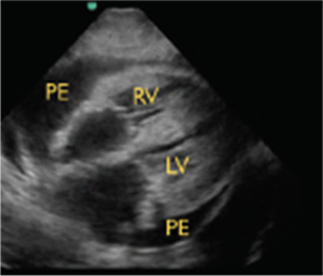
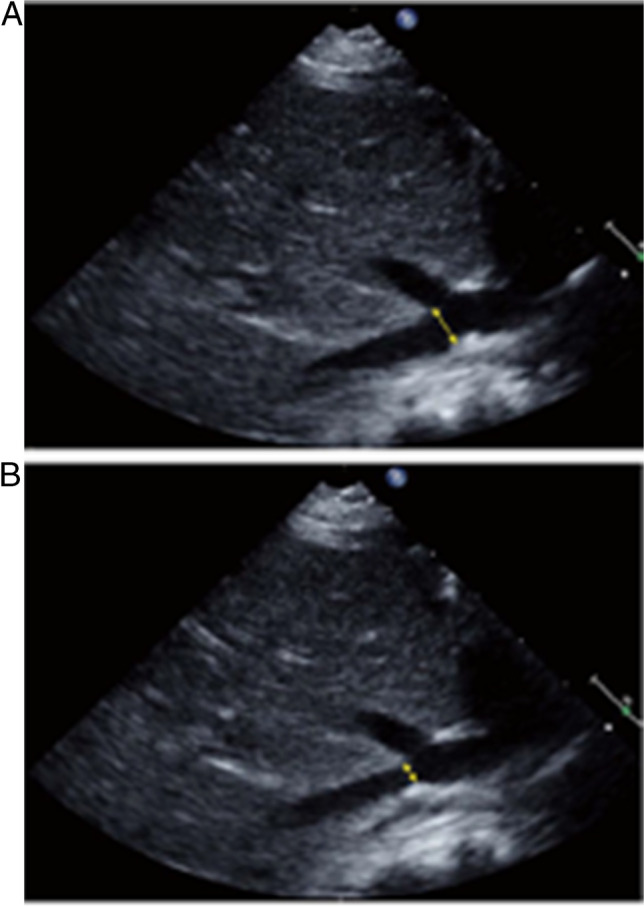
Recent findings: The use of POCUS in critically ill pediatric patients has emerged as an essential diagnostic tool that enhances the physical examination and influences delivery of care. Assessment of a wide range of body systems and pathologies has been impacted by the use of POCUS. Recent studies have demonstrated the use of POCUS for evaluation of cardiac tamponade, pneumonia, vocal cord function, and loss of muscle mass in critically ill children (Hamilton et al. Pediatr Crit Care Med 22(10):e532-e539, 2021; Hoffmann et al. Pediatr Crit Care Med 22(10):889-897, 2021; Najgrodzka et al. Ultrasound Q 35(2):157 163, 2019; Alerhand et al. Pediatr Ann 50(10):e424-e431, 2021).
Summary: POCUS is a non-invasive, low-risk, imaging modality that can be used to diagnose and help guide management of critically ill children in the cardiac intensive care unit. POCUS can be performed by an intensivist at the patient's bedside with real-time interpretation, leading to rapid clinical decision-making and the hope of improving patient outcomes.
Supplementary information: The online version contains supplementary material available at 10.1007/s40746-022-00250-1.
Keywords: Congenital heart disease; Diagnostic ultrasound; PICU; POCUS; Pediatric cardiac critical care; Point-of-care ultrasound.
© The Author(s), under exclusive licence to Springer Nature Switzerland AG 2022.
Conflict of interest statement
Conflict of InterestJessica N. Persson declares that she has no conflict of interest. John S. Kim declares that he has no conflict of interest. Ryan J. Good declares that he has no conflict of interest.
Figures


Similar articles
-
The Utility of Point-of-Care Ultrasound in the Pediatric Intensive Care Unit.J Intensive Care Med. 2022 Aug;37(8):1029-1036. doi: 10.1177/08850666211047824. Epub 2021 Oct 9. J Intensive Care Med. 2022. PMID: 34632837
-
Evolving the Scope of Cardiac Point-of-Care Ultrasound in the Current Era.Cureus. 2024 Feb 10;16(2):e53985. doi: 10.7759/cureus.53985. eCollection 2024 Feb. Cureus. 2024. PMID: 38476776 Free PMC article. Review.
-
Cardiac Ultrasound for Pediatric Emergencies.Pediatr Ann. 2021 Oct;50(10):e424-e431. doi: 10.3928/19382359-20210913-01. Epub 2021 Oct 1. Pediatr Ann. 2021. PMID: 34617847
-
Cardiac point-of-care ultrasound: Practical integration in the pediatric and neonatal intensive care settings.Eur J Pediatr. 2024 Apr;183(4):1525-1541. doi: 10.1007/s00431-023-05409-y. Epub 2024 Jan 18. Eur J Pediatr. 2024. PMID: 38236402 Review.
-
Point-of-care abdominal ultrasound in pediatric and neonatal intensive care units.Eur J Pediatr. 2024 May;183(5):2059-2069. doi: 10.1007/s00431-024-05443-4. Epub 2024 Mar 8. Eur J Pediatr. 2024. PMID: 38459132 Review.
Cited by
-
Point-of-Care Ultrasound for the Diagnosis of Frequent Cardiovascular Diseases: A Review.Cureus. 2023 Dec 24;15(12):e51032. doi: 10.7759/cureus.51032. eCollection 2023 Dec. Cureus. 2023. PMID: 38264374 Free PMC article. Review.
-
Inferior vena cava-aortic ratio measurement as a promising modality in assessing intravascular volume in children with sepsis.Pediatr Nephrol. 2024 Nov;39(11):3339-3346. doi: 10.1007/s00467-024-06446-x. Epub 2024 Jul 8. Pediatr Nephrol. 2024. PMID: 38977444
References
References and Recommended Reading
Papers of particular interest, published recently, have been highlighted as: • Of importance •• Of major importance
-
- Adler AC, et al. Cardiac and lung point-of-care ultrasound in pediatric anesthesia and critical care medicine: uses, pitfalls, and future directions to optimize pediatric care. Paediatr Anaesth. 2019;29(8):790–798. - PubMed
-
- Conlon TW, Nishisaki A, Singh Y, et al. Moving beyond the stethoscope: diagnostic point-of-care ultrasound in Pediatric Practice. Pediatrics. 2019;144(4):e20191402. 10.1542/peds.2019-1402. - PubMed
Publication types
LinkOut - more resources
Full Text Sources
Medical
Research Materials