

A Case Report of Pseudomyxoma Peritonei Arising From Primary Mucinous Ovarian Neoplasms
- PMID: 36277572
- PMCID: PMC9579829
- DOI: 10.7759/cureus.29309
A Case Report of Pseudomyxoma Peritonei Arising From Primary Mucinous Ovarian Neoplasms
Abstract
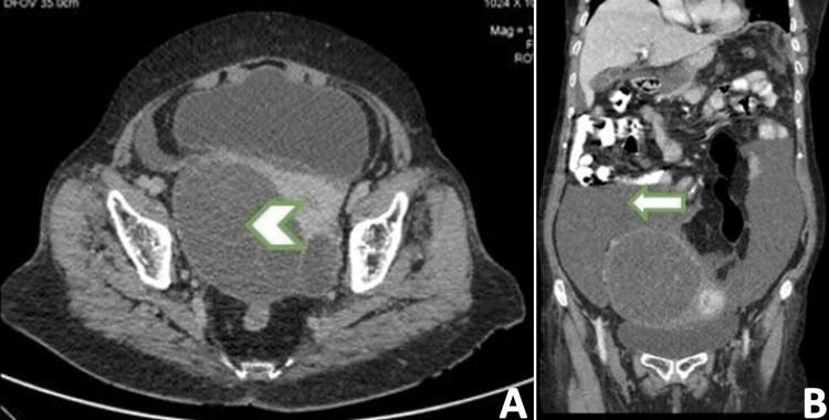
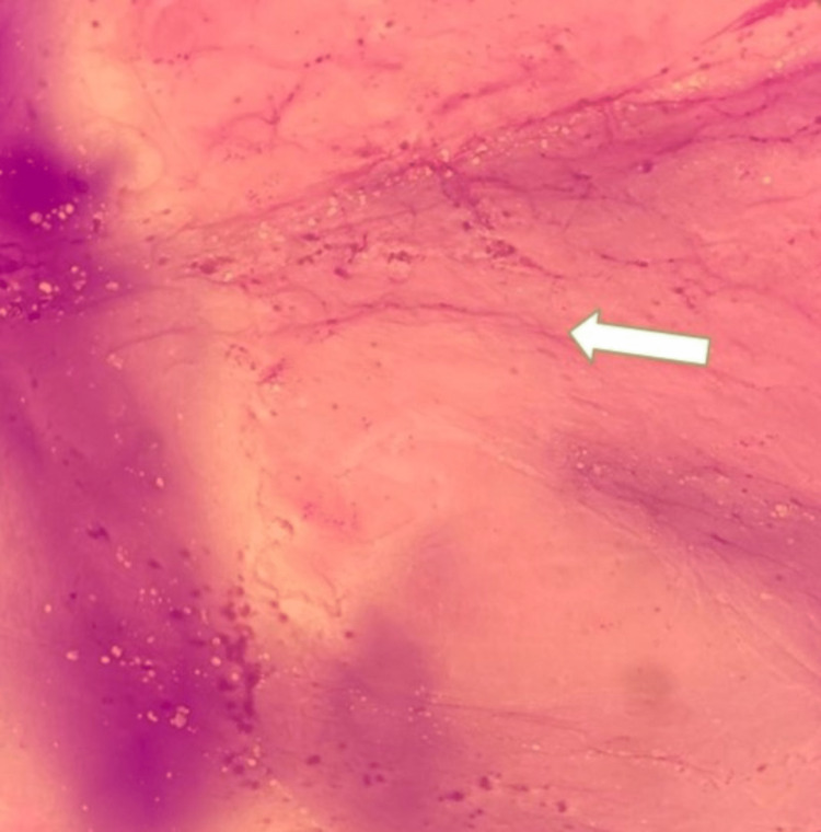
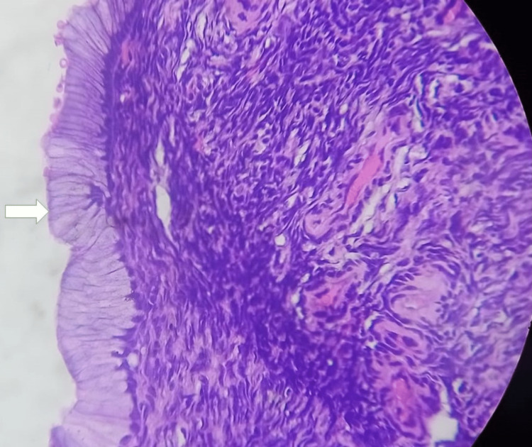
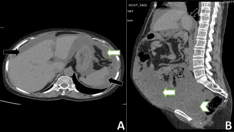
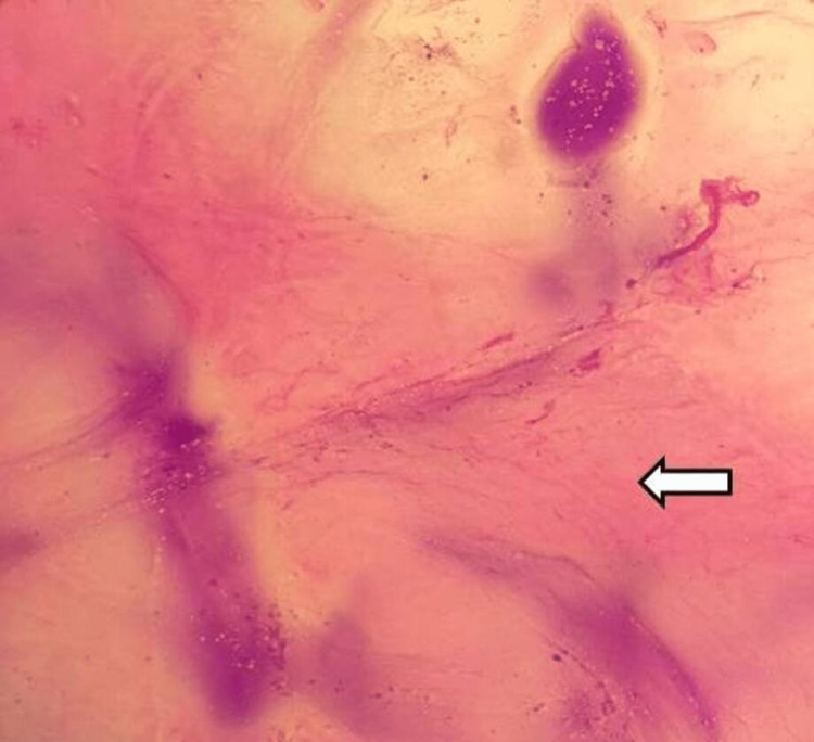
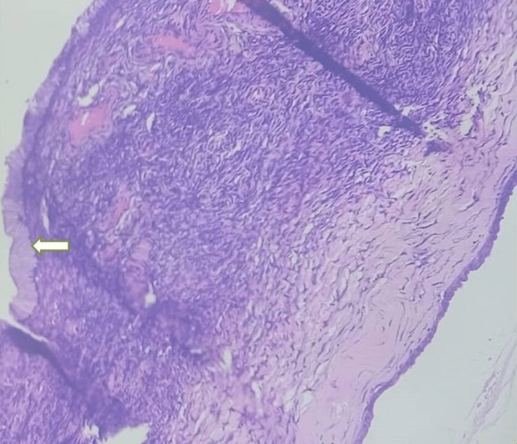
Pseudomyxoma peritonei (PMP) is a rare manifestation of primary mucinous neoplasms. We report two rare cases of PMP originating from mucinous primary ovarian neoplasms. The case series discusses the cases of female patients aged 86 and 52 years who presented with worsening dyspepsia, abdominal distension, pelvic pain, and altered bowel habits. Both of the patients underwent evaluation comprising cancer antigen-125 (CA-125) levels, ultrasound (US) examination of the abdomen and the pelvis, tumor markers, cytological evaluation, and contrast-enhanced computed tomography (CECT) of the pelvis and abdomen. Patients were diagnosed to have pseudomyxoma peritonei arising from mucinous ovarian tumors. Patients were referred to the surgical department and were successfully managed with repeated removal of mucinous material. The present case report highlights the significant radio-pathologic characteristics of PMP, which originated from mucinous ovarian tumors.
Keywords: abdominal radiology; academic radiology; appendix; cystic mass; mucinous neoplasms; ovary; pseudomyxoma peritonei.
Copyright © 2022, Joseph et al.
Conflict of interest statement
The authors have declared that no competing interests exist.
Figures
Similar articles
-
An unusual case of pseudomyxoma peritonei associated with synchronous primary mucinous tumors of the ovary and appendix: A case report.Oncol Lett. 2017 Jun;13(6):4813-4817. doi: 10.3892/ol.2017.6079. Epub 2017 Apr 24. Oncol Lett. 2017. PMID: 28599482 Free PMC article.
-
[Pseudomyxoma peritonei (PMP) secondary to mucinous carcinoma of the ovary: a case study].Pan Afr Med J. 2019 Aug 2;33:283. doi: 10.11604/pamj.2019.33.283.17203. eCollection 2019. Pan Afr Med J. 2019. PMID: 31692896 Free PMC article. French.
-
Ovarian mature teratomas with mucinous epithelial neoplasms: morphologic heterogeneity and association with pseudomyxoma peritonei.Am J Surg Pathol. 2008 May;32(5):645-55. doi: 10.1097/PAS.0b013e31815b486d. Am J Surg Pathol. 2008. PMID: 18344868
-
Mucinous cystadenocarcinoma of the appendix. The controversy persists: a review.Hepatogastroenterology. 2003 Mar-Apr;50(50):432-7. Hepatogastroenterology. 2003. PMID: 12749241 Review.
-
Ovarian Causes of Pseudomyxoma Peritonei (PMP)-A Literature Review.Cancers (Basel). 2024 Apr 9;16(8):1446. doi: 10.3390/cancers16081446. Cancers (Basel). 2024. PMID: 38672528 Free PMC article. Review.
Cited by
-
Pseudomyxoma Peritonei Misdiagnosed as Liver Cirrhosis: A Case Report and Literature Review.Cureus. 2024 Apr 8;16(4):e57857. doi: 10.7759/cureus.57857. eCollection 2024 Apr. Cureus. 2024. PMID: 38721212 Free PMC article.
References
-
- A consensus for classification and pathologic reporting of pseudomyxoma peritonei and associated appendiceal neoplasia: the results of the peritoneal surface oncology group international (PSOGI) modified Delphi process. Carr NJ, Cecil TD, Mohamed F, et al. Am J Surg Pathol. 2016;40:14–26. - PubMed
-
- Pseudomyxoma peritonei: natural history and treatment. Mittal R, Chandramohan A, Moran B. Int J Hyperthermia. 2017;33:511–519. - PubMed
Publication types
LinkOut - more resources
Full Text Sources
Research Materials
Miscellaneous