. 2022 Aug 5;13(35):10406-10427.
doi: 10.1039/d2sc00345g. eCollection 2022 Sep 14.

Structural and dynamical determinants of a β-sheet-enriched intermediate involved in amyloid fibrillar assembly of human prion protein

Affiliations

Structural and dynamical determinants of a β-sheet-enriched intermediate involved in amyloid fibrillar assembly of human prion protein

Luigi Russo et al. Chem Sci. .

Abstract

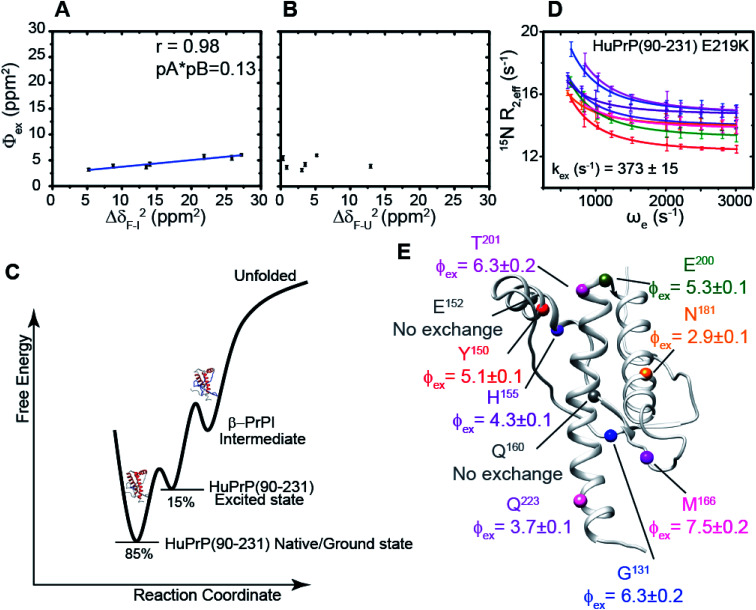
The conformational conversion of the cellular prion protein (PrPC) into a misfolded, aggregated and infectious scrapie isoform is associated with prion disease pathology and neurodegeneration. Despite the significant number of experimental and theoretical studies the molecular mechanism regulating this structural transition is still poorly understood. Here, via Nuclear Magnetic Resonance (NMR) methodologies we investigate at the atomic level the mechanism of the human HuPrP(90-231) thermal unfolding and characterize the conformational equilibrium between its native structure and a β-enriched intermediate state, named β-PrPI. By comparing the folding mechanisms of metal-free and Cu2+-bound HuPrP(23-231) and HuPrP(90-231) we show that the coupling between the N- and C-terminal domains, through transient electrostatic interactions, is the key molecular process in tuning long-range correlated μs-ms dynamics that in turn modulate the folding process. Moreover, via thioflavin T (ThT)-fluorescence fibrillization assays we show that β-PrPI is involved in the initial stages of PrP fibrillation, overall providing a clear molecular description of the initial phases of prion misfolding. Finally, we show by using Real-Time Quaking-Induced Conversion (RT-QuIC) that the β-PrPI acts as a seed for the formation of amyloid aggregates with a seeding activity comparable to that of human infectious prions.


Conflict of interest statement

There are no conflicts to declare.

Figures

Fig. 1
Fig. 1. NMR thermal unfolding of the C-terminal HuPrP domain. (A) Series of 1H–15N HSQC NMR spectra of HuPrP(90–231) as a function of temperature for selected residues (Gly131, Thr199, Ile139, Phe141). Temperature increase is indicated by a gradual change in the color of the resonances from light cyan (15 °C) to red (61 °C). (B) Plot of normalized chemical shifts against temperature for 10 representative Hα protons showing mean-like behaviour in the 15–61 °C range. (C) 1H–15N HSQC spectrum of HuPrP(90–231) acquired at 600 MHz at 61 °C and pH 5.5. (D) 1H–15N HSQC spectrum of the unfolded HuPrP(90–231) at 61 °C. The NMR spectrum was reconstructed (see the ESI†) using the random coil 1H and 15N chemical shifts predicted for HuPrP(90–231) at 61 °C and pH 5.5 through a specific algorithm suitable for IDPs (Intrinsically Disordered Proteins). (E–G) Ribbon drawing representation of the HuPrP(90–231) NMR structure showing the thermal stability for HN (E), Hα (F) and both (G) protons mapped on their corresponding heavy atoms. The inset indicates the Tm scale. The Tm histogram is also reported.
Fig. 2
Fig. 2. Structural details of the HuPrP(90–231) conformational intermediate state. (A and B) Mapping of the SSP scores onto the representative NMR structure of HuPrP(90–231). The α-helix and β-strand populations are reported in red and blue, respectively. The insets report the increase and the reduction of β-sheet and α1 helix populations, respectively. The disulphide bridge, formed by Cys179 and Cys214, is also reported as a yellow stick. (C) Normalized distribution of 1HN temperature coefficient values (ΔδHNT) for each of the three α-helices of the β-PrPI intermediate state.
Fig. 3
Fig. 3. The mechanism driving the inter-domain coupling. (A) (upper) Electrostatic surface potential of the N-terminal domain (23–89) of HuPrP(23–231) reported as a histogram. The OR region is also indicated as a light blue box. (lower) C-terminal domain electrostatic surface potential, HuPrP(90–231), at pH 5.5 depicted from electropositive (blue; 10 kcal mol−1) to electronegative (red; −10 kcal mol−1). The negatively charged residues are indicated. (B) Comparison of 1H–15N HSQC NMR spectra used to detect the chemical shift variations for HuPrP(90–231) (blue) and HuPrP(23–231) (red) upon addition of 50 and 200 mM NaCl. (C) (ΔδHN,N23–231 − ΔδHN,N90–231)2 values calculated for 50 and 200 mM plotted versus the primary sequence. The cyan and light green lines indicate the average value for 50 and 200 mM, respectively. (D) Mapping of the residues showing significant HN and N chemical shift changes upon addition of 50 and 200 mM NaCl, respectively.
Fig. 4
Fig. 4. Copper binding effects on the prion protein unfolding mechanisms. (A) 1H–15N signal intensity changes shown as a fraction of the starting values for the addition of 1, 4 and 6 equivalent(s) of Cu2+. I0 and I are intensities of 1H–15N cross-peaks of HuPrP(23–231) in the absence and presence of Cu2+. (B) CSPs of HuPrP(23–231) upon binding to 1 and 4 equivalent(s) of Cu2+ mapped onto the NMR structure of the C-terminal domain in two orientations. Residues for which CSPHN,N > mean + SD are shown in light green and orange for HuPrP(23–231)/Cu2+ (1 : 1) and (1 : 4), respectively. In the case of HuPrP(23–231)/Cu2+ (1 : 6) the residues for which the HN/N signals are not detected are reported as violet. (C) Cα secondary chemical shifts observed for the copper induced intermediate state for HuPrP(23–231) upon binding to 1 and 4 equivalent(s) Cu2+. The Cα secondary chemical shifts related to the β-PrPI intermediate state are also reported (light grey). (D) Mapping of the SSP scores obtained for HuPrP(23–231)/Cu2+ (1 : 1) and HuPrP(23–231)/Cu2+ (1 : 1) at 61 °C onto the representative NMR structure of HuPrP(90–231). The α-helix and β-strand populations are reported in red and blue, respectively. (E and F) Thermal stability of HN protons for HuPrP(23–231)/Cu2+ (1 : 1) (E) and (1 : 4) (F) mapped on their corresponding heavy atoms.
Fig. 5
Fig. 5. Conformational exchange processes of HuPrP(90–231) and HuPrP(23–231) implicating the N-terminal domain as a dynamic switch. (A) Close-up view of HuPrP(90–231) regions showing a significant increase in Rex upon deletion of the N-terminal domain. The residues investigated by 15N R1ρ RD experiments are labelled. (B) Best fit curves to a two-site exchange model assuming a common motion for residues of HuPrP(90–231) (blue) and HuPrP(23–231) (red). (C) Residue-specific Φex (×103 (s−1)2) parameters obtained from the global fit of the HuPrP(90–231) cluster A residues (blue spheres); the sites reporting different exchange processes are reported in yellow ((*) indicates data extracted from the individual fit). (D) Residue-specific Φex parameters extracted for HuPrP(23–231) from the global fit (yellow spheres). The residues that do not show any RD profile are reported (red spheres).
Fig. 6
Fig. 6. Correlation between relaxation dispersion/thermal unfolding data. (A and B) Φex parameters (ppm2) derived from the global fit of cluster A (kex = 2502 ± 106 s−1) of HuPrP(90–231) plotted as a function of the squared differences in chemical shift between the native and the intermediate state (ΔδF–I)2 (ppm2) (A) and between the native and the denaturated state (ΔδF–U)2 (ppm2) (B). (C) Conformational landscape of HuPrP(90–231) revealed by NMR relaxation dispersion and thermal melt data. (D) Best fit curves to a two-site exchange model assuming a common motion for residues of HuPrP(90–231) E219K. (E) Mapping of the residue-specific Φex parameters extracted for HuPrP(90–231) E219K from the global fit. The residues that do not show any RD profile are reported as dark grey spheres.
Fig. 7
Fig. 7. Role of the β-PrPI intermediate state in amyloid fibril formation and in prion conversion. (A) HuPrP(90–231) and HuPrP(23–231) were induced to aggregate by alternating cycles of incubation and shaking at 55 °C. Average ThT fluorescence intensity was plotted against time. (B) Serial dilutions of the artificial β-PrPI(o) seeds, previously produced by incubating HuPrP(90–231) at 61 °C, were used to promote the aggregation of HuPrP(23–231). Average ThT fluorescence intensity was plotted against time. (C and D) RT-QuIC experiments monitoring the seeding activity of the β-PrPI oligomers. The experiments were conducted collecting from patients the following biological samples: brain homogenates (dilution 10−5 v/v) sCJD-129MM1 (), sCJD-129MM2 (), sCJD-129MV1 (), sCJD-129MV2 (), sCJD-129VV1 (), sCJD-129VV2 (), Alzheimer's disease and non-neurodegenerative disorder (); amyloid fibrillary assemblies (ng mL−1) β-PrPI amyloid fibrils pH 6.8 () and 5.5 (), monomer HuPrP(90–231) pH 6.8 and 5.5 (); cerebrospinal fluid sCJD-129MM (), sCJD-129MV (), sCJD-129VV (), Alzheimer's disease and hydrocephalus (). (E and F) Aggregation kinetics of HuPrP(23–231) at 25 °C (E) and 61 °C (F) upon addition of 1 (light green) and 4 (light orange) equivalent(s) of Cu2+ monitored by ThT fluorescence.

Similar articles

Cited by

References

    1. Weissmann C. Enari M. Klöhn P. C. Rossi D. Flechsig E. Proc. Natl. Acad. Sci. U. S. A. 2002;99(Suppl. 4):16378–16383. doi: 10.1073/pnas.172403799. - DOI - PMC - PubMed
    1. Moser M. Colello R. J. Pott U. Oesch B. Neuron. 1995;14:509–517. doi: 10.1016/0896-6273(95)90307-0. - DOI - PubMed
    1. Stahl N. Borchelt D. R. Hsiao K. Prusiner S. B. Cell. 1987;51:229–240. doi: 10.1016/0092-8674(87)90150-4. - DOI - PubMed
    1. Caiati M. D. Safiulina V. F. Fattorini G. Sivakumaran S. Legname G. Cherubini E. J. Neurosci. 2013;33:2973–2983. doi: 10.1523/JNEUROSCI.4149-12.2013. - DOI - PMC - PubMed
    1. Santuccione A. Sytnyk V. Leshchyns'ka I. Schachner M. J. Cell Biol. 2005;169:341–354. doi: 10.1083/jcb.200409127. - DOI - PMC - PubMed