A Clinical Study on the Treatment of Recurrent Chiari (Type I) Malformation with Syringomyelia Based on the Dynamics of Cerebrospinal Fluid
- PMID: 36277900
- PMCID: PMC9584687
- DOI: 10.1155/2022/9770323
A Clinical Study on the Treatment of Recurrent Chiari (Type I) Malformation with Syringomyelia Based on the Dynamics of Cerebrospinal Fluid
Abstract
Objective: Combining the dynamics of cerebrospinal fluid, our study investigates the clinical effects of syringomyelia after the combination of fourth ventricle-subarachnoid shunt (FVSS) for recurrent Chiari (type I) malformations after cranial fossa decompression (foramen magnum decompression (FMD)).
Methods: From December 2018 to December 2020, 15 patients with recurrent syringomyelia following posterior fossa decompression had FVSS surgery. Before and after the procedure, the clinical and imaging data of these individuals were retrospectively examined.
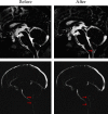
Results: Following FVSS, none of the 15 patients experienced infection, nerve injury, shunt loss, or obstruction. 13 patients improved dramatically after surgery, while 2 patients improved significantly in the early postoperative period, but the primary symptoms returned 2 months later. The Japanese Orthopedic Association (JOA) score was 12.67 ± 3.95, which was considerably better than preoperatively (t = 3.69, P0.001). The MRI results revealed that the cavities in 13 patients were reduced by at least 50% compared to the cavities measured preoperatively. The shrinkage rate of syringomyelia was 86.67% (13/15). One patient's cavities nearly vanished following syringomyelia. The size of the cavity in the patient remain unchanged, and the cavity's maximal diameter was significantly smaller than the size measured preoperatively (P < 0.001) PC-MRI results indicated that the peak flow rate of cerebrospinal fluid at the central segment of the midbrain aqueduct and the foramen magnum in patients during systole and diastole were significantly reduced after surgery (P < 0.05).
Conclusion: After posterior fossa decompression, FVSS can effectively restore the smooth circulation of cerebrospinal fluid and alleviate clinical symptoms in patients with recurrent Chiari (type I) malformation and syringomyelia. It is a highly effective way of treatment.
Copyright © 2022 Yongli Lou et al.
Conflict of interest statement
The authors declare that there is no conflict of interest regarding the publication of this paper.
Figures


References
-
- Hofkes S. K., Iskandar B. J., Turski P. A., Gentry L. R., Mccue J. B., Haughton V. M. Differentiation between symptomatic Chiari I malformation and asymptomatic tonsilar ectopia by using cerebrospinal fluid flow imaging: initial estimate of imaging accuracy. Radiology . 2007;245(2):532–540. doi: 10.1148/radiol.2452061096. - DOI - PubMed
MeSH terms
LinkOut - more resources
Full Text Sources
Medical

