

Retinal nerve fiber layer in frontotemporal lobar degeneration and amyotrophic lateral sclerosis
- PMID: 36278002
- PMCID: PMC9583385
- DOI: 10.3389/fnins.2022.964715
Retinal nerve fiber layer in frontotemporal lobar degeneration and amyotrophic lateral sclerosis
Abstract
Purpose: Tauopathy and transactive response DNA binding protein 43 (TDP-43) proteinopathy are associated with neurodegenerative diseases. These proteinopathies are difficult to detect in vivo. This study examined if spectral-domain optical coherence tomography (SD-OCT) can differentiate in vivo the difference in peripapillary retinal nerve fibre layer (pRNFL) thickness and macular retinal thickness between participants with presumed tauopathy (progressive supranuclear palsy) and those with presumed TDP-43 proteinopathy (amyotrophic lateral sclerosis and semantic variant primary progressive aphasia).
Study design: Prospective, multi-centre, observational study.
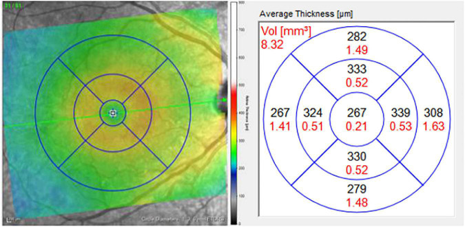
Materials and methods: pRNFL and macular SD-OCT images were acquired in both eyes of each participant using Heidelberg Spectralis SD-OCT. Global and pRNFL thickness in 6 sectors were analyzed, as well as macular thickness in a central 1 mm diameter zone and 4 surrounding sectors. Linear mixed model methods adjusting for baseline differences between groups were used to compare the two groups with respect to pRNFL and macular thickness.
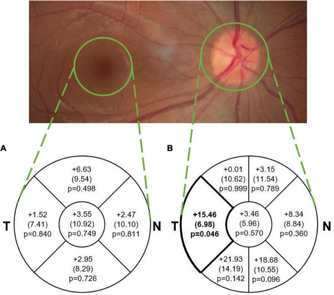
Results: A significant difference was found in mean pRNFL thickness between groups, with the TDP-43 group (n = 28 eyes) having a significantly thinner pRNFL in the temporal sector than the tauopathy group (n = 9 eyes; mean difference = 15.46 μm, SE = 6.98, p = 0.046), which was not significant after adjusting for multiple comparisons. No other significant differences were found between groups for pRNFL or macular thickness.
Conclusion: The finding that the temporal pRNFL in the TDP-43 group was on average 15.46 μm thinner could potentially have clinical significance. Future work with larger sample sizes, longitudinal studies, and at the level of retinal sublayers will help to determine the utility of SD-OCT to differentiate between these two proteinopathies.
Keywords: TDP-43 proteinopathy; amyotrophic lateral sclerosis; frontotemporal lobar degeneration; optical coherence tomography; retinal nerve fibre layer; tauopathy.
Copyright © 2022 Wong, Hudson, Snook, Tayyari, Jung, Binns, Samet, Cheng, Balian, Mandelcorn, Margolin, Finger, Black, Tang-Wai, Zinman, Tan, Lou, Masellis, Abrahao, Frank, Beaton, Sunderland, Arnott, ONDRI Investigators, Tartaglia and Hatch.
Conflict of interest statement
EM received honoraria for speaking engagements from Bayer and Novartis, none of which are relevant to this study. SB was a consultant for Roche and Pfizer. SB obtained funding and has funding pending (through institutions) from GE Healthcare, Eli Lilly, Biogen Idec, Genentech, Optina, Roche, and Novartis. SA was a consultant for Indoc Research Canada (not relevant to this study). The remaining authors declare that the research was conducted in the absence of any commercial or financial relationships that could be construed as a potential conflict of interest.
Figures
References
LinkOut - more resources
Full Text Sources