. 2022 Oct 4;7(41):36543-36550.
doi: 10.1021/acsomega.2c04442. eCollection 2022 Oct 18.

Multifunctional Core@Satellite Magnetic Particles for Magnetoresistive Biosensors

Affiliations

Multifunctional Core@Satellite Magnetic Particles for Magnetoresistive Biosensors

Raffaele Campanile et al. ACS Omega. .

Abstract

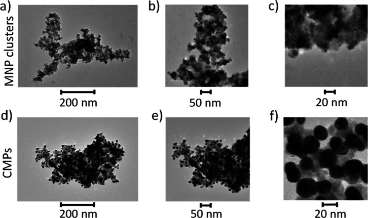
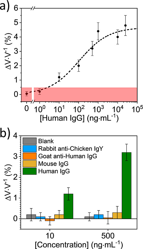
Magnetoresistive (MR) biosensors combine distinctive features such as small size, low cost, good sensitivity, and propensity to be arrayed to perform multiplexed analysis. Magnetic nanoparticles (MNPs) are the ideal target for this platform, especially if modified not only to overcome their intrinsic tendency to aggregate and lack of stability but also to realize an interacting surface suitable for biofunctionalization without strongly losing their magnetic response. Here, we describe an MR biosensor in which commercial MNP clusters were coated with gold nanoparticles (AuNPs) and used to detect human IgG in water using an MR biochip that comprises six sensing regions, each one containing five U-shaped spin valve sensors. The isolated AuNPs (satellites) were stuck onto an aggregate of individual iron oxide crystals (core) so that the resulting core@satellite magnetic particles (CSMPs) could be functionalized by the photochemical immobilization technique-an easy procedure that leads to oriented antibodies immobilized upright onto gold. The morphological, optical, hydrodynamic, magnetic, and surface charge properties of CSMPs were compared with those exhibited by the commercial MNP clusters showing that the proposed coating procedure endows the MNP clusters with stability and ductility without being detrimental to magnetic properties. Eventually, the high-performance MR biosensor allowed us to detect human IgG in water with a detection limit of 13 pM (2 ng mL-1). Given its portability, the biosensor described in this paper lends itself to a point-of-care device; moreover, the features of the MR biochip also make it suitable for multiplexed analysis.


Conflict of interest statement

The authors declare no competing financial interest.

Figures

Figure 1
Figure 1
a) Commercial MNP (Fe3O4 MNP) clusters (diameter ≈ 250 nm) were coated with smaller AuNPs (diameter approximately 15–20 nm). The resulting CSMPs were functionalized by means of the PIT. (b) Schematic representation (not in scale) of the MR chip detection system. The gold pad over the SV sensor was functionalized by PIT as well. The Abs on the chip surface captured the targets in solution. Subsequently, functionalized CSMPs recognized the target, and a “sandwich” was formed. The fringe magnetic field due to CSMPs changed the magnetization of the free layer of the SV sensor, and the resulting flux difference was detected as a resistance change.
Figure 2
Figure 2
TEM micrographs at different magnification of (a–c) MNP clusters from the stock and (d–f) CSMPs. AuNPs with diameter approximately 15–20 nm are clearly visible on the surface of MNP clusters whose length scale is approximately 250 nm [panels (c,f)].
Figure 3
Figure 3
Black and red colors refer to the aqueous solutions of MNP clusters and CSMPs, respectively. (a) Size distributions retrieved from DLS measurements. (b) Zeta potential distributions. (c) Experimental (solid lines) and simulated (dashed lines) extinction spectra of MNP clusters and CSMPs, respectively. The extinction peak at 567 nm signaled the growth of AuNPs onto the MNP cluster surfaces after the coating process. (d) Hysteresis loops obtained by a VSM. Data are normalized to the total mass without subtracting the gold contribution.
Figure 4
Figure 4
(a) Dose–response curve. Experimental data were fitted to eq 1. Each data point was duplicated with a different biochip. (b) Sensor specificity. The signal obtained with Abs other than human IgG is compatible with blank.
Figure 5
Figure 5
Typical dynamic response of the MR biosensor (sensorgram). The y-axis shows the potential difference across the chip, and the x-axis shows the time interval. The green line represents the response of the sensor in each of the following steps: (I) voltage baseline acquisition (flow rate 50 μL min–1), (II) CSMPs interacting with the sensor (static), and (III) washing step (flow rate 150 μL min–1).

References

    1. Baselt D. R.; Lee G. U.; Natesan M.; Metzger S. W.; Sheehan P. E.; Colton R. J. A Biosensor Based on Magnetoresistance Technology. Biosens. Bioelectron. 1998, 13, 731–739. 10.1016/S0956-5663(98)00037-2. - DOI - PubMed
    1. Graham D. L.; Ferreira H. A.; Freitas P. P. Magnetoresistive-Based Biosensors and Biochips. Trends Biotechnol. 2004, 22, 455–462. 10.1016/j.tibtech.2004.06.006. - DOI - PubMed
    1. Lim B.; Mahfoud M.; Das P. T.; Jeon T.; Jeon C.; Kim M.; Nguyen T.-K.; Tran Q.-H.; Terki F.; Kim C. Advances and Key Technologies in Magnetoresistive Sensors with High Thermal Stabilities and Low Field Detectivities. APL Mater. 2022, 10, 051108.10.1063/5.0087311. - DOI
    1. Hall D. A.; Gaster R. S.; Makinwa K. A. A.; Wang S. X.; Murmann B. A. A 256 Pixel Magnetoresistive Biosensor Microarray in 0.18 μm CMOS. IEEE J. Solid-State Circuits 2013, 48, 1290–1301. 10.1109/JSSC.2013.2245058. - DOI - PMC - PubMed
    1. Wu K.; Tonini D.; Liang S.; Saha R.; Chugh V. K.; Wang J. P. Giant Magnetoresistance Biosensors in Biomedical Applications. ACS Appl. Mater. Interfaces 2022, 14, 9945–9969. 10.1021/acsami.1c20141. - DOI - PMC - PubMed

LinkOut - more resources