. 2022 Dec;21(12):e13734.
doi: 10.1111/acel.13734. Epub 2022 Oct 24.

Advanced glycation end products induce senescence of atrial myocytes and increase susceptibility of atrial fibrillation in diabetic mice

Affiliations

Advanced glycation end products induce senescence of atrial myocytes and increase susceptibility of atrial fibrillation in diabetic mice

Dan-Lin Zheng et al. Aging Cell. 2022 Dec.

Abstract

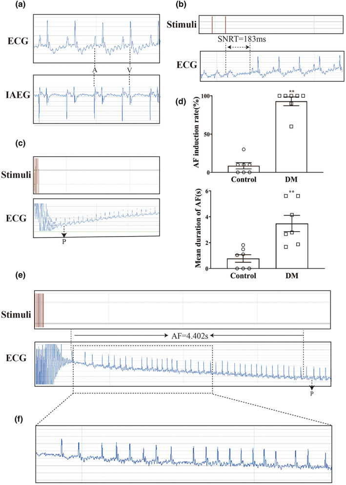
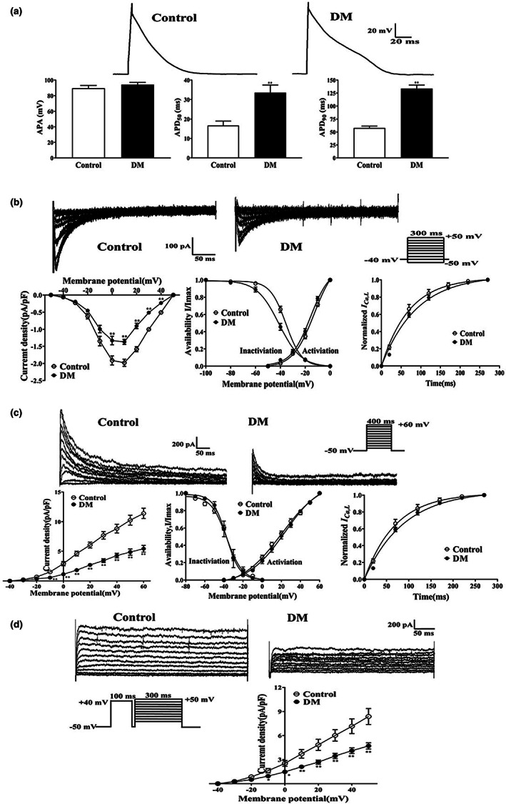
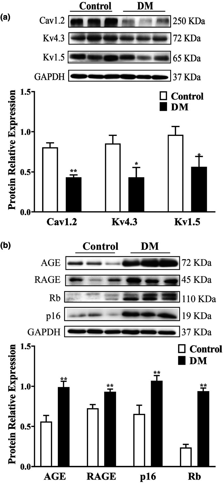
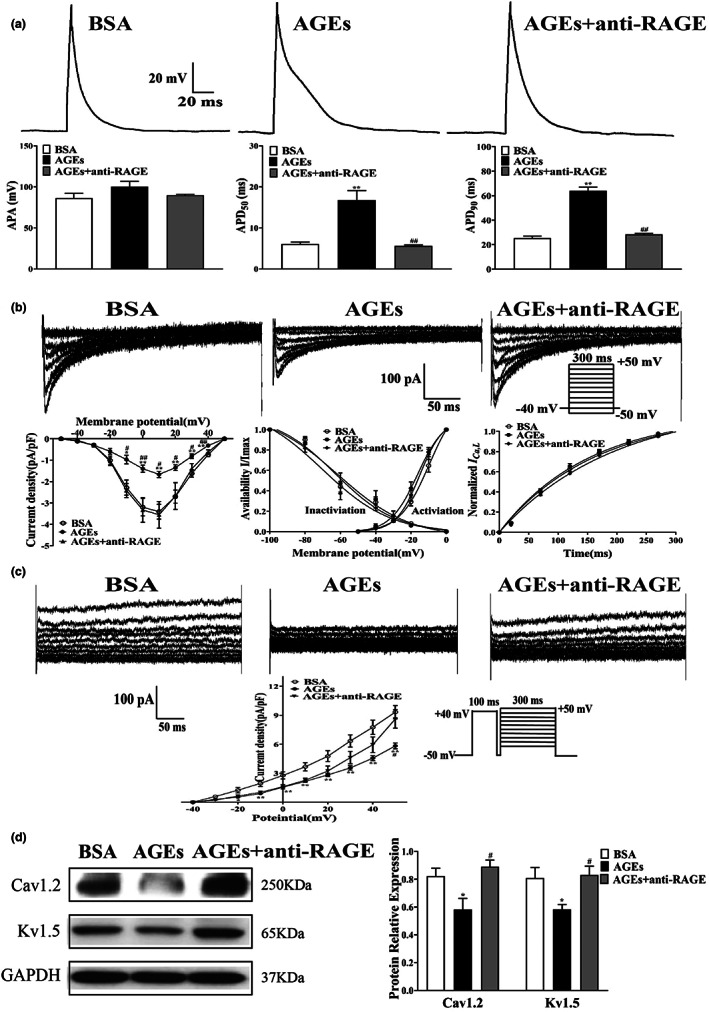
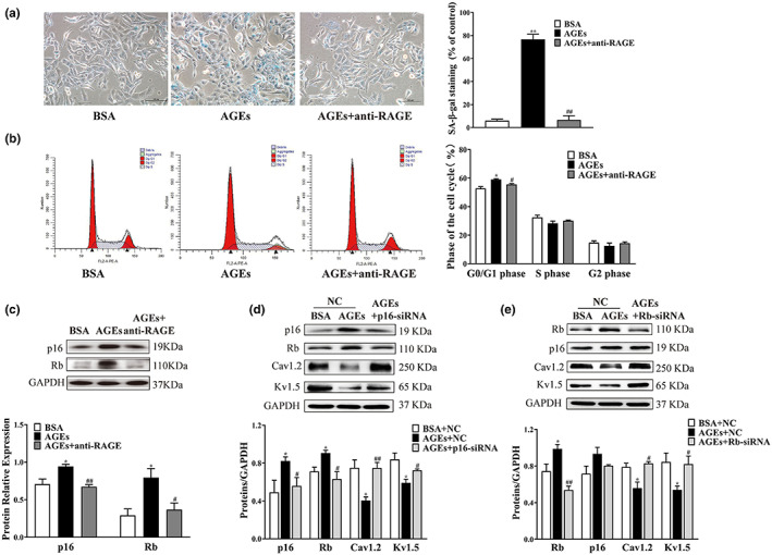
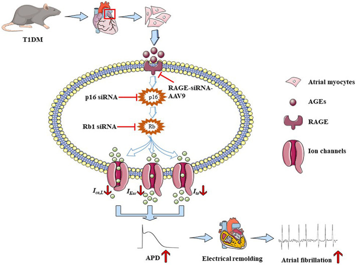
Diabetes mellitus (DM) is a common chronic metabolic disease caused by significant accumulation of advanced glycation end products (AGEs). Atrial fibrillation (AF) is a common cardiovascular complication of DM. Here, we aim to clarify the role and mechanism of atrial myocyte senescence in the susceptibility of AF in diabetes. Rapid transesophageal atrial pacing was used to monitor the susceptibility of mice to AF. Whole-cell patch-clamp was employed to record the action potential (AP) and ion channels in single HL-1 cell and mouse atrial myocytes. More importantly, anti-RAGE antibody and RAGE-siRNA AAV9 were used to investigate the relationship among diabetes, aging, and AF. The results showed that elevated levels of p16 and retinoblastoma (Rb) protein in the atrium were associated with increased susceptibility to AF in diabetic mice. Mechanistically, AGEs increased p16/Rb protein expression and the number of SA-β-gal-positive cells, prolonged the action potential duration (APD), reduced protein levels of Cav1.2, Kv1.5, and current density of ICa,L , IKur in HL-1 cells. Anti-RAGE antibody or RAGE-siRNA AAV9 reversed these effects in vitro and in vivo, respectively. Furthermore, downregulating p16 or Rb by siRNA prevented AGEs-mediated reduction of Cav1.2 and Kv1.5 proteins expression. In conclusion, AGEs accelerated atrial electrical remodeling and cellular senescence, contributing to increased AF susceptibility by activating the p16/Rb pathway. Inhibition of RAGE or the p16/Rb pathway may be a potential therapeutic target for AF in diabetes.

Keywords: AGEs; atrial fibrillation; cell senescence; diabetes; electrical remodeling; p16 and Rb.


Conflict of interest statement

The authors declare that they have no conflict of interest. All institutional and national guidelines for the care and use of laboratory animals were followed.

Figures

FIGURE 1
FIGURE 1
Representative electrophysiology results for atrial rapid pacing in diabetic mice. (a) Typical baseline surface electrocardiogram (ECG) and intra atrial electrocardiogram (IAEG). (b) Typical surface ECG recordings of sinus node recovery time (SNRT) following a 6 s pacing train. (c) Typical surface ECG recordings of rats maintaining SR after 15 s of atrial burst pacing. (d) AF induction rate and mean duration of AF in Control and DM mice (n = 7). (e) Typical surface ECG recordings of diabetic mice with AF that spontaneously reverted to SR. (f) Typical disorganized atrial wave (f wave). ** p < 0.01 vs. Control group.
FIGURE 2
FIGURE 2
Effects of APD and ion channel currents in atrial myocytes of diabetic mice. (a) Representative traces of APD [n = 9–11/6 (myocytes/mice)] in atrial myocytes from Control and DM groups. (b) Representative traces (pulse protocol, inset), corresponding current–voltage (IV) relationship, mean data for voltage dependence activation, inactivation, and time course of recovery current for I Ca,L [n = 9–16/6 (myocytes/mice)] in atrial myocytes from Control and DM mice. (c) Representative traces (pulse protocol, inset), corresponding current–voltage (IV) relationship, mean data for voltage dependence activation, inactivation, and time course of recovery current for I to [n = 8–12/6 (myocytes/mice)] in atrial myocytes from Control and DM mice. (d) Representative traces and current–voltage (IV) relationship for I Kur [n = 8–9/6 (myocytes/mice)] in atrial myocytes from Control and DM mice. ** p < 0.01 vs. Control group.
FIGURE 3
FIGURE 3
Expression of ion channels, AGE, RAGE and p16/Rb proteins in atrium of diabetic mice. (a) Representative blots and densitometry analysis of Cav1.2, K4.3, and Kv1.5 proteins in atrial tissues from Control and DM groups (n = 7–12). (b) Representative blots and densitometry analysis of AGE, RAGE, p16, Rb proteins in atrial tissues from Control and DM groups (n = 6–12). *p < 0.05, **p < 0.01 vs. Control group.
FIGURE 4
FIGURE 4
Effects of APD and ion channel currents on HL‐1 cells treated with AGEs or anti‐RAGE antibody. (a) Representative traces of APD (n = 10–12) in HL‐1 cells treated with AGEs and anti‐RAGE antibody. (b) Representative traces (pulse protocol, inset), corresponding current–voltage (IV) relationship, mean data for voltage dependence activation, inactivation, and time course of recovery current for I Ca,L (n = 6–12) in HL‐1 cells treated with AGEs or anti‐RAGE antibody. (c) Representative traces and current–voltage (IV) relationship for I Kur (n = 5–8) in HL‐1 cells treated with AGEs or anti‐RAGE antibody. (d) Representative blots and densitometry analysis of Cav1.2 and Kv1.5 proteins in HL‐1 cells treated with AGEs or anti‐RAGE antibody (n = 4). *p < 0.05 vs. BSA group. # p < 0.05 vs. AGEs group.
FIGURE 5
FIGURE 5
Alterations in senescence phenotype and expression levels of HL‐1 cells treated with AGEs, anti‐RAGE antibody, plasmid transfection. (a) SA‐β‐gal staining was used to elevate the positive rate of senescent cells treated with AGEs or anti‐RAGE antibody (n = 4). (b) Flow cytometry was used to detect cell cycle distribution in HL‐1 cells treated with AGEs or anti‐RAGE antibody (n = 4). (c) Representative blots and densitometry analysis of p16 and Rb proteins in HL‐1 cells treated with AGEs or anti‐RAGE antibody (n = 4). (d) Representative blots and densitometry analysis of Cav1.2 and Kv1.5 proteins in HL‐1 cells intervene with p16 protein (n = 3). (e) Representative blots and densitometry analysis of Cav1.2 and Kv1.5 proteins in HL‐1 cells intervene with Rb protein (n = 3–5). *p < 0.05, **p < 0.01 vs. BSA group or BSA + NC group. # p < 0.05, ## p < 0.01 vs. AGEs group or AGEs + NC group.
FIGURE 6
FIGURE 6
Schematic diagrams depicting proposed the mechanism of AGEs can accelerate cellular senescence of atrial myocytes through the p16/Rb pathways and increase the susceptibility to atrial fibrillation in diabetes. AGE and RAGE are accumulated in diabetes mellitus, which can accelerate cellular senescence and electrical remodeling of atrial myocytes through the p16/Rb signaling pathway. Meanwhile, reducing I Ca,L , I to and I Kur current density, extending the APD, and finally leading to atrial fibrillation. Mechanically, inhibition of RAGE can improve cellular senescence and atrial electrical remodeling; what is more, silencing p16 or Rb protein will promote atrial electrical remodeling by increasing the level of Cav1.2 and Kv1.5 proteins, revealing that RAGE or the p16/Rb pathway may be a potential therapeutic target for AF in diabetes.

References

    1. Alam, P. , Haile, B. , Arif, M. , Pandey, R. , Rokvic, M. , Nieman, M. , Maliken, B. D. , Paul, A. , Wang, Y. G. , Sadayappan, S. , Ahmed, R. P. H. , & Kanisicak, O. (2019). Inhibition of Senescence‐Associated Genes Rb1 and Meis2 in Adult Cardiomyocytes Results in Cell Cycle Reentry and Cardiac Repair Post‐Myocardial Infarction. Journal of the American Heart Association, 8, e012089. 10.1161/jaha.119.012089 - DOI - PMC - PubMed
    1. Andrade, J. , Khairy, P. , Dobrev, D. , & Nattel, S. (2014). The clinical profile and pathophysiology of atrial fibrillation: relationships among clinical features, epidemiology, and mechanisms. Circulation Research, 114, 1453–1468. 10.1161/circresaha.114.303211 - DOI - PubMed
    1. Baker, D. J. , Childs, B. G. , Durik, M. , Wijers, M. E. , Sieben, C. J. , Zhong, J. , Saltness, R. A. , Jeganathan, K. B. , Verzosa, G. C. , Pezeshki, A. , Khazaie, K. , Miller, J. D. , & van Deursen, J. M. (2016). Naturally occurring p16(Ink4a)‐positive cells shorten healthy lifespan. Nature, 530, 184–189. 10.1038/nature16932 - DOI - PMC - PubMed
    1. Barana, A. , Matamoros, M. , Dolz‐Gaitón, P. , Pérez‐Hernández, M. , Amorós, I. , Núñez, M. , Sacristán, S. , Pedraz, Á. , Pinto, Á. , Fernández‐Avilés, F. , Tamargo, J. , Delpón, E. , & Caballero, R. (2014). Chronic atrial fibrillation increases microRNA‐21 in human atrial myocytes decreasing L‐type calcium current. Circulation: Arrhythmia and Electrophysiology, 7, 861–868. 10.1161/circep.114.001709 - DOI - PubMed
    1. Campisi, J. , & d'Adda di Fagagna, F. (2007). Cellular senescence: when bad things happen to good cells. Nature Reviews Molecular Cell Biology, 8, 729–740. 10.1038/nrm2233 - DOI - PubMed

MeSH terms

Substances