. 2022 Nov 15;149(22):dev201016.
doi: 10.1242/dev.201016. Epub 2022 Nov 16.

Shared cis-regulatory modules control expression of the tandem paralogs midline and H15 in the follicular epithelium

Affiliations

Shared cis-regulatory modules control expression of the tandem paralogs midline and H15 in the follicular epithelium

Cody A Stevens et al. Development. .

Abstract

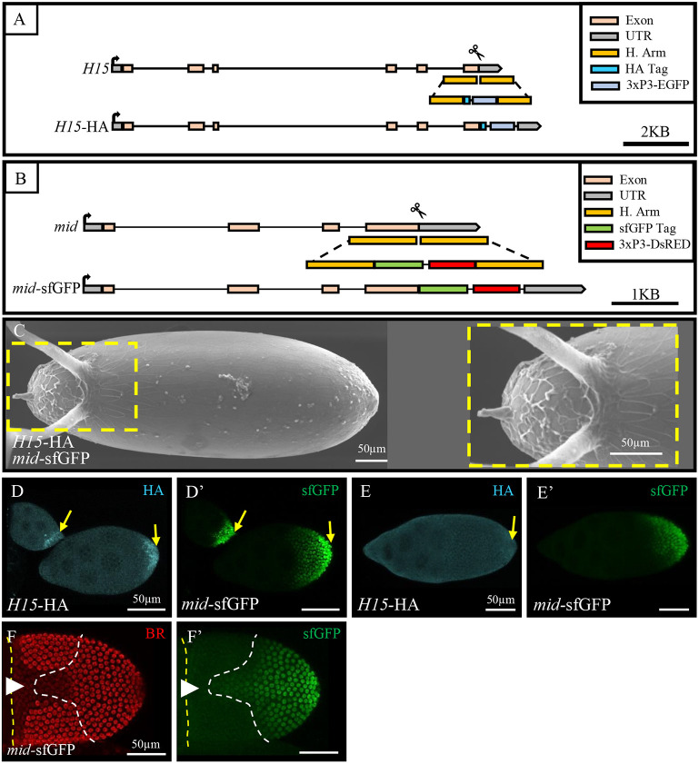
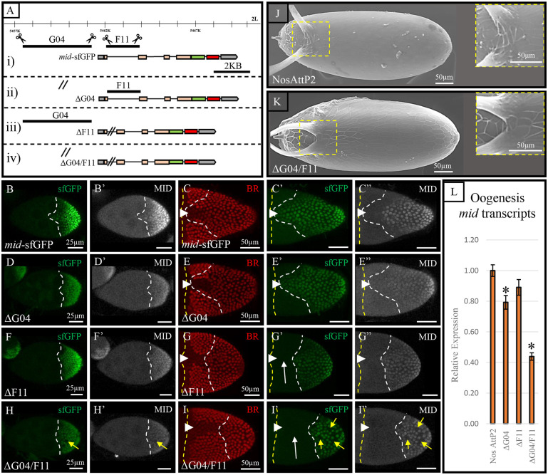
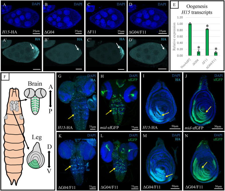
The posterior end of the follicular epithelium is patterned by midline (MID) and its paralog H15, the Drosophila homologs of the mammalian Tbx20 transcription factor. We have previously identified two cis-regulatory modules (CRMs) that recapitulate the endogenous pattern of mid in the follicular epithelium. Here, using CRISPR/Cas9 genome editing, we demonstrate redundant activity of these mid CRMs. Although the deletion of either CRM alone generated marginal change in mid expression, the deletion of both CRMs reduced expression by 60%. Unexpectedly, the deletion of the 5' proximal CRM of mid eliminated H15 expression. Interestingly, expression of these paralogs in other tissues remained unaffected in the CRM deletion backgrounds. These results suggest that the paralogs are regulated by a shared CRM that coordinates gene expression during posterior fate determination. The consistent overlapping expression of mid and H15 in various tissues may indicate that the paralogs could also be under shared regulation by other CRMs in these tissues.

Keywords: Cis-regulation; Anterior-posterior axis coordination; Developmental genes; Gene regulatory network; Genes tagging; Shared enhancers.


Conflict of interest statement

Competing interests The authors declare no competing or financial interests.

Figures

Fig. 1.
Fig. 1.
CRISPR/Cas9 endogenous tagging of H15 and mid. (A,B) Schematic diagram of CRISPR/Cas9 genome editing of H15 and mid loci. H. Arm, homology arm. Black arrows indicate the direction of transcription; scissors indicate the location of a guide RNA double-stranded break. (C) H15-HA and mid-sfGFP eggshell (n=12) exhibiting wild-type eggshell morphology (compare with Fig. 2J). Dorsal view. Dashed yellow line indicates the area shown on the right. (D-E′) The patterns of H15-HA and MID-sfGFP at stages 7 and 8 (D,D′) (n=15) and stage 9 (E,E′) (n=8), yellow arrows mark the posterior end of the egg chamber. (F,F′) The pattern of MID-sfGFP at stage 10B. White dashed line indicates the boundary of MID-sfGFP pattern. Broad (BR, red) (n=8). Yellow dashed lines indicate the anterior boundary; white arrowheads indicate the dorsal midline. All images are shown with anterior towards the left; n, number of images with same results. Scale bars: 50 µm.
Fig. 2.
Fig. 2.
Testing the function of G04 and F11 CRMs in the regulation of mid expression. (A) Schematic diagram of CRISPR/Cas9 genome editing of CRM deletions in the mid locus; horizontal line with small vertical lines marks the scaffold location on left arm 2nd chromosome. Scissors indicate the location of guide RNAs. Double diagonal lines indicate CRM deletion (Δ) regions. (B-C″) MID-sfGFP within the control at stage 8 (B,B′) (n=8) and stage 10B (C-C″) (n=16). (D-E″) MID-sfGFP within the G04 CRM deletion (ΔG04) at stage 8 (D,D′) (n=18) and stage 10B (E-E″) (n=10). (F-G″) MID-sfGFP within the F11 CRM deletion (ΔF11) at stage 8 (F,F′) (n=11) and stage 10B (G-G″) (n=6). White arrows in G′,I′ indicate loss of MID-sfGFP in dorsal midline domain. MID-sfGFP within G04 and F11 CRM deletion (ΔG04/F11) at stage 8 (n=6) and stage 10B (G,G′,I,I′) (n=5). Yellow arrows indicate loss of observable MID-sfGFP and MID. (C,E,G,I) Broad (BR, red). Yellow dashed line marks the anterior boundary; white arrowheads mark the dorsal midline; white dashed lines mark the boundary of the MID-sfGFP pattern. (B-I″) Using the antiGFP antibody together with the anti-MID increases the detection sensitivity of the anti-GFP antibody. (J) Eggshell of control (n=23) and (K) ΔG04/F11 line (n=70). The areas outlined by the dashed yellow boxes at the base of the two dorsal appendages are shown on the right. (L) qPCR for relative expression of mid transcripts compared with control NosAttP2 for each respective background. mid is reduced compared with control NosAttP2 expression (*P≤0.05; unpaired Student's t-test). All images are shown with anterior towards the left; n, number of images with same results. Scale bars: 25 µm (B,B′,D,D′,F,F′,H,H′); 50 µm (C-C″,E-E″,G-G″,I-K).
Fig. 3.
Fig. 3.
The G04 CRM co-regulates mid and H15 expression. The pattern of H15-HA in the control (A,A′) (n=21), (B,B′) ΔG04 (n=11), (C,C′) ΔF11 (n=12), (D,D′) ΔG04/F11 (n=10). (A,B,C,D) Corresponding DAPI staining of nuclei (blue). (E) qPCR for relative expression of H15 transcripts compared with control NosAttP2 during oogenesis in each background. H15 transcripts are significantly reduced compared with the control NosAttP2 (*P≤0.05; unpaired Student's t-test). (F) Schematic diagram of 3rd instar larval brain and leg imaginal discs. Green indicates known mid/H15 expression. A, anterior, P, posterior, D, dorsal, V, ventral. (G-J) Control line exhibiting the patterns of H15-HA and MID-sfGFP in larval brain ventral nerve cord and leg imaginal disc (yellow arrows) (n=7, 5, 10 and 8, respectively). (K-N) ΔG04/F11 line exhibiting the pattern of H15-HA and MID-sfGFP as in G-J (n=3, 4, 14 and 8, respectively). For egg chambers, anterior is towards the left. n, number of images with same results. Yellow arrow indicates the protein pattern. Scale bars: 25 µm in A-D′; 50 µm in I,J,M,N; 75 µm in G,H,K,L.

References

    1. Altschul, S. F., Madden, T. L., Schaffer, A. A., Zhang, J., Zhang, Z., Miller, W. and Lipman, D. J. (1997). Gapped BLAST and PSI-BLAST: a new generation of protein database search programs. Nucleic Acids Res. 25, 3389-3402. 10.1093/nar/25.17.3389 - DOI - PMC - PubMed
    1. Andersson, R. and Sandelin, A. (2020). Determinants of enhancer and promoter activities of regulatory elements. Nat. Rev. Genet. 21, 71-87. 10.1038/s41576-019-0173-8 - DOI - PubMed
    1. Bastock, R. and St Johnston, D. (2008). Drosophila oogenesis. Curr. Biol. 18, R1082-R1087. 10.1016/j.cub.2008.09.011 - DOI - PubMed
    1. Baudouin-Gonzalez, L., Santos, M. A., Tempesta, C., Sucena, E., Roch, F. and Tanaka, K. (2017). Diverse Cis-Regulatory mechanisms contribute to expression evolution of tandem gene duplicates. Mol. Biol. Evol. 34, 3132-3147. 10.1093/molbev/msx237 - DOI - PMC - PubMed
    1. Berg, C. A. (2005). The Drosophila shell game: patterning genes and morphological change. Trends Genet. 21, 346-355. 10.1016/j.tig.2005.04.010 - DOI - PubMed

Publication types

MeSH terms

LinkOut - more resources