Review
. 2022 Oct;30(4):231-262.
doi: 10.4250/jcvi.2022.0058.

Aortic Arch Variants and Anomalies: Embryology, Imaging Findings, and Clinical Considerations

Affiliations
Review

Aortic Arch Variants and Anomalies: Embryology, Imaging Findings, and Clinical Considerations

Sang Bin Bae et al. J Cardiovasc Imaging. 2022 Oct.

Abstract

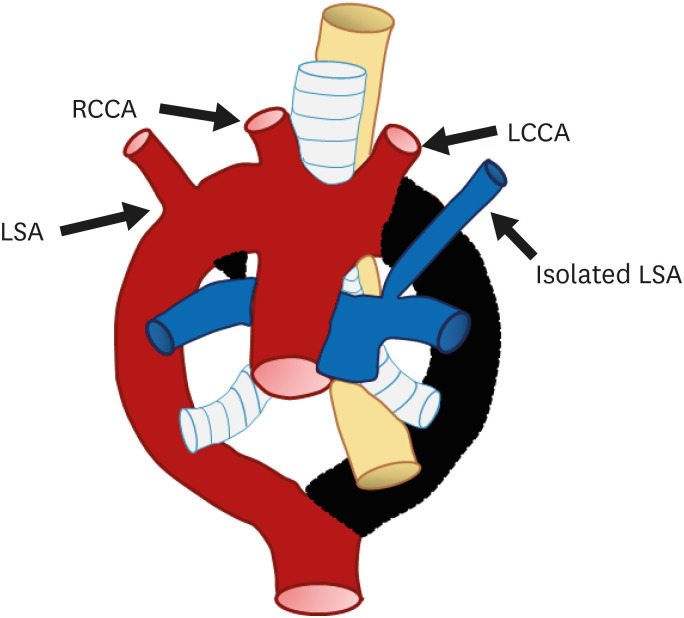
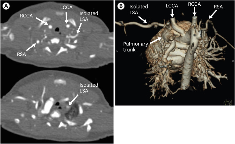
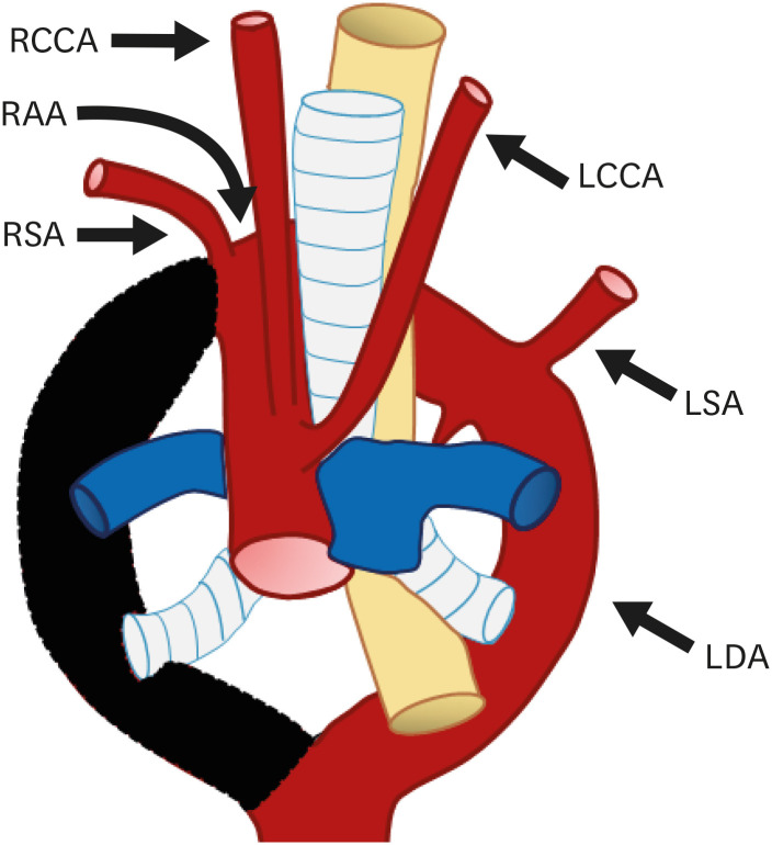
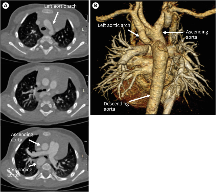
There is a wide spectrum of congenital anomalies or variations of the aortic arch, ranging from non-symptomatic variations that are mostly detected incidentally to clinically symptomatic variations that cause severe respiratory distress or esophageal compression. Some of these may be accompanied by other congenital heart diseases or chromosomal anomalies. The widespread use of multidetector computed tomography (CT) in clinical practice has resulted in incidental detection of several variations of the aortic arch in adults. Thus, radiologists and clinicians should be aware of the classification of aortic arch anomalies and carefully look for imaging features associated with a high risk of clinical symptoms. Understanding the embryological development of the aortic arch aids in the classification of various subtypes of aortic arch anomalies and variants. For accurate diagnosis and precise evaluation of aortic arch anomalies, cross-sectional imaging modalities, such as multidetector CT or magnetic resonance imaging, play an important role by providing three-dimensional reconstructed images. In this review, we describe the embryological development of the thoracic aorta and discuss variations and anomalies of the aortic arch along with their clinical implications.

Keywords: Aorta; Congenital abnormalities; Multidetector computed tomography; Technology.


Conflict of interest statement

The authors have no financial conflicts of interest.

Figures

Figure 1
Figure 1. (A) Schematic representation of Rathke’s diagram. Six paired branchial arches (numbered 1 to 6) connect the paired ventral aortae (arrowheads) and dorsal aortae (curved arrows). A pair of intersegmental arteries (straight arrows) arises from the aortic sac. (B) Schematic representation of the development of the normal aortic arch and branch vessels. Selective involution of the branchial arch segments results in the final adult aortic arch and is thought to be the main mechanism underlying arch and branch vessel anomalies.
BCA: brachiocephalic artery, CCA: common carotid artery, LECA: left external carotid artery, LICA: left internal carotid artery, LSA: left subclavian artery, RECA: right external carotid artery, RICA: right internal carotid artery, RSA: right subclavian artery.
Figure 2
Figure 2. (A) Schematic figure and (B) 3D volume-rendered image of a normal left aortic arch based on Edward’s hypothetical double arch. Black-shaded area represents involuted segments in the hypothetical double arch. A normal left aortic arch results from involution of the right arch between the right subclavian artery and descending aorta.
LCCA: left common carotid artery, LSA: left subclavian artery, RBCA: right brachiocephalic artery.
Figure 3
Figure 3. Common origin of the right brachiocephalic trunk and left common carotid artery in a 83-year-old man. Oblique coronal maximum intensity projection computed tomography angiographic image (A) and 3D volume-rendered image (B) show the common origin of the right brachiocephalic artery and left common carotid artery from the left aortic arch.
LCCA: left common carotid artery, LSA: left subclavian artery, RBCA: right brachiocephalic artery.
Figure 4
Figure 4. Direct origin of the left vertebral artery from the aortic arch in a 53-year-old man. Oblique sagittal 3D volume-rendered computed tomography angiographic image shows the direct origin of the LVA from the left aortic arch.
LCCA: left common carotid artery, LSA: left subclavian artery, LVA: left vertebral artery, RBCA: right brachiocephalic artery.
Figure 5
Figure 5. Origin of the right brachiocephalic artery in a 1-year-old boy. (A) Serial axial CT angiographic images and (B) 3D volume-rendered CT angiographic image show the origin of the brachiocephalic artery (RBCA, arrows) just left of midline on the aortic arch.
CT: computed tomography, RBCA: right brachiocephalic artery.
Figure 6
Figure 6. Classification of aortic arch variants and anomalies based on arch-sidedness and mirror imaging.
ALSA: aberrant left subclavian artery, ARSA: aberrant right subclavian artery, LAA: left-sided aortic arch, LSA: left subclavian artery, RAA: right-sided aortic arch, RSA: right subclavian artery.
Figure 7
Figure 7. Schematic figure of a left aortic arch with an ARSA. Black-shaded area represents involuted segments in a hypothetical double arch. This anomaly results from regression of the right arch between the right subclavian artery and right common carotid artery.
ARSA: aberrant right subclavian artery, LCCA: left common carotid artery, LSA: left subclavian artery, RCCA: right common carotid artery.
Figure 8
Figure 8. Left aortic arch with ARSA without Kommerell diverticulum in a 45-year-old man. (A, B) Transaxial and coronal CT angiographic images show an ARSA with a posterior mediastinal course. (C) Oblique coronal 3D volume-rendered CT angiographic image shows the branch arteries arising from the left aortic arch in order of RCCA, LCCA, left subclavian artery, and aberrant right subclavian artery.
ARSA: aberrant right subclavian artery, CT: computed tomography, LCCA: left common carotid artery, LSA: left subclavian artery, RCCA: right common carotid artery.
Figure 9
Figure 9. Left aortic arch with an ARSA with Kommerell diverticulum in a 63-year-old woman. (A, B) Axial and coronal CT angiographic images show an ARSA with Kommerell diverticulum (asterisk). (C) Sagittal CT angiographic image shows the Kommerell diverticulum causing esophageal compression (arrows). (D) 3D volume-rendered CT angiographic image shows the retro-tracheal course of the ARSA with diverticulum of Kommerell.
ARSA: aberrant right subclavian artery, CT: computed tomography.
Figure 10
Figure 10. Schematic figure of the left aortic arch with an isolated right subclavian artery. Black-shaded area represents involuted segments in a hypothetical double arch. This anomaly results from (i) regression of the right arch between the origin of the right common carotid artery and right subclavian artery and (ii) distal to the origin of the right ductus and right subclavian artery. Thus, the right subclavian artery is connected to the pulmonary artery instead of the aortic arch.
LCCA: left common carotid artery, LSA: left subclavian artery, RCCA: right common carotid artery, RSA: right subclavian artery.
Figure 11
Figure 11. Schematic figure of the left circumflex aorta. Black-shaded area represents involuted segments in a hypothetical double arch. This anomaly results from regression of the right fourth arch between the RCCA and right subclavian artery and persistence of the right-sided sixth arch component.
ARSA: aberrant right subclavian artery, LAA: left aortic arch, LCCA: left common carotid artery, LSA: left subclavian artery, RCCA: right common carotid artery, RDA: right descending aorta.
Figure 12
Figure 12. Schematic figure of the right aortic arch with mirror image branching. Black-shaded area represents involuted segments in a hypothetical double arch. This anomaly results from regression of the left aortic arch between the left subclavian artery and descending aorta.
LBCA: left brachiocephalic artery, RCCA: right common carotid artery, RSA: right subclavian artery.
Figure 13
Figure 13. Right aortic arch with mirror image branching in a 75-year-old woman. (A) Axial and coronal CT angiographic images show the left brachiocephalic artery arising from the aortic arch and then branching into the left common carotid and subclavian arteries. (C) Coronal 3D volume-rendered CT angiographic image shows branch arteries arising from the right aortic arch in order of left brachiocephalic artery, right common carotid artery, and right subclavian artery.
CT: computed tomography, LBCA: left brachiocephalic artery, LCCA: left common carotid artery, LSA: left subclavian artery, RCCA: right common carotid artery, RSA: right subclavian artery.
Figure 14
Figure 14. Schematic figure of a right aortic arch with an ALSA. Black-shaded area represents involuted segments in a hypothetical double arch. This anomaly results from regression of the left arch between the left subclavian artery and left common carotid artery.
ALSA: aberrant left subclavian artery, LCCA: left common carotid artery, RCCA: right common carotid artery, RSA: right subclavian artery.
Figure 15
Figure 15. Right aortic arch with an ALSA with Kommerell diverticulum in a 59-year-old man. (A, B) Axial and coronal CT angiography images show an ALSA with dilated Kommerell diverticulum (asterisk). The branch arteries arise in order of LCCA, RCCA, RSA, and ALSA. (C) 3D volume-rendered CT angiographic image shows the retro-tracheal course of the ALSA with diverticulum of Kommerell.
ALSA: aberrant left subclavian artery, CT: computed tomography, LCCA: left common carotid artery, RCCA: right common carotid artery, RSA: right subclavian artery.
Figure 16
Figure 16. Right aortic arch with an ALSA without Kommerell diverticulum in a 20-year-old man who underwent total correction surgery for Tetralogy of Fallot. (A-C) Serial axial and coronal computed tomography angiographic images show a right sided aortic arch with a non-dilated ALSA. The branch arteries arise from the right aortic arch in order of LCCA, RCCA, RSA, and ALSA.
ALSA: aberrant left subclavian artery, LCCA: left common carotid artery, RCCA: right common carotid artery, RSA: right subclavian artery.
Figure 17
Figure 17. Schematic figure of a right aortic arch with an isolated LSA. Black-shaded area represents involuted segments in a hypothetical double arch. This anomaly results from regression of the left arch i) between the origin of the LCCA and LSA and ii) distal to the origin of the left ductus and LSA.
LCCA: left common carotid artery, RCCA: right common carotid artery, LSA: left subclavian artery.
Figure 18
Figure 18. Isolated LSA in a newborn boy. (A) Serial axial CT angiographic images and (B) 3D volume-rendered CT angiographic image show the isolated LSA arising from the pulmonary trunk.
CT: computed tomography, LCCA: left common carotid artery, LSA: left subclavian artery, RCCA: right common carotid artery, RSA: right subclavian artery.
Figure 19
Figure 19. Schematic figure of the right circumflex aorta. Black-shaded area represents involuted segments of a hypothetical double arch. This anomaly results from regression of the left fourth arch between the LCCA and LSA and persistence of the left-sided sixth arch component.
LCCA: left common carotid artery, LDA: left descending aorta, LSA: left subclavian artery, RAA: right aortic arch, RCCA: right common carotid artery, RSA: right subclavian artery.
Figure 20
Figure 20. Left circumflex aorta in a 3-year-old boy. (A) Serial axial CT angiographic images and (B) 3D volume-rendered CT angiographic image show a left aortic arch crossing the midline posterior to the trachea and further descending on the right side of spine. Additionally, aortic arch hypoplasia and coarctation of the left aortic arch are present.
CT: computed tomography.
Figure 21
Figure 21. Schematic figure of a double aortic arch with ductus arteriosus on each side. This anomaly results from abnormal persistence of both aortic arches with absence of physiological regression.
LCCA: left common carotid artery, LSA: left subclavian artery, RCCA: right common carotid artery, RSA: right subclavian artery.
Figure 22
Figure 22. Double aortic arch in a 68-year-old man. (A, B) Serial transaxial and 3D volume-rendered computed tomography angiographic images show a double aortic arch with symmetric bilateral common carotid arteries and subclavian arteries also referred to as the “four vessels” sign. Double aortic arch encircling the trachea forms a complete vascular ring.
CT: computed tomography, LCCA: left common carotid artery, LSA: left subclavian artery, RCCA: right common carotid artery, RSA: right subclavian artery.
Figure 23
Figure 23. Schematic figure of the left cervical aortic arch. There are two hypothetical pathophysiological causes of a cervical aortic arch based on embryology: i) abnormal involution of the fourth branchial arches and persistence of the second or third arch instead, or ii) failure of caudal migration of the immature fourth arch.
LCCA: left common carotid artery, LSA: left subclavian artery, RCCA: right common carotid artery, RSA: right subclavian artery.
Figure 24
Figure 24. Schematic figure of a persistent fifth aortic arch. Remnant of the aortic arch to the descending aorta or pulmonary artery.
LCCA: left common carotid artery, LSA: left subclavian artery, RCCA: right common carotid artery, RSA: right subclavian artery.
Figure 25
Figure 25. Schematic figure of the three subtypes of IAA based on the location of the interrupted segment: Type A, interruption distal to the origin of the left subclavian artery; Type B, interruption between the origins of the left common carotid artery and left subclavian artery; Type C, interruption between the origins of the right brachiocephalic artery and left common carotid artery.
IAA: interrupted aortic arch.
Figure 26
Figure 26. Type B interrupted aortic arch in a newborn girl. (A) Axial CT image shows interruption of the aortic arch distal to left CCA, and the descending aorta is continuous with the main pulmonary trunk. Additionally, an aberrant right SCA is noted in this case. (B) 3D volume-rendered CT angiographic image of another type B interrupted aortic arch patient.
ARSA: aberrant right subclavian artery, CT: computed tomography, LCCA: left common carotid artery, LSA: left subclavian artery, RCCA: right common carotid artery, RSA: right subclavian artery, SVC: superior vena cava.
Figure 27
Figure 27. Coarctation of the aorta with a left aortic arch in a 52-year-old man. (A, B) Serial axial images and 3D volume-rendered CT angiographic images showing focal stenosis of the descending thoracic aorta (arrow). Additionally, hypertrophy of both internal mammary, superior epigastric, intercostal, and lateral thoracic arteries (arrowheads) is visible in the axial CT angiographic images. (C) Echocardiography shows a typical bicuspid aortic valve with small raphe (arrows) between the right and left coronary cuspid valves.
CT: computed tomography.
Figure 28
Figure 28. Pseudocoarctation of the aorta with a left aortic arch in a 69-year-old woman. (A, B) Serial axial images and 3D volume-rendered computed tomography angiographic images show focal narrowing of the proximal descending thoracic aorta (arrows) with aneurysmal dilatation (arrowheads) distal to the narrowed portion. Aortic arch shows an elongated course without abnormal collateral vessel formation.

Similar articles

Cited by

References

    1. Goldmuntz E. The epidemiology and genetics of congenital heart disease. Clin Perinatol. 2001;28:1–10. - PubMed
    1. Kocis KC, Midgley FM, Ruckman RN. Aortic arch complex anomalies: 20-year experience with symptoms, diagnosis, associated cardiac defects, and surgical repair. Pediatr Cardiol. 1997;18:127–132. - PubMed
    1. Jakanani GC, Adair W. Frequency of variations in aortic arch anatomy depicted on multidetector CT. Clin Radiol. 2010;65:481–487. - PubMed
    1. Schleich JM. Development of the human heart: days 15–21. Heart. 2002;87:487. - PMC - PubMed
    1. Kau T, Sinzig M, Gasser J, et al. Aortic development and anomalies. Semin Intervent Radiol. 2007;24:141–152. - PMC - PubMed

Grants and funding