

Evaluation of Cardiac Mechanical Dyssynchrony in Heart Failure Patients Using Current Echo-Doppler Modalities
- PMID: 36280273
- PMCID: PMC9592249
- DOI: 10.4250/jcvi.2022.0061
Evaluation of Cardiac Mechanical Dyssynchrony in Heart Failure Patients Using Current Echo-Doppler Modalities
Abstract
Background: Current guidelines indicate electrical dyssynchrony as the major criteria for selecting patients for cardiac resynchronization therapy, and 25-35% of patients exhibit unfavorable responses to cardiac resynchronization therapy (CRT). We aimed to evaluate different cardiac mechanical dyssynchrony parameters in heart failure patients using current echo-Doppler modalities and we analyzed their association with electrical dyssynchrony.
Methods: The study included 120 heart failure with reduced ejection fraction (HFrEF) who underwent assessments for left ventricular mechanical dyssynchrony (LVMD) and interventricular mechanical dyssynchrony (IVMD).
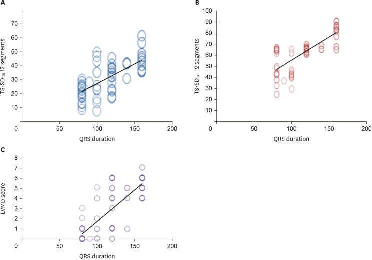
Results: Patients were classified according to QRS duration: group I with QRS < 120 ms, group II with QRS 120-149 ms, and group III with QRS ≥ 150 ms. Group III had significantly higher IVMD, LVMD indices, TS-SD speckle-tracking echocardiography (STE) 12 segments (standard deviation of time to peak longitudinal strain speckle tracking echocardiography in 12 LV-segments), and LVMD score compared with group I and group II. Group II and group III were classified according to QRS morphology into left bundle branch block (LBBB) and non-LBBB subgroups. LVMD score, TS-SD 12 TDI, and TS-SD 12 STE had good correlations with QRS duration.
Conclusions: HFrEF patients with wide QRS duration (> 150 ms) had more evident LVMD compared with patients with narrow or intermediate QRS. Those patients with intermediate QRS duration (120-150 ms) had substantial LVMD assessed by both TDI and 2D STE, regardless of QRS morphology. Subsequently, we suggest that LVMD indices might be employed as additive criteria to predict CRT response in that patient subgroup. Electrical and mechanical dyssynchrony were strongly correlated in HFrEF patients.
Keywords: Heart failure; Interventricuar mechanical delay; Left ventricular mechanical dyssynchrony score; Speckle tracking echocardiography.
Copyright © 2022 Korean Society of Echocardiography.
Conflict of interest statement
The authors have no financial conflicts of interest.
Figures
Similar articles
-
Frequency of inter- and intraventricular dyssynchrony in patients with heart failure according to QRS width.Europace. 2007 Dec;9(12):1171-6. doi: 10.1093/europace/eum234. Epub 2007 Oct 19. Europace. 2007. PMID: 17951575
-
Assessment of mechanical dyssynchrony in cardiac resynchronization therapy.Dan Med J. 2014 Dec;61(12):B4981. Dan Med J. 2014. PMID: 25441737 Review.
-
Cardiac resynchronization therapy in patients with heart failure and narrow QRS complexes (≤ 130 ms): role of speckle tracking echocardiography and different interventricular (VV) pacing intervals.J Interv Card Electrophysiol. 2022 Mar;63(2):369-377. doi: 10.1007/s10840-021-01021-y. Epub 2021 Jun 17. J Interv Card Electrophysiol. 2022. PMID: 34138397
-
Association of intraventricular mechanical dyssynchrony with response to cardiac resynchronization therapy in heart failure patients with a narrow QRS complex.Eur Heart J. 2010 Dec;31(24):3054-62. doi: 10.1093/eurheartj/ehq334. Epub 2010 Sep 23. Eur Heart J. 2010. PMID: 20864484 Free PMC article.
-
Role of echocardiography before cardiac resynchronization therapy: new advances and current developments.Echocardiography. 2016 Nov;33(11):1745-1752. doi: 10.1111/echo.13334. Epub 2016 Aug 25. Echocardiography. 2016. PMID: 27562174 Review.
Cited by
-
The Role of Echocardiography in Evaluating Cardiovascular Diseases in Patients with Diabetes Mellitus.Diabetes Metab J. 2023 Jul;47(4):470-483. doi: 10.4093/dmj.2023.0036. Epub 2023 Jul 27. Diabetes Metab J. 2023. PMID: 37533197 Free PMC article. Review.
-
Association between ultrasound-quantified cardiac mechanical dyssynchrony and left heart function and remodeling.Quant Imaging Med Surg. 2025 May 1;15(5):4454-4469. doi: 10.21037/qims-2024-2554. Epub 2025 Apr 28. Quant Imaging Med Surg. 2025. PMID: 40384660 Free PMC article.
-
Parameters of the mitral apparatus in patients with ischemic and nonischemic dilated cardiomyopathy.J Int Med Res. 2023 Dec;51(12):3000605231218645. doi: 10.1177/03000605231218645. J Int Med Res. 2023. PMID: 38150557 Free PMC article. Review.
-
Correlation Between Electrical and Mechanical Dyssynchrony in Patients With Heart Failure With Reduced Ejection Fraction.J Cardiovasc Imaging. 2022 Oct;30(4):320-321. doi: 10.4250/jcvi.2022.0095. J Cardiovasc Imaging. 2022. PMID: 36280274 Free PMC article. No abstract available.
-
Refining cardiac resynchronization therapy: a comprehensive review on the role of advanced multimodality imaging.Front Cardiovasc Med. 2024 Dec 18;11:1406899. doi: 10.3389/fcvm.2024.1406899. eCollection 2024. Front Cardiovasc Med. 2024. PMID: 39744205 Free PMC article. Review.
References
-
- Glikson M, Nielsen JC, Kronborg MB, et al. 2021 ESC Guidelines on cardiac pacing and cardiac resynchronization therapy. Eur Heart J. 2021;42:3427–3520. - PubMed
-
- Bank AJ, Burns KV, Gage RM. Echocardiographic measurement of mechanical dyssynchrony in heart failure and cardiac resynchronization therapy. US Cardiol Rev. 2010;7:24–32.
-
- Kleijn SA, Aly MF, Knol DL, et al. A meta-analysis of left ventricular dyssynchrony assessment and prediction of response to cardiac resynchronization therapy by three-dimensional echocardiography. Eur Heart J Cardiovasc Imaging. 2012;13:763–775. - PubMed
-
- Mor-Avi V, Lang RM, Badano LP, et al. Current and evolving echocardiographic techniques for the quantitative evaluation of cardiac mechanics: ASE/EAE consensus statement on methodology and indications endorsed by the Japanese Society of Echocardiography. J Am Soc Echocardiogr. 2011;24:277–313. - PubMed
-
- Dillikar MV, Venkateshvaran A, Barooah B, Varyani R, Kini P. Three-dimensional versus two - dimensional strain for the assessment of myocardial function: a case series. J Ind Acad Echocardiogr Cardiovasc Imaging. 2017;1:18–23.
LinkOut - more resources
Full Text Sources
Research Materials
