Review
. 2023 Jul:95:101133.
doi: 10.1016/j.preteyeres.2022.101133. Epub 2022 Oct 22.

Congenital aniridia beyond black eyes: From phenotype and novel genetic mechanisms to innovative therapeutic approaches

Affiliations
Review

Congenital aniridia beyond black eyes: From phenotype and novel genetic mechanisms to innovative therapeutic approaches

Alejandra Daruich et al. Prog Retin Eye Res. 2023 Jul.

Abstract

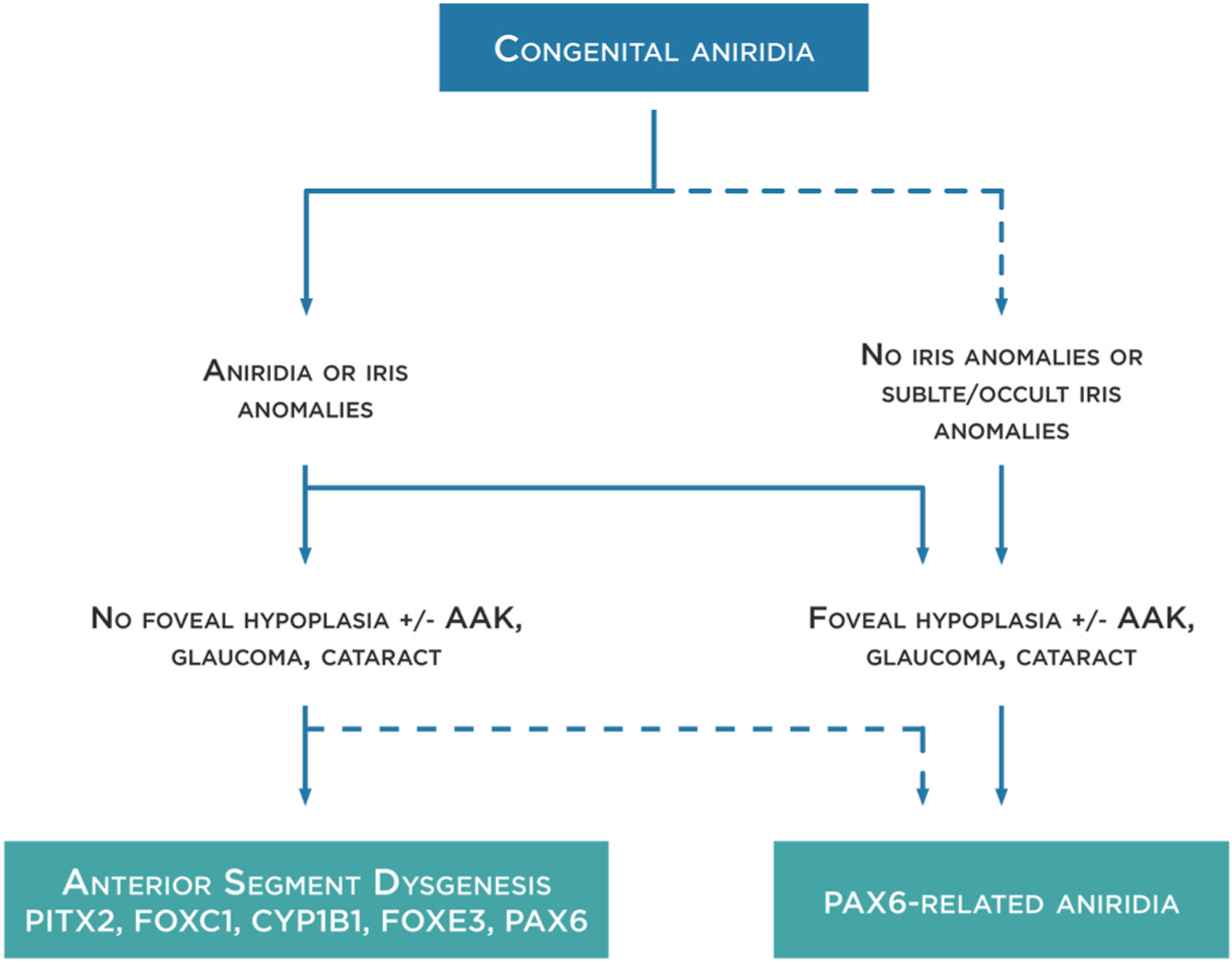
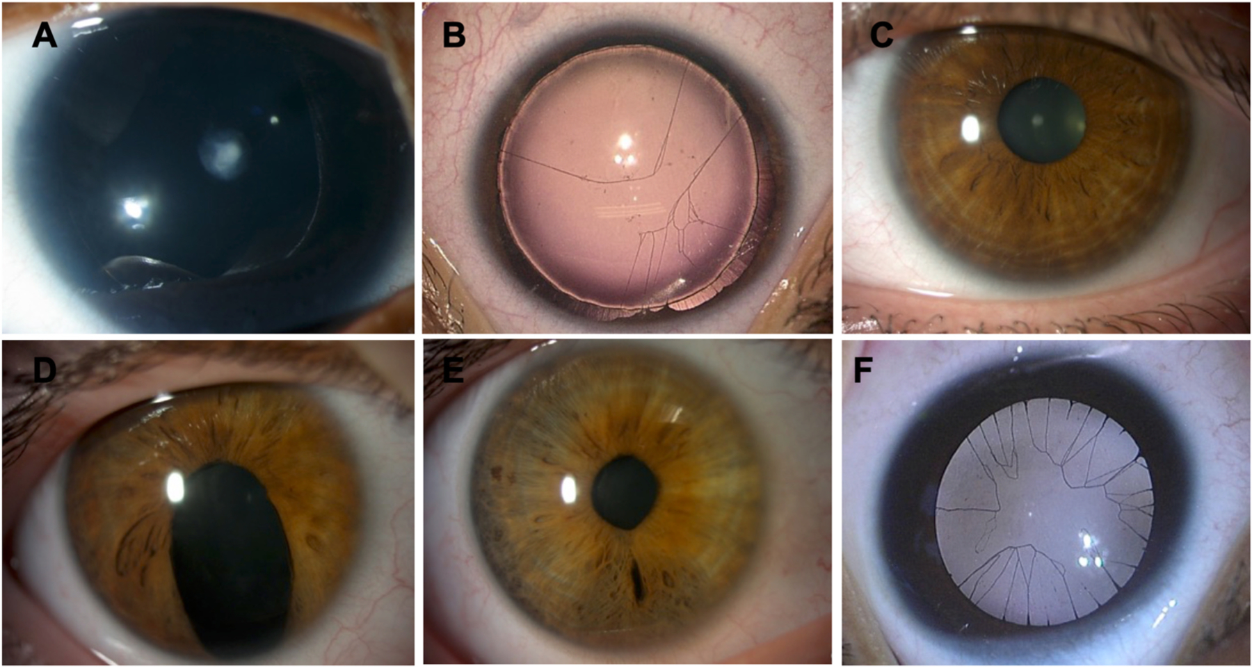
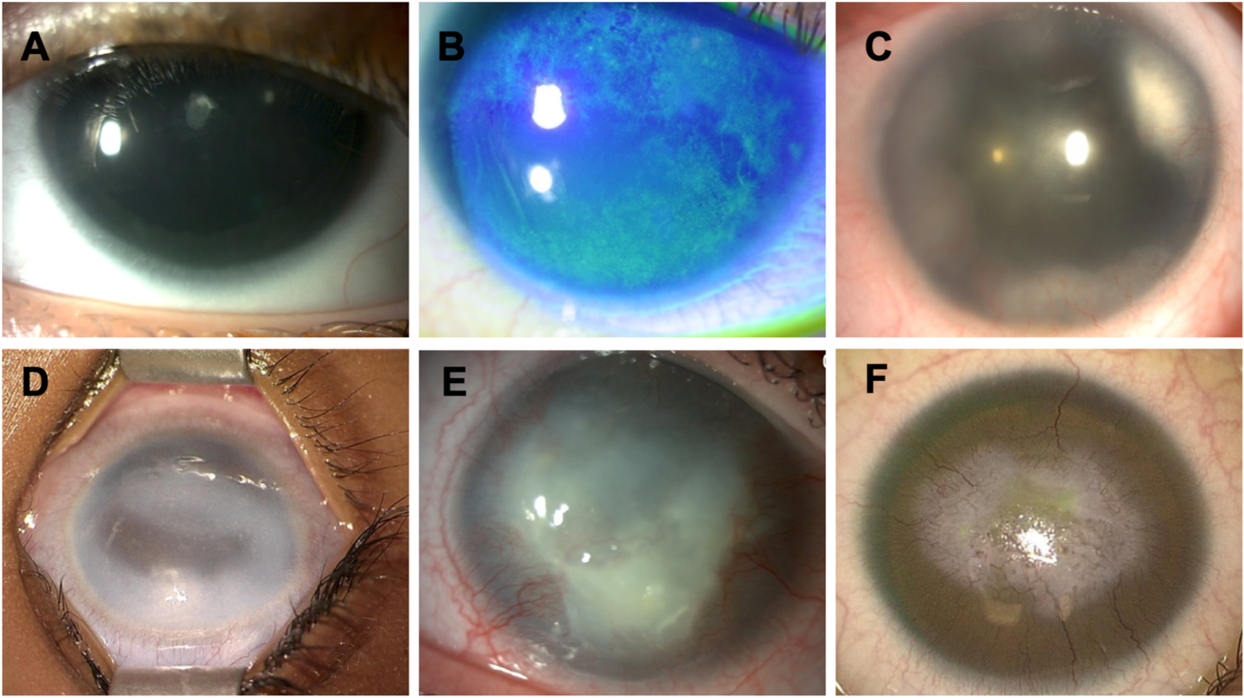
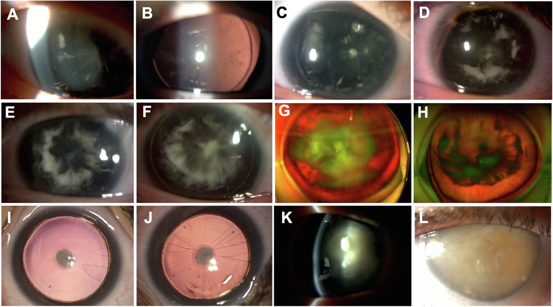
Congenital PAX6-aniridia, initially characterized by the absence of the iris, has progressively been shown to be associated with other developmental ocular abnormalities and systemic features making congenital aniridia a complex syndromic disorder rather than a simple isolated disease of the iris. Moreover, foveal hypoplasia is now recognized as a more frequent feature than complete iris hypoplasia and a major visual prognosis determinant, reversing the classical clinical picture of this disease. Conversely, iris malformation is also a feature of various anterior segment dysgenesis disorders caused by PAX6-related developmental genes, adding a level of genetic complexity for accurate molecular diagnosis of aniridia. Therefore, the clinical recognition and differential genetic diagnosis of PAX6-related aniridia has been revealed to be much more challenging than initially thought, and still remains under-investigated. Here, we update specific clinical features of aniridia, with emphasis on their genotype correlations, as well as provide new knowledge regarding the PAX6 gene and its mutational spectrum, and highlight the beneficial utility of clinically implementing targeted Next-Generation Sequencing combined with Whole-Genome Sequencing to increase the genetic diagnostic yield of aniridia. We also present new molecular mechanisms underlying aniridia and aniridia-like phenotypes. Finally, we discuss the appropriate medical and surgical management of aniridic eyes, as well as innovative therapeutic options. Altogether, these combined clinical-genetic approaches will help to accelerate time to diagnosis, provide better determination of the disease prognosis and management, and confirm eligibility for future clinical trials or genetic-specific therapies.

Keywords: Congenital aniridia; Foveal hypoplasia; Gene therapy; Next-generation sequencing; PAX6; Whole-genome sequencing.


Conflict of interest statement

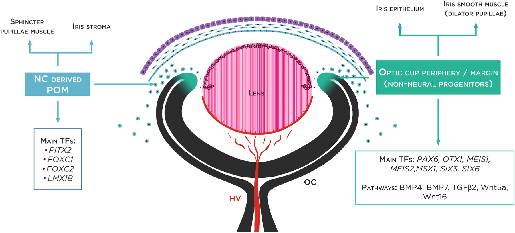
Declarations of competing interest None. The three layers of the cornea are defined: the corneal endothelium (CEnd) deriving from the first set of neural crest cells/POM cells, the corneal stroma (CS) deriving from the second wave of NC cells/POM cells and the corneal epithelium (CE) developing from the outer surface ectoderm. The third wave of NC cells/POM cells arrives at the angle of the future cornea and the periphery of the optic cup and contributes to pupillary membrane and iris stroma development. From the periphery of the optic cup will then originate the pigmented epithelium and the smooth muscle of the iris. HV (hyaloid vessels, tunica vasculosa lentis).

Figures

Fig. 1.
Fig. 1.. Congenital aniridia definition.
PAX6-related aniridia mostly presents with complete or partial iris hypoplasia (or aniridia) and foveal hypoplasia. However, PAX6-related aniridia could be also observed in the absence of iris anomalies. In these cases, foveal hypoplasia is usually present, as well as other aniridia-related findings such as aniridia-associated keratopathy (AAK), glaucoma and cataracts. Iris anomalies could be also part of an anterior segment dysgenesis phenotype where foveal hypoplasia and other aniridia-related findings are not present. Although PAX6 anomalies could be responsible for these phenotypes, others genes such as PITX2, FOXC1, CYP1B1 and FOXE3 are most frequently detected. Dot lines represent less frequent manifestations.
Fig. 2.
Fig. 2.
Major genes involved in iris development. Schematic overview of the developing optic cup (8 week old human embryo). Iris development relies on interactions between the periphery of optic cup (OC) or ciliary margin zone (neuroectoderm) and the periocular mesenchyme (POM), which receives contributions from the neural crest (NC) and mesoderm. Main transcription factors (TF) specifically expressed in the optic periphery cup and POM are represented.
Fig. 3.
Fig. 3.. Congenital aniridia workup.
Differentiation of isolated or syndromic congenital aniridia is essential for appropriate management and follow-up. Prompt diagnosis of WAGR or WAGRO is critical to avoid vital risks. An extensive ophthalmological workup needs to be performed to characterize the ocular phenotype, especially foveal hypoplasia through spectral-domain optical coherence tomography (SD-OCT). A genetic workup should first include comparative genomic hybridization arrays to allow for the quick identification of isolated deletions on the PAX6 gene or deletions involving the WT1 gene. In sporadic cases, and until the results are from comparative genomic hybridization arrays, a kidney ultrasound should be performed every 3 months for early detection of a Wilms’ tumor. In case of WT1 deletion, a kidney ultrasound should be continued every 3 months until the age of 7. If no deletion is found, a molecular genetic test (NGS and/or WGS) will be performed to confirm the clinical diagnosis and identify the pathogenic variants allowing phenotype-genotype correlations. It is to be noted that NGS-based protocol has the potential of becoming the one-for-all test for molecular diagnosis of aniridia (see section 4.3 and 4.4), replacing the standard strategy represented here. BCVA: best-corrected visual acuity, ON: optic nerve, ICA: iridocorneal angle, RNFL: retinal nerve fiber layer.
Fig. 4.
Fig. 4.. Genetic characteristics of the aniridia cohort from BaMaRa,
the French national database for rare diseases. Schematic representation of the cohort of 475 patients with iris anomalies. Distribution of genes found to be mutated among 382 patients who presented with aniridia phenotype after excluding patients presenting iris coloboma.
Fig. 5
Fig. 5. Variability of iris hypoplasia in congenital aniridia and genotype correlation.
PAX6-related congenital aniridia is associated to various iris phenotypes such as complete aniridia with subcapsular anterior cataracts (A), iris root and iris membrane remnants (B), almost normal iris (C), iris pseudocoloboma (D), discrete iris defects in the contralateral eye of the same patient (E) and congenital aniridia in Gillespie syndrome with iris membrane remnants (F).
Fig. 6.
Fig. 6.. Aniridia-associated keratopathy (AAK).
Grade 1 limbal insufficiency with mild anterior subcapsular cataracts (A) or with irregular ocular surface after fluoresceine instillation (B). Grade 2 limbal insufficiency (C). Grade 3 limbal insufficiency with corneal opacity. Note the isolated corneal fibrotic reaction without neovascularization (D). Grade 4 limbal insufficiency with corneal opacity and progressive neovascularization (E). Grade 4 limbal insufficiency with fibrotic reaction and extended corneal neovascularization (F). AAK terminal forms may be distinguished into two types, one with classical limbal insufficiency with central opacities and neovascularization (E, F) and a less common type with dense fibrotic whitish central opacities without neovascularization (D).
Fig. 7.
Fig. 7.. Cataract-related congenital aniridia. A-H. Cartwheel cataract pathognomonic of a PAX6 gene anomaly.
Early onset (AB) and progressive evolution of cartwheel cataracts with posterior subcapsular opacities (C–H). Note the asymmetrical severity of cartwheel cataracts between the eyes of the same patient (G and H). I-J. Anterior subcapsular congenital cataracts with pupillary membrane remnants. K. Anterior subcapsular cataract with marked posterior cartwheel cataract. L. Total Morgagnian cataract.
Fig. 8.
Fig. 8.. PAX6 role in pancreas organogenesis, particularly in islet development and in mature islets cells.
PAX6 is involved in pancreatic endocrine cells development. PAX6 regulates directly expression of insulin genes and of somatostatin in delta-cells in mature cells.
Fig. 9.
Fig. 9.. Schematic representation of the human PAX6 locus.
Representation of the PAX6 locus in chromosomal 11p13 region (A). Representation of human PAX6 and its surrounding 3′downstream genes. The downstream regulatory region (DDR) is represented within the neighboring ELP4 gene, with the SIMO enhancer highlighting by a black star. Each grey box represents a gene with its respective DNA strand location, and the arrows above each gene indicate the orientation of transcription. The long non-coding RNA, PAX6-AS1/PAUPAR, is located upstream and in the opposite orientation to PAX6 as indicated by the arrow (B). The human PAX6 gene is represented with its coding exons appearing as black boxes and non-coding exons as white boxes. The coding alternative spliced exon 5a is also represented between exons 5 and 6. The three alternative promoters of PAX6 are indicated by black arrows (C). tel: telomere, cen: centromere, 5′ UTR: 5′ untranslated region, 3′ UTR: 3′ untranslated region.
Fig. 10.
Fig. 10.. Innovative therapeutic approaches for aniridia-associated keratopathy (AAK).
To overcome the limitations of current therapeutic options for AAK, innovative approaches are under evaluation including gene therapy (adeno-associated virus (AAV) delivery, in vivo gene editing through clustered regularly interspaced short palindromic repeats (CRISPR)/CRISPR-associated protein 9 (Cas9) and nonsense mutation suppression) as well as drug repurposing.

Similar articles

Cited by

References

    1. Aalfs CM, Fantes JA, Wenniger-Prick LJ, Sluijter S, Hennekam RC, van Heyningen V, Hoovers JM, 1997. Tandem duplication of 11p12-p13 in a child with borderline development delay and eye abnormalities: dose effect of the PAX6 gene product? Am. J. Med. Genet. 73, 267–271. 10.1002/(sici)1096-8628(19971219)73:3<267::aid-ajmg7>3.0.co, 2. - DOI - PubMed
    1. Abouzeid H, Youssef MA, ElShakankiri N, Hauser P, Munier FL, Schorderet DF, 2009. PAX6 aniridia and interhemispheric brain anomalies. Mol. Vis. 15, 2074–2083. - PMC - PubMed
    1. Acampora D, Mazan S, Avantaggiato V, Barone P, Tuorto F, Lallemand Y, Brûlet P, Simeone A, 1996. Epilepsy and brain abnormalities in mice lacking the Otx1 gene. Nat. Genet. 14, 218–222. 10.1038/ng1096-218. - DOI - PubMed
    1. Acuna-Hidalgo R, Veltman JA, Hoischen A, 2016. New insights into the generation and role of de novo mutations in health and disease. Genome Biol. 17, 241. 10.1186/s13059-016-1110-1. - DOI - PMC - PubMed
    1. Adachi M, Dickens CJ, Hetherington J, Hoskins HD, Iwach AG, Wong PC, Nguyen N, Ma AS, 1997. Clinical experience of trabeculotomy for the surgical treatment of aniridic glaucoma. Ophthalmology 104, 2121–2125. 10.1016/s0161-6420(97)30041-4. - DOI - PubMed

Supplementary concepts