

Mitral Annular Disjunction Assessed Using CMR Imaging: Insights From the UK Biobank Population Study
- PMID: 36280553
- PMCID: PMC9640354
- DOI: 10.1016/j.jcmg.2022.07.015
Mitral Annular Disjunction Assessed Using CMR Imaging: Insights From the UK Biobank Population Study
Abstract
Background: Mitral annular disjunction is the atrial displacement of the mural mitral valve leaflet hinge point within the atrioventricular junction. Said to be associated with malignant ventricular arrhythmias and sudden death, its prevalence in the general population is not known.
Objectives: The purpose of this study was to assess the frequency of occurrence and extent of mitral annular disjunction in a large population cohort.
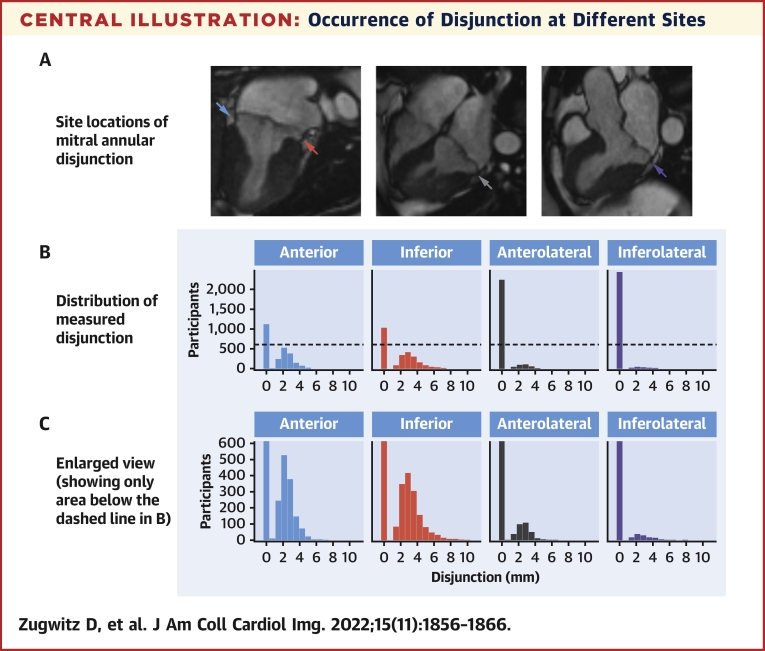
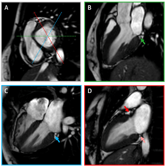
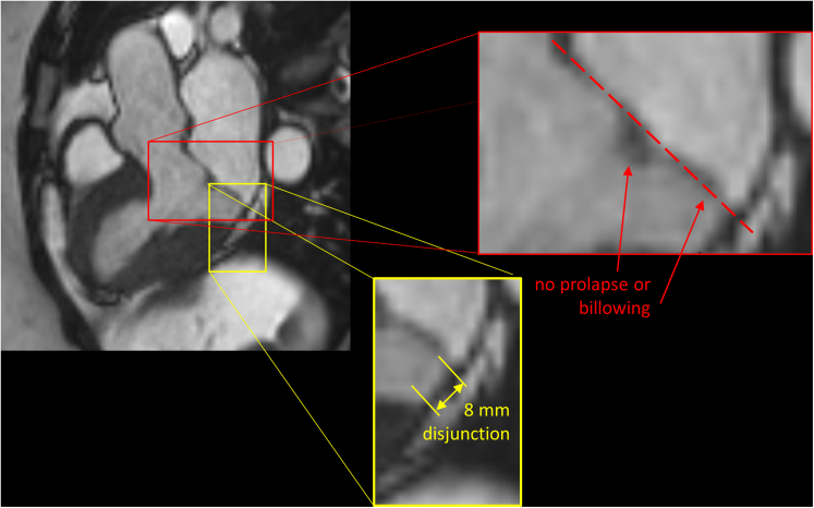
Methods: The authors assessed the cardiac magnetic resonance (CMR) images in 2,646 Caucasian subjects enrolled in the UK Biobank imaging study, measuring the length of disjunction at 4 points around the mitral annulus, assessing for presence of prolapse or billowing of the leaflets, and for curling motion of the inferolateral left ventricular wall.
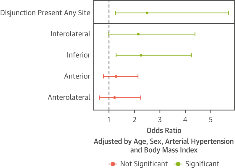
Results: From 2,607 included participants, the authors found disjunction in 1,990 (76%) cases, most commonly at the anterior and inferior ventricular wall. The authors found inferolateral disjunction, reported as clinically important, in 134 (5%) cases. Prolapse was more frequent in subjects with disjunction (odds ratio [OR]: 2.5; P = 0.02), with positive associations found between systolic curling and disjunction at any site (OR: 3.6; P < 0.01), and systolic curling and prolapse (OR: 71.9; P < 0.01).
Conclusions: This large-scale study shows that disjunction is a common finding when using CMR. Disjunction at the inferolateral ventricular wall, however, was rare. The authors found associations between disjunction and both prolapse and billowing of the mural mitral valve leaflet. These findings support the notion that only extensive inferolateral disjunction, when found, warrants consideration of further investigation, but disjunction elsewhere in the annulus should be considered a normal finding.
Keywords: cardiac magnetic resonance; mitral annular disjunction; mitral valve prolapse.
Copyright © 2022 The Authors. Published by Elsevier Inc. All rights reserved.
Conflict of interest statement
Funding Support and Author Disclosures This work was partly funded by the European Union’s Horizon 2020 research and innovation program under grant agreement number 825903 (euCanSHare project, Dr Petersen). Dr Petersen acknowledges support from the National Institute for Health Research (NIHR) Biomedical Research Centre at Barts, London, United Kingdom. Drs Petersen, Neubauer, and Piechnik acknowledge the British Heart Foundation, London, United Kingdom, for funding the manual analysis to create a cardiovascular magnetic resonance imaging reference standard for the UK Biobank imaging resource in 5000 CMR scans (PG/14/89/31194). This project was enabled through access to the Medical Research Council eMedLab Medical Bioinformatics infrastructure, supported by the Medical Research Council (MR/L016311/1). Dr Zugwitz acknowledges funding received from the European Society of Cardiology, Sophia Antipolis Cedex, France, in the form of an European Society of Cardiology Training Grant. Dr Neubauer acknowledges support from the Oxford NIHR Biomedical Research Centre and the Oxford British Heart Foundation Centre of Research Excellence. Dr Aung recognizes the NIHR Integrated Academic Training program, which supports his Academic Clinical Lectureship post. Drs McCracken and Neubauer are supported by the Oxford NIHR Biomedical Research Centre. Drs Petersen and Rauseo acknowledge support by the London Medical Imaging and Artificial Intelligence Centre for Value Based Healthcare (AI4VBH), which is funded from the Data to Early Diagnosis and Precision Medicine strand of the government’s Industrial Strategy Challenge Fund, managed and delivered by Innovate UK on behalf of United Kingdom Research and Innovation (UKRI). Dr Nijveldt has received research grants from Philips Volcano and Biotronik. Dr Petersen provides consultancy to Circle Cardiovascular Imaging, Inc. All other authors have reported that they have no relationships relevant to the contents of this paper to disclose.
Figures

Comment in
-
Mitral Annular Disjunction: Normal or Abnormal: It Is All About Location.JACC Cardiovasc Imaging. 2022 Nov;15(11):1867-1869. doi: 10.1016/j.jcmg.2022.08.002. Epub 2022 Oct 19. JACC Cardiovasc Imaging. 2022. PMID: 36357129 No abstract available.
References
-
- Henle J. Handbuch der Systematischen Anatomie des Menschen. Nature. 1871;4:101.
-
- Hutchins G.M., Moore G.W., Skoog D.K. The association of floppy mitral valve with disjunction of the mitral annulus fibrosus. N Engl J Med. 1986;314:535–540. - PubMed
-
- Angelini A., Ho S.Y., Anderson R.H., Becker A.E., Davies M.J., Hutchins G.M., et al. Disjunction of the mitral annulus in floppy mitral valve. N Engl J Med. 1988;318:188–189. - PubMed
Publication types
MeSH terms
Grants and funding
LinkOut - more resources
Full Text Sources
Medical
