

Dehydrozingerone promotes healing of diabetic foot ulcers: a molecular insight
- PMID: 36280629
- PMCID: PMC10409929
- DOI: 10.1007/s12079-022-00703-0
Dehydrozingerone promotes healing of diabetic foot ulcers: a molecular insight
Abstract
Introduction: One of the most common problems of diabetes are diabetic foot ulcers (DFUs). According to National Institute for Health, initial management of DFUs can decrease the complication of limb amputations and can improve the patient's quality of life. DFU treatment can be optimized with the help of multidisciplinary approach. Based on many studies, control of glucose levels in blood, antioxidant activity, reduction in cytokine levels, re-epithelialization, collagen formation, migration of fibroblasts are major phases involved in managing DFU. Dehydrozingerone (DHZ), has been known for its anti-inflammatory, antioxidant and wound healing properties.
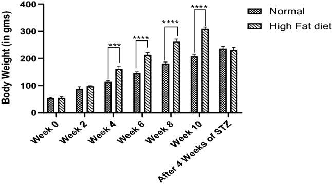
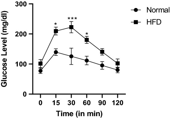
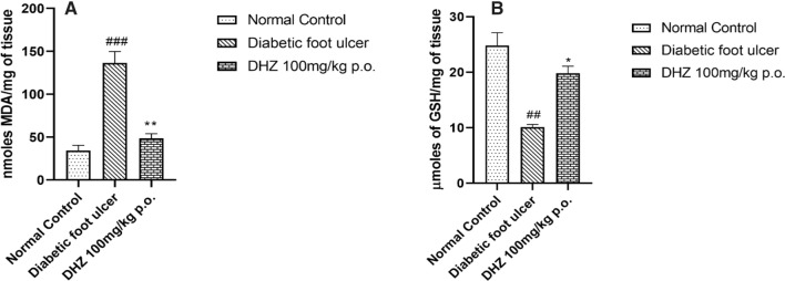
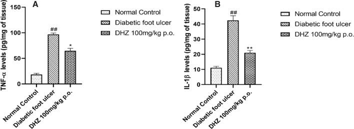
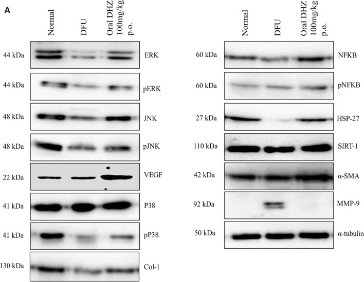
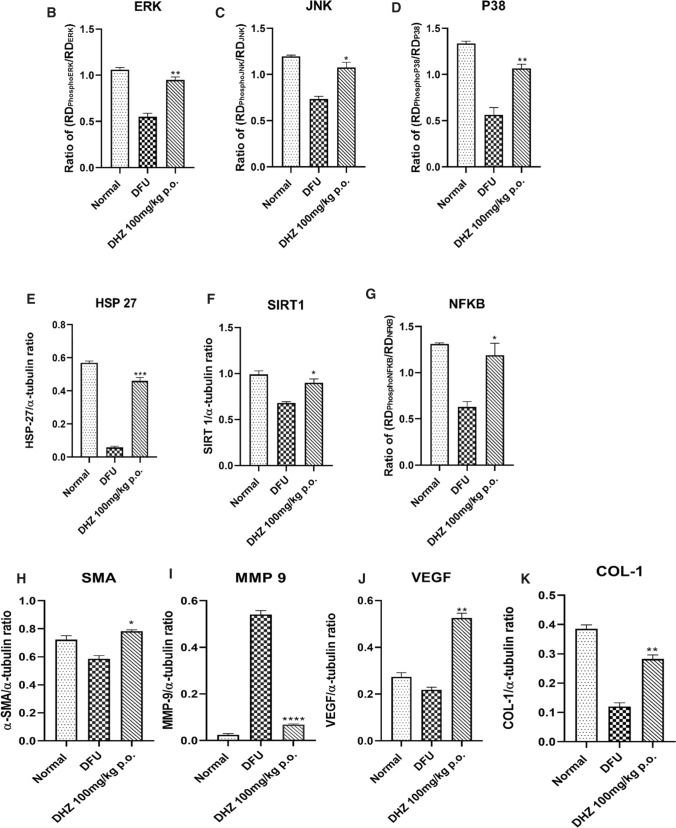
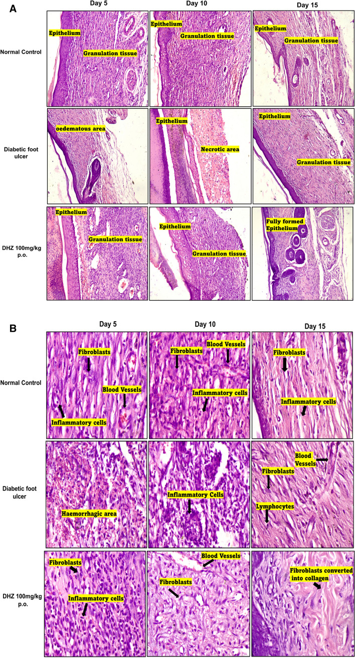
Methodology: Three months high-fat diet and low dose of streptozotocin-induced type-II diabetic foot ulcer model was used to evaluate the effectiveness of dehydrozingerone. DHZ was given orally to rats for 15 days post wounding. TNF-α, IL-1β and antioxidant parameters like lipid peroxidation, glutathione reductase were estimated. Immunoblotting was done to investigate the effect of DHZ on the expression of ERK, JNK, HSP-27, P38, SIRT-1, NFκB, SMA, VEGF and MMP-9 in skin tissue. Histopathology was performed for analyzing DHZ effect on migration of fibroblasts, formation of epithelium, granulation tissue formation, angiogenesis and collagen formation.
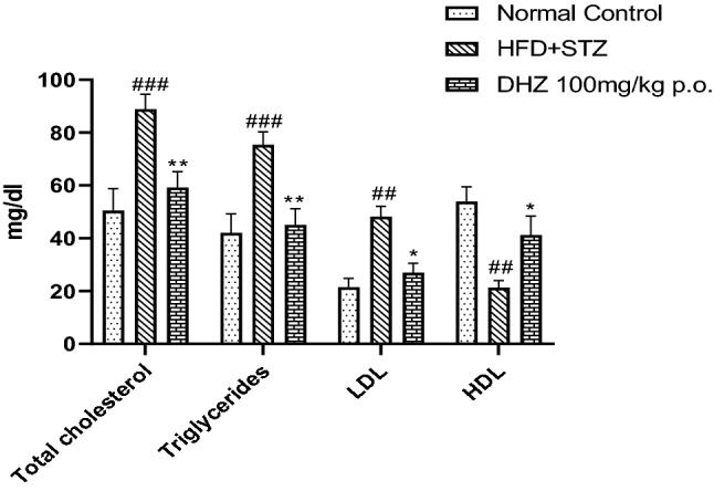
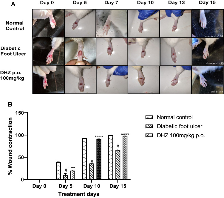
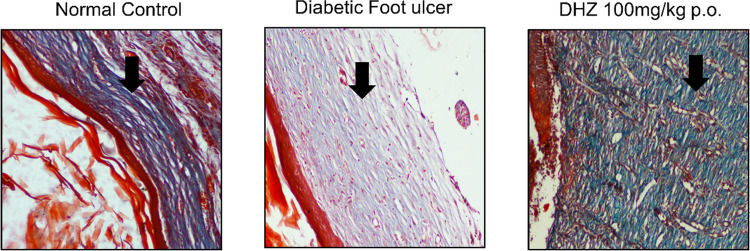
Results: DHZ decreased the levels of malondialdehyde, TNF-α, IL-1β and increased glutathione levels in wound tissue. Western blotting results suggested that DHZ activated ERK1/2/JNK/p38 signaling, increased expression of HSP-27, SIRT-1, VEGF, SMA thus facilitating the migration and proliferation of fibroblasts, angiogenesis and decreased inflammation. Masson Trichrome & histopathology showed an increase in collagen, epithelial and granulation tissue formation.
Conclusion: DHZ significantly accelerates the healing of diabetic foot ulcers in high fat diet fed plus low dose streptozotocin induced type-II diabetic Wistar rats.
Keywords: Cellular mechanism; Dehydrozingerone; Diabetic foot ulcers; High fat diet; Inflammation.
© 2022. The Author(s).
Conflict of interest statement
Authors declare no conflict of interest.
Figures
Similar articles
-
Investigation of the cellular and molecular effects of dehydrozingerone formulation on various days of diabetic wound repair.3 Biotech. 2024 Apr;14(4):124. doi: 10.1007/s13205-024-03963-2. Epub 2024 Apr 1. 3 Biotech. 2024. PMID: 38566928 Free PMC article.
-
Sesamol-Loaded PLGA Nanosuspension for Accelerating Wound Healing in Diabetic Foot Ulcer in Rats.Int J Nanomedicine. 2020 Nov 23;15:9265-9282. doi: 10.2147/IJN.S268941. eCollection 2020. Int J Nanomedicine. 2020. PMID: 33262587 Free PMC article.
-
Neurotensin-loaded collagen dressings reduce inflammation and improve wound healing in diabetic mice.Biochim Biophys Acta. 2014 Jan;1842(1):32-43. doi: 10.1016/j.bbadis.2013.10.009. Epub 2013 Oct 23. Biochim Biophys Acta. 2014. PMID: 24161538
-
Role of fibroblast plasticity and heterogeneity in modulating angiogenesis and healing in the diabetic foot ulcer.Mol Biol Rep. 2023 Feb;50(2):1913-1929. doi: 10.1007/s11033-022-08107-4. Epub 2022 Dec 17. Mol Biol Rep. 2023. PMID: 36528662 Review.
-
The role of CXCL8 in chronic nonhealing diabetic foot ulcers and phenotypic changes in fibroblasts: a molecular perspective.Mol Biol Rep. 2022 Feb;49(2):1565-1572. doi: 10.1007/s11033-022-07144-3. Epub 2022 Jan 19. Mol Biol Rep. 2022. PMID: 35044539 Review.
Cited by
-
Dang-Gui-Si-Ni decoction facilitates wound healing in diabetic foot ulcers by regulating expression of AGEs/RAGE/TGF-β/Smad2/3.Arch Dermatol Res. 2024 Jun 7;316(7):338. doi: 10.1007/s00403-024-03021-0. Arch Dermatol Res. 2024. PMID: 38847916
-
Wound management, healing, and early prosthetic rehabilitation: Part 1 - A scoping review of healing and non-healing definitions.Can Prosthet Orthot J. 2024 Nov 15;7(2):43715. doi: 10.33137/cpoj.v7i2.43715. eCollection 2024. Can Prosthet Orthot J. 2024. PMID: 39990241 Free PMC article. Review.
-
Dehydrozingerone Improves Mood and Memory in Diabetic Mice via Modulating Core Neuroimmune Genes and Their Associated Proteins.ACS Pharmacol Transl Sci. 2025 May 19;8(6):1694-1710. doi: 10.1021/acsptsci.5c00046. eCollection 2025 Jun 13. ACS Pharmacol Transl Sci. 2025. PMID: 40534667
-
Neuroprotective Effects of the Nutraceutical Dehydrozingerone and Its C2-Symmetric Dimer in a Drosophila Model of Parkinson's Disease.Biomolecules. 2024 Feb 24;14(3):273. doi: 10.3390/biom14030273. Biomolecules. 2024. PMID: 38540694 Free PMC article.
-
Voglibose Attenuates Amyloid Beta-Induced Memory Deficits in a Rodent Model: A Potential Alzheimer's Therapy via Wnt Signaling Modulation.Mol Neurobiol. 2025 Sep;62(9):12184-12204. doi: 10.1007/s12035-025-05047-5. Epub 2025 May 17. Mol Neurobiol. 2025. PMID: 40381169
References
-
- Agyare C, Osafo N, Boakye YD (2018a) Biomarkers of Wound Healing. Wound Heal Curr Perspect
-
- Aktunc E, Ozacmak VH, Ozacmak HS, Barut F, Buyukates M, Kandemir O, et al. N-acetyl cysteine promotes angiogenesis and clearance of free oxygen radicals, thus improving wound healing in an alloxan-induced diabetic mouse model of incisional wound. Clin Exp Dermatol. 2010;35(8):902–909. doi: 10.1111/j.1365-2230.2010.03823.x. - DOI - PubMed
LinkOut - more resources
Full Text Sources
Research Materials
Miscellaneous
