

Fidelity of 3D Printed Brains from MRI Scan in Children with Pathology (Prior Hypoxic Ischemic Injury)
- PMID: 36280655
- PMCID: PMC9984578
- DOI: 10.1007/s10278-022-00723-7
Fidelity of 3D Printed Brains from MRI Scan in Children with Pathology (Prior Hypoxic Ischemic Injury)
Abstract
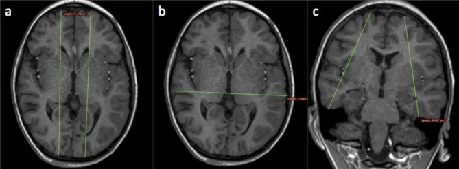
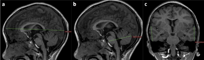
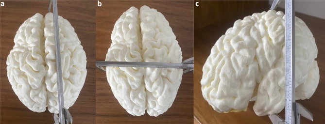
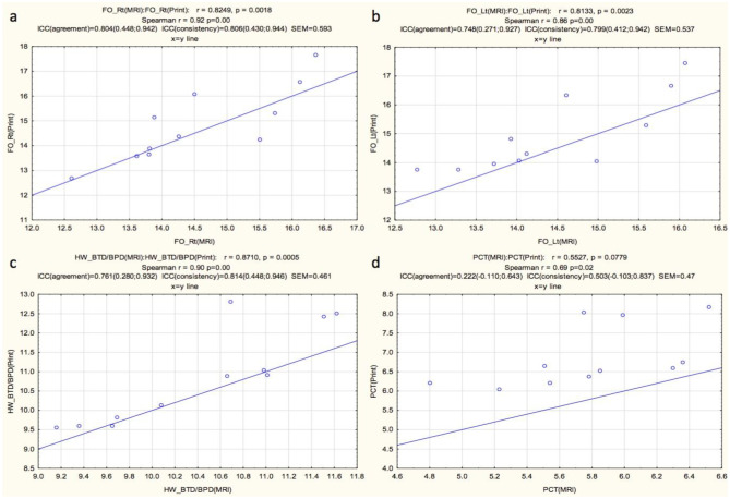
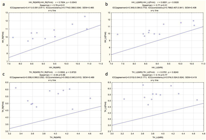
Cortical injury on the surface of the brain in children with hypoxic ischemic injury (HII) can be difficult to demonstrate to non-radiologists and lay people using brain images alone. Three-dimensional (3D) printing is helpful to communicate the volume loss and pathology due to HII in children's brains. 3D printed models represent the brain to scale and can be held up against models of normal brains for appreciation of volume loss. If 3D printed brains are to be used for formal communication, e.g., with medical colleagues or in court, they should have high fidelity of reproduction of the actual size of patients' brains. Here, we evaluate the size fidelity of 3D printed models from MRI scans of the brain, in children with prior HII. Twelve 3D prints of the brain were created from MRI scans of children with HII and selected to represent a variety of cortical pathologies. Specific predetermined measures of the 3D prints were made and compared to measures in matched planes on MRI. Fronto-occipital length (FOL) and bi-temporal/bi-parietal diameters (BTD/BPD) demonstrated high interclass correlations (ICC). Correlations were moderate to weak for hemispheric height, temporal height, and pons-cerebellar thickness. The average standard error of measurement (SEM) was 0.48 cm. Our results demonstrate high correlations in overall measurements of each 3D printed model derived from brain MRI scans versus the original MRI, evidenced by high ICC values for FOL and BTD/BPD. Measures with low correlation values can be explained by variability in matching the plane of measurement to the MRI slice orientation.
Keywords: 3D printing; Cerebral palsy; Fidelity; Hypoxic ischemic injury; Magnetic resonance imaging; Pediatric brain.
© 2022. The Author(s).
Conflict of interest statement
The authors declare no competing interests.
Figures


Similar articles
-
Accuracy of non-medical and medical individuals in identifying cerebral cortical abnormality from three-dimensional printed models of magnetic resonance images in children with hypoxic ischemic injury.Pediatr Radiol. 2024 Mar;54(3):450-456. doi: 10.1007/s00247-023-05653-2. Epub 2023 Apr 11. Pediatr Radiol. 2024. PMID: 37039912 Free PMC article. Review.
-
Technical report: 3D printing of the brain for use as a visual-aid tool to communicate MR imaging features of hypoxic ischaemic injury at term with non-physicians.Childs Nerv Syst. 2018 Aug;34(8):1573-1577. doi: 10.1007/s00381-018-3838-2. Epub 2018 May 26. Childs Nerv Syst. 2018. PMID: 29804212 Free PMC article.
-
Frequency of Cerebellar Abnormalities Associated With the Differing Magnetic Resonance Imaging Patterns of Term Hypoxic-Ischemic Injury in Children.Pediatr Neurol. 2024 Mar;152:73-78. doi: 10.1016/j.pediatrneurol.2023.12.023. Epub 2024 Jan 1. Pediatr Neurol. 2024. PMID: 38232653
-
Frequency of ulegyria on delayed MRI scans in children with term hypoxic-ischemic injury.Pediatr Radiol. 2023 Jan;53(1):104-111. doi: 10.1007/s00247-022-05445-0. Epub 2022 Jul 27. Pediatr Radiol. 2023. PMID: 35882664
-
Accuracy of 3D Printed Models Created by Two Technologies of Printers with Different Designs of Model Base.J Prosthodont. 2020 Feb;29(2):124-128. doi: 10.1111/jopr.13107. Epub 2019 Sep 9. J Prosthodont. 2020. PMID: 31498957 Review.
Cited by
-
3-Dimensional printing and bioprinting in neurological sciences: applications in surgery, imaging, tissue engineering, and pharmacology and therapeutics.J Mater Sci Mater Med. 2025 Apr 9;36(1):32. doi: 10.1007/s10856-025-06877-4. J Mater Sci Mater Med. 2025. PMID: 40205004 Free PMC article. Review.
-
Pixels to precision: Neuroradiology's leap into 3D printing for personalized medicine.J Clin Imaging Sci. 2024 Dec 17;14:49. doi: 10.25259/JCIS_119_2024. eCollection 2024. J Clin Imaging Sci. 2024. PMID: 39777212 Free PMC article. Review.
References
-
- Chacko A, Vedajallam S, Andronikou S, Simpson E, Thai NJ. Accuracy of radiologists, nonradiologists, and laypeople for identifying children with cerebral cortical atrophy from “Mercator map” curved reconstructions of MRIs of the brain. Indian Journal of Radiology and Imaging. 2020;30(2):111–115. doi: 10.4103/ijri.IJRI_130_20. - DOI - PMC - PubMed
-
- Andronikou S, Simpson E, Klemm M, Vedajallam S, Chacko A, Thai NJ. Technical report: 3D printing of the brain for use as a visual aid tool to communicate MR imaging features of hypoxic ischaemic injury at term with non-physicians. Child's Nervous System. 2018;34(8):1573–1577. doi: 10.1007/s00381-018-3838-2. - DOI - PMC - PubMed
MeSH terms
LinkOut - more resources
Full Text Sources
Medical
Miscellaneous
