. 2022 Oct 25;7(1):64.
doi: 10.1038/s41536-022-00261-4.

Loss of Mst1/2 activity promotes non-mitotic hair cell generation in the neonatal organ of Corti

Affiliations

Loss of Mst1/2 activity promotes non-mitotic hair cell generation in the neonatal organ of Corti

Xiaoling Lu et al. NPJ Regen Med. .

Abstract

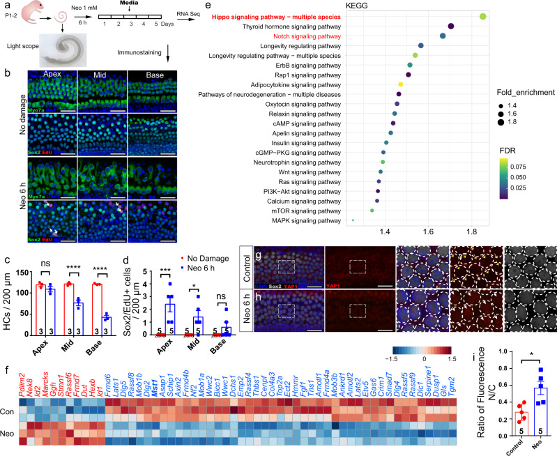
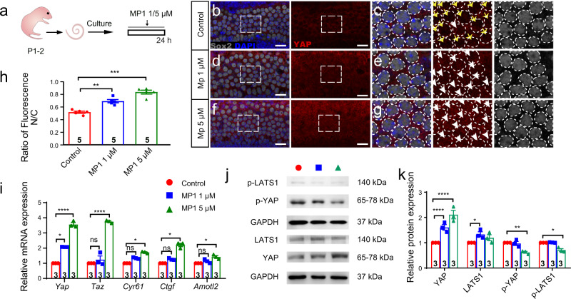
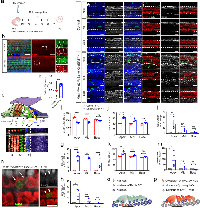
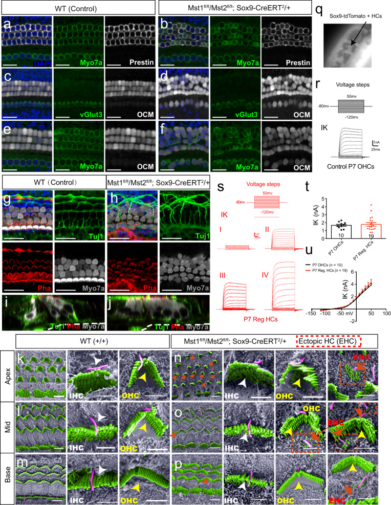
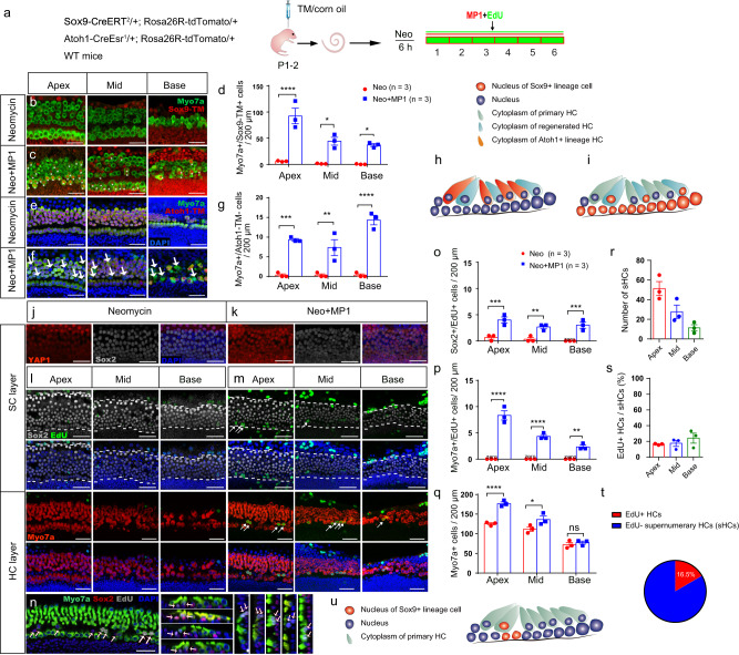
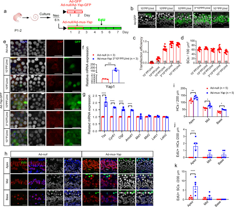
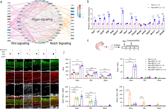
Mammalian sensory hair cells (HCs) have limited capacity for regeneration, which leads to permanent hearing loss after HC death. Here, we used in vitro RNA-sequencing to show that the Hippo signaling pathway is involved in HC damage and self-repair processes. Turning off Hippo signaling through Mst1/2 inhibition or Yap overexpression induces YAP nuclear accumulation, especially in supporting cells, which induces supernumerary HC production and HC regeneration after injury. Mechanistically, these effects of Hippo signaling work synergistically with the Notch pathway. Importantly, the supernumerary HCs not only express HC markers, but also have cilia structures that are able to form neural connections to auditory regions in vivo. Taken together, regulating Hippo suggests new strategies for promoting cochlear supporting cell proliferation, HC regeneration, and reconnection with neurons in mammals.


Conflict of interest statement

The authors declare no competing interests.

Figures

Fig. 1
Fig. 1. Hippo signaling is involved during post-injury and self-repair processes in damaged neonatal mammalian cochleae in vitro.
a Cochleae of P2 neonatal mice were isolated and cultured for 12 h, then damaged with 1 mM neomycin for 6 h and cultured for another 5 days with 10 μM EdU. b, c After neomycin damage, HC death increased from the apex to the base of the cochlea (Myosin7a+ HCs, apex: 109.0 ± 6.4; middle: 76.7 ± 6.7; base: 43.0 ± 4.6), and two-way ANOVA followed by Sidak’s multiple comparison test showed that there was a significant difference in the middle and basal turns. d Very few proliferative SCs were observed after HC damage (Sox2/EdU double-positive SCs, apex: 2.4 ± 0.6; middle: 1.4 ± 0.5; base: 0.6 ± 0.4), although two-way ANOVA followed by Sidak’s multiple comparison test showed the significant difference in the apical and middle turns. e Kyoto Encyclopedia of Genes and Genomes (KEGG) pathway analysis of the transcriptome data. f Heatmap showing relative expression levels of the differentially expressed genes associated with the Hippo signaling pathway after HC damage. (FDR < 0.01; n = 3 for each condition). Higher expressed genes are shown in red, while the genes with relatively lower levels are depicted in blue. gi Cochleae of P2 mice were dissected and cultured for 12 h and then treated with 1 mM neomycin for 6 h. Immunohistochemical staining was conducted after another 24 h culture. Compared with control groups, immunohistochemical staining and relative fluorescence quantitative analysis results analyzed with two-tailed, unpaired Student’s t tests showed that YAP (red) nuclear accumulation in Sox2+ SCs (white) was increased after neomycin injury. *p < 0.05, **p < 0.01, ***p < 0.001, ****p < 0.0001. Data are shown as the mean ± SEM. Scale bars = 20 μm.
Fig. 2
Fig. 2. Turning off Hippo signaling induces YAP nuclear accumulation and subsequent interaction with its target genes.
a Cochleae of P2 neonatal mice were isolated and cultured. After 12 h, 1 μM or 5 μM XMU-MP-1 was added for 24 h. bg The distribution of YAP (red) in Sox2+ SCs (white) from P2 neonatal control and 1 µM or 5 μM XMU-MP-1-treated mouse cochleae, respectively. The yellow arrows indicate that the YAP nuclear accumulation was very low in the control group, while the white arrows indicate that the YAP nuclear accumulation was increased in the XMU-MP-1 groups. h The relative florescence of YAP in the nuclei and cytoplasm of SCs in the control and XMU-MP-1-treated groups was analyzed with one-way ANOVA followed by Dunnett’s multiple comparisons test, indicating that XMU-MP-1 significantly promoted the translocation of YAP into the nucleus. i The relative mRNA expression levels of Hippo pathway-related genes and downstream genes after exogenous XMU-MP-1 treatment analyzed with one-way ANOVA followed by Tukey’s multiple comparisons test. j, k The western blots and the relative fluorescence quantification of p-YAP, p-LATS, YAP, and LATS in the control group and the XMU-MP-1 groups analyzed with two-way ANOVA followed by Dunnett’s multiple comparisons test. *p < 0.05, **p < 0.01, ***p < 0.001, ****p < 0.0001. Data are shown as the mean ± SEM. Scale bars = 20 μm.
Fig. 3
Fig. 3. Turning off Hippo signaling initiates the proliferation of SCs and the direct differentiation of HCs from SCs in vivo.
a Mst1f1/f1/Mst2fl/fl; Sox9-CreERT2/+ mice were used to knock out Mst1/2 in Sox9+ SCs and thus turn-off Hippo signaling to promote YAP nuclear translocation. Tamoxifen was injected at P1–2, and EdU was injected from P2–7 and the cochleae were harvested at P7. b Compared with the control groups, there was much more YAP nuclear translocation in Mst1f1/f1/Mst2fl/fl; Sox9-CreERT2/+ mice. The nuclei are circled in white. Compared with controls, more YAP was transferred into the cell nucleus in the Mst1/2 knockout group. c The relative florescence of YAP in the nuclei and cytoplasm of SCs in control and Mst1/2 knockout mice was analyzed with two-tailed, unpaired Student’s t tests, indicating that Mst1/2 knockout promoted the translocation of YAP into the nucleus. d The schematic diagram of the sectional views of the organ of Corti. DC, Deiters’ cell; PC, pillar cell; IPhC, inner phalangeal cell; IBC, inner border cell; GER, greater epithelial ridge. eh Very few EdU+ SCs were detected in control mice, and two-way ANOVA followed by Sidak’s multiple comparison test showed that there were significantly greater numbers of Sox2+/EdU+ SCs in the SR and GER with the total amount of Sox2+ SCs decreasing in Mst1f1/f1/Mst2fl/fl; Sox9-CreERT2/+ mice. im There were large numbers of newly generated HCs among both the IHCs and OHCs, and many Sox2+/Myosin7a+ HCs were detected in both the IHCs and OHCs of Mst1f1/f1/Mst2fl/fl; Sox9-CreERT2/+ mice (i), and two-way ANOVA followed by Sidak’s multiple comparison test was performed to show the significant differences. n Very few Myosin7a+/EdU+ HCs, about 0–2 per cochlea, were observed in Mst1f1/f1/Mst2fl/fl; Sox9-CreERT2/+ mice. o The schematic diagram of SC proliferation in Mst1/2 knockout mice in vivo. p The schematic diagram of supernumerary HCs in Mst1/2 knockout mice in vivo. *p < 0.05, **p < 0.01, ***p < 0.001, ****p < 0.0001. Data are shown as the mean ± SEM. Scale bars = 20 μm.
Fig. 4
Fig. 4. The supernumerary HCs induced by YAP nuclear translocation are functional but immature in vivo.
af The supernumerary ectopic HCs in Mst1fl/fl/Mst2fl/fl; Sox9-CreERT2/+ mice were stained with the IHC marker vGlut3 and the OHC marker Prestin. Compared with WT mice, the newly generated HCs were labeled with OCM despite being located in the IHC and OHC region. g Phalloidin was used to label the stereocilia structure, and Tuj1 was used to label the nerve fiber. A “V”-shaped structure in OHCs and a “–”-shaped structure in IHCs labeled by phalloidin were observed and surrounded by orderly nerve fiber connections that were labeled by Tuj1 in P7 control mice. h In P7 Mst1fl/fl/Mst2fl/fl; Sox9-CreERT2/+ mice, almost all HCs—including regenerated HCs—had similarly shaped “V” and “–” structures labeled by phalloidin in the OHCs and IHCs, respectively. Tuj1 labeling showed 1–2 rows of inner HCs and 3–5 rows of outer HCs surrounded by irregular nerve fibers. i, j The sectional views of phalloidin and Tuj1 staining in control and transgenic mice at P7. km The representative view of ciliary structures from the apical to basal turn of the organ of Corti in WT mice under an electron microscope. np In littermate P7 Mst1fl/fl/Mst2fl/fl; Sox9-CreERT2/+ mice, immature hair bundles were detected with a decrease from the apical to basal turn. The enlarged views of IHC and OHC bundles in situ in control and Mst1/2 knockout mice and the immature ectopic hair bundles with slightly longer microvilli and a kinocilium from the apical to basal turn are shown on the right. q Mst1fl/fl/Mst2fl/fl; Sox9-CreERT2/+; Rosa26-tdTomato/+ mice were used to conduct the patch-clamp experiment. After tamoxifen injection at P1-2, cochleae were harvested at P7, thus the Sox9-tdTomato+ HCs could be observed under a microscope. r Representative outward K+ currents of WT P7 OHCs evoked by the voltage steps shown above. s Representative outward K+ currents of regenerated HCs in transgenic mice evoked by the same voltage steps shown above. t Mean ± SEM IK (Delayed rectifier potassium current) in response to 0 mV depolarization of WT P7 OHCs (n = 10) and regenerated HCs (n = 19). u Mean ± SEM peak current–voltage relations of WT P7 OHCs and regenerated HCs. Data are shown as the mean ± SEM. Scale bars = 20 μm in (af, g, h). Scale bars = 5 μm in the low magnification and 2 μm in the high magnification, respectively in (kp).
Fig. 5
Fig. 5. YAP nuclear translocation enhances HC regeneration after neomycin damage in vitro.
a Sox9-CreERT2/+; Rosa26R-tdTomato/+ and Atoh1-CreEsr1/+; Rosa26R-tdTomato/+ mouse models were used to trace the SC and HC lineages. Tamoxifen was injected at P1 to activate tdTomato expression overnight, and the cochleae were harvested at P2 and then cultured in vitro. After neomycin exposure for 6 h, the cochlear explants were cultured with exogenous XMU-MP-1 or 0.5% DMSO for 5 days. bd Compared with controls, the numbers of Myosin7a+/Sox9-tdTomato+ HCs were largely increased from the apical to basal turn in the XMU-MP-1-treated group analyzed with two-way ANOVA followed by Sidak’s multiple comparison test. eg There were large numbers of Myosin7a+/Atoh1-tdTomato– HCs in the XMU-MP-1-treated groups compared to controls analyzed with two-way ANOVA followed by Sidak’s multiple comparison test. h The schematic diagram of Myosin7a+/Sox9-tdTomato+ HCs after XMU-MP-1 treatment. i The schematic diagram of Myosin7a+/Atoh1-tdTomato– HCs after XMU-MP-1 treatment. j, k The cochlear explants from P2 WT mice were harvested and then cultured. After neomycin exposure for 6 h, the explants were cultured with exogenous XMU-MP-1 or 0.5% DMSO for 24 h. Immunohistochemical staining results showed that the addition of XMU-MP-1 significantly increased the nuclear accumulation of YAP1 in Sox2+ SCs after neomycin injury. l, m After neomycin injury, the cochleae were cultured with DMSO or XMU-MP-1 for another 5 days, and 10 μM EdU was added the whole time to trace proliferating cells. In the control group with DMSO treatment, no proliferating HCs and very few SCs were detected. In the XMU-MP-1-treated group, there were some EdU+ SCs and HCs from the apical to basal turn. n The EdU+ HCs were mostly distributed in the tunnel between the inner and outer HCs, and they mostly appeared in pairs and stained with Sox2. oq Compared with the control groups, there was a significant increase in the number of proliferating SCs and HCs after XMU-MP-1 addition in vitro analyzed with two-way ANOVA followed by Sidak’s multiple comparison test. The total number of HCs was significantly increased. rt Only about 16% of HCs were EdU+ when compared with the increased proportion of HCs in the apical turn; sHC: supernumerary HC. u The schematic diagram of the mitotically regenerated HCs and proliferating SCs. *p < 0.05, **p < 0.01, ***p < 0.001, ****p < 0.0001. Data are shown as mean ± SEM. Scale bars = 20 μm.
Fig. 6
Fig. 6. Overexpression of YAP initiated supernumerary HCs.
a After neomycin exposure for 6 h, the cochlear explants were cultured with exogenous Ad-GFP/Ad-Yap-GFP for 48 h, and the transfection efficiency based on the GFP fluorescence was analyzed in Sox2+ SCs in the SR. b, c GFP+/Sox2+ cells were counted, and the results showed that the transfection efficiency increased with the increasing adenovirus concentrations (107, 108, 109, 3 × 10⁹, and 1010 PFU/ml). d The numbers of Sox2+ SCs treated with different concentrations of adenovirus were calculated. It was found that the number of SCs decreased slightly at high concentration. e, f To upregulate the expression of Yap, the cochlear explants after neomycin insult for 6 h were treated with Ad-Yap-GFP for 48 h. One-sample t test was performed and showed that YAP was significantly increased with a concentration of 3 × 10⁹ PFU/ml adenovirus, and the distribution of YAP in the nucleus was significantly increased. g The relative mRNA expression level of Hippo signaling and downstream target genes after Ad-Yap-GFP treatment, and two-way ANOVA followed by Sidak’s multiple comparison test was performed to show the significant differences. hk After neomycin exposure for 6 h, the cochlear explants were cultured with Ad-mus-Yap1 or Ad-null for 7 days, and 10 μM EdU was added the whole time to trace proliferating cells. In the control group with Ad-null treatment, no proliferating HCs or SCs were detected. In the Ad-mus-Yap1 treatment group, there were significantly more EdU+ SCs and HCs from the apical to basal turn analyzed with two-way ANOVA followed by Sidak’s multiple comparison test. Compared with controls, the number of HCs was significantly increased from the apical to the basal turn analyzed with two-way ANOVA followed by Sidak’s multiple comparison test. *p < 0.05, **p < 0.01, ***p < 0.001, ****p < 0.0001. Data are shown as the mean ± SEM. Scale bars = 20 μm in (b) and (h), Scale bars = 10 μm in (e).
Fig. 7
Fig. 7. Notch pathway inhibition works synergistically with Hippo-off to promote HC regeneration.
a Protein–protein interaction network analysis of differentially expressed genes of the Hippo, Notch, and Wnt signaling pathways. Purple lines indicate interactions between Hippo and Wnt genes, and orange lines indicate interactions between Hippo and Notch genes. The color of the nodes indicates the gene expression fold change. b The cochlear explants from P2 wild-type mice were harvested and then cultured. After neomycin exposure for 6 h, the explants were cultured with XMU-MP-1 or DMSO for 24 h. Related mRNA expression levels between XMU-MP-1 treated groups and controls are shown, and two-way ANOVA followed by Sidak’s multiple comparison test was performed to show the significant differences. c The cochlear explants from P2 WT mice were harvested and then cultured. After neomycin exposure for 6 h, the explants were cultured with exogenous compounds (including XMU-MP-1, BIO, or DAPT) or 0.5% DMSO for 3 days, and 10 μM EdU was added the whole time to trace proliferating cells. dg The total numbers of HCs and EdU+ HCs were not significantly different between the XMU-MP-1-treated and XMU-MP-1/BIO-treated groups. e, h, i The number of regenerated HCs was significantly increased in the XMU-MP-1/DAPT-treated group when compared to the XMU-MP-1-treated group. j Two-way ANOVA followed by Dunnett’s multiple comparisons test was performed and showed that the upregulation of Wnt signaling by BIO could promote the proliferation of SCs. However, XMU-MP-1/DAPT co-regulation generated more HCs, including EdU+ HCs, and large numbers of proliferating SCs were found in the GER. *p < 0.05, **p < 0.01, ***p < 0.001, ****p < 0.0001. Data are shown as the mean ± SEM. Scale bars = 20 μm.

Similar articles

Cited by

References

    1. Lasak JM, Allen P, McVay T, Lewis D. Hearing loss: diagnosis and management. Prim. Care. 2014;41:19–31. - PubMed
    1. Cunningham LL, Tucci DL. Hearing loss in adults. N. Engl. J. Med. 2017;377:2465–2473. - PMC - PubMed
    1. Warchol ME. Sensory regeneration in the vertebrate inner ear: differences at the levels of cells and species. Hear Res. 2011;273:72–79. - PubMed
    1. Bermingham-McDonogh O, Reh TA. Regulated reprogramming in the regeneration of sensory receptor cells. Neuron. 2011;71:389–405. - PMC - PubMed
    1. Chai R, et al. Wnt signaling induces proliferation of sensory precursors in the postnatal mouse cochlea. Proc. Natl Acad. Sci. USA. 2012;109:8167–8172. - PMC - PubMed