Review
. 2022 Dec;18(12):707-722.
doi: 10.1038/s41582-022-00727-5. Epub 2022 Oct 24.

Astrocytes in the initiation and progression of epilepsy

Affiliations
Review

Astrocytes in the initiation and progression of epilepsy

Annamaria Vezzani et al. Nat Rev Neurol. 2022 Dec.

Abstract

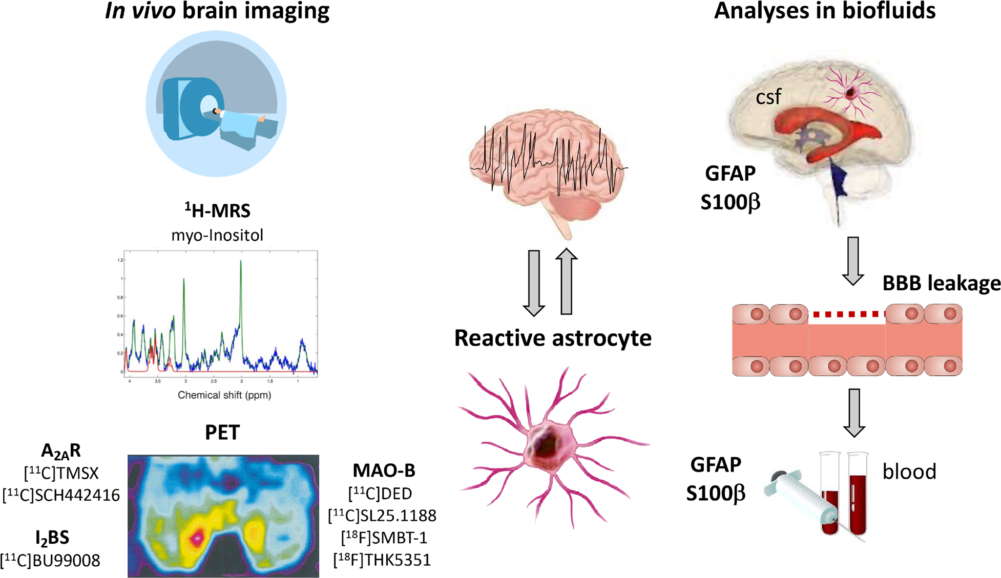
Epilepsy affects ~65 million people worldwide. First-line treatment options include >20 antiseizure medications, but seizure control is not achieved in approximately one-third of patients. Antiseizure medications act primarily on neurons and can provide symptomatic control of seizures, but do not alter the onset and progression of epilepsy and can cause serious adverse effects. Therefore, medications with new cellular and molecular targets and mechanisms of action are needed. Accumulating evidence indicates that astrocytes are crucial to the pathophysiological mechanisms of epilepsy, raising the possibility that these cells could be novel therapeutic targets. In this Review, we discuss how dysregulation of key astrocyte functions - gliotransmission, cell metabolism and immune function - contribute to the development and progression of hyperexcitability in epilepsy. We consider strategies to mitigate astrocyte dysfunction in each of these areas, and provide an overview of how astrocyte activation states can be monitored in vivo not only to assess their contribution to disease but also to identify markers of disease processes and treatment effects. Improved understanding of the roles of astrocytes in epilepsy has the potential to lead to novel therapies to prevent the initiation and progression of epilepsy.


Conflict of interest statement

Competing interests

The authors declare no competing interests

Figures

Figure 1.
Figure 1.. Astrocytes and their interactions with neurons and blood vessels
Photomicrographs depict GFAP-positive immunostaining in human astrocytes. A,B: Protoplasmic astrocytes morphology in normal human temporal cortex depicting highly branched bushy processes; cells are widely distributed between neurons. An astrocyte embracing a neuron is shown in B (magnification in inset). Arrows indicate a large astroglia process extending its foot along a blood vessel. C,D: Chronic dense fibrillary gliosis in human TLE sclerotic hippocampus. Arrow in C indicates a surviving CA1 neuron entrapped in dense glial meshwork. Scale bars: 50 μm. There are different splice variants of GFAP (β, γ, δ, κ) including the predominant GFAPα form. The minor isoforms are increased in subpopulations of astrocytes in focal lesions associated with epilepsy, however, their functional consequences for the epileptic network are unclear. E: Schematic representation of the shift of astrocytic functional states (in healthy conditions, blue color) to reactive pathologic states (orange color). Reactive astrocytes, due to impairment of their homeostatic functions (blue circles), cause several alterations in surrounding brain tissue (orange shape) that contribute to epilepsy initiation and progression.
Figure 2.
Figure 2.. Gliotransmission in the epileptic brain
(1) Glutamate or ATP derived from neurons, astrocytes or microglia trigger an increase in the intracellular Ca2+ concentration [Ca2+]i in astrocytes, which promotes the release of gliotransmitters. (2) Glutamate released by astrocytes activates extrasynaptic neuronal GluNRs and mGluRs, thereby increasing presynaptic release probability (Pr) and postsynaptic excitability. (3) Astrocytic ATP release evoked by elevated [Ca2+]i levels or activation of cytokine receptors lead to autocrine or paracrine activation of P2YRs, resulting in potentiation and intercellular propagation of astrocytic Ca2+ signals. It also triggers microglial activation and release of pro-inflammatory cytokines through activation of microglial P2X7 and/or P2Y1Rs. (4) Extracellular ATP is rapidly hydrolysed by ectonucleotidases to adenosine, with the latter targeting pre- and postsynaptic A1Rs to decrease neuronal excitability. (5) Epileptic activity triggers astrocytic GABA production via decarboxylation of glutamate by glutamate decarboxylase (GAD) and degradation of putrescine by monoamine oxidase B (MAO-B). Upon release into the extracellular space, GABA activates extrasynaptic GABAARs on excitatory neurons and elicits tonic inhibitory Cl currents, which attenuates neuronal excitability. Abbreviations: GluAR – α-amino-3-hydroxy-5-methyl-4-isoxazolepropionic acid receptor; ER – endoplasmic reticulum; GABAAR – γ-aminobutyric acid receptor; IL– interleukin; IP3 – inositol 1,4,5-trisphosphate; NLRP3 – NLR family pyrin domain containing 3; GluNR – N-methyl-d-aspartate receptor; TGF-β – transforming growth factor β; TNFα – tumor necrosis factor α; TNFR1 – tumor necrosis factor α receptor 1.
Figure 3.
Figure 3.. Key mechanisms through which metabolic dysfunction in astrocytes contributes to increased neuronal excitability
Changes in key metabolic enzymes or transporters lead to an imbalance in the key neurotransmitters and neuromodulators glutamate, glutamine, GABA, and adenosine. Glycolysis can lead to the formation of lactate, which is a key energy metabolite for neurons supplied by the astrocyte neuron lactate shuttle. Adenosine in the cell nucleus, regulated by ADK-L contributes to epigenetic reprogramming as driver of the epileptogenic process. Consequently, metabolic therapies, such as ketogenic diet, glucose restriction, adenosine, and ketones can reduce epileptic activity through multiple mechanisms. Abbreviations: ADO – adenosine; ADK-L – long isoform of adenosine kinase; ADK-S – short isoform of adenosine kinase; ANLS – astrocyte neuron lactate shuttle; A1R – adenosine A1 receptor; DNA-CH3 – DNA methylation; Gln – glutamine; GLT-1 – astroglial glutamate transporter 1; Glu – glutamate; GS – glutamine synthetase; KD – ketogenic diet; LDH – lactate dehydrogenase.
Figure 4.
Figure 4.. Imaging modalities and biofluids fingerprint of astrocyte reactivity
Brain imaging using proton magnetic resonance spectroscopy (1H-MRS) and positron emission tomography (PET) are minimally invasive tools to monitor astrocyte activation in vivo by targeting molecular markers enriched in reactive astrocytes, namely myo-Inositol, adenosine 2A receptors (A2AR), imidazoline2 binding sites (I2BS), monoamine-oxidase B (MAO-B). Additionally, analysis of circulating markers of astrocytes such as GFAP and S100β in blood or CSF may provide an index of astrocyte reactivity or injury. These astrocyte-related biomarkers may help to identify epileptic foci, to monitor epileptogenesis or the effect of therapies.

References

    1. Khakh BS & Deneen B The Emerging Nature of Astrocyte Diversity. Annu Rev Neurosci 42, 187–207 (2019).

      *This review emphasizes that astrocytes represent a diverse population of cells and that they display brain area- and disease-specific properties and functions. Knowledge gaps are identified that need to be addressed in order to elucidate astrocyte diversity and its physiological relevance in the CNS.

    1. Archie SR, Al Shoyaib A & Cucullo L Blood-Brain Barrier Dysfunction in CNS Disorders and Putative Therapeutic Targets: An Overview. Pharmaceutics 13, 1779 (2021). - PMC - PubMed
    1. MacVicar BA & Newman EA Astrocyte regulation of blood flow in the brain. Cold Spring Harb Perspect Biol 7, a020388 (2015). - PMC - PubMed
    1. Araque A, Parpura V, Sanzgiri RP & Haydon PG Tripartite synapses: glia, the unacknowledged partner. Trends Neurosci 22, 208–215 (1999). - PubMed
    1. Devinsky O, Vezzani A, Najjar S, De Lanerolle NC & Rogawski MA Glia and epilepsy: excitability and inflammation. Trends Neurosci 36, 174–84 (2013). - PubMed

Publication types