. 2022 Nov;19(11):1367-1370.
doi: 10.1038/s41592-022-01624-x. Epub 2022 Oct 24.

SyConn2: dense synaptic connectivity inference for volume electron microscopy

Affiliations

SyConn2: dense synaptic connectivity inference for volume electron microscopy

Philipp J Schubert et al. Nat Methods. 2022 Nov.

Abstract

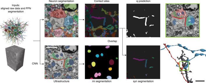
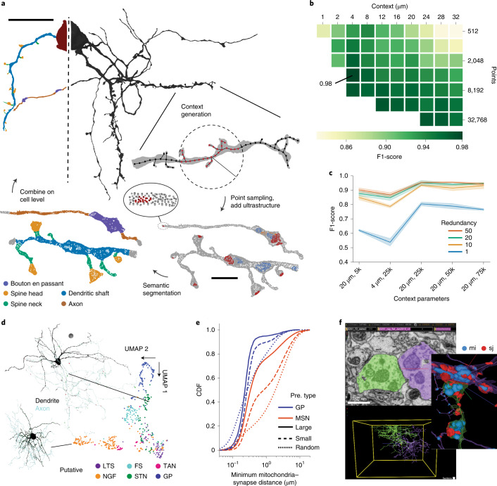
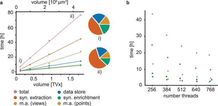
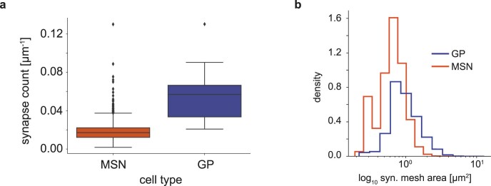
The ability to acquire ever larger datasets of brain tissue using volume electron microscopy leads to an increasing demand for the automated extraction of connectomic information. We introduce SyConn2, an open-source connectome analysis toolkit, which works with both on-site high-performance compute environments and rentable cloud computing clusters. SyConn2 was tested on connectomic datasets with more than 10 million synapses, provides a web-based visualization interface and makes these data amenable to complex anatomical and neuronal connectivity queries.


Conflict of interest statement

F.S. and J.K. disclose financial interests in ariadne.ai ag. S.D., M.J. and V.J. are employees of Google LLC, which sells cloud computing services. The remaining authors declare no competing interests.

Figures

Fig. 1
Fig. 1. SyConn2 processing on the voxel level.
Neuron segmentation and ultrastructure prediction (synaptic junctions (sj) in red; mitochondria (mi) in blue; vesicle clouds in green) derived from raw data. Contact sites and synaptic junction are assigned as queryable instances to neuron reconstructions. 1, MSN dendrite; 2, excitatory axon (EA); 3, inhibitory axon (IA). The scale bar is 1 µm and refers to all electron microscopy images in the figure. CNN: convolutional neural network ; FFN: flood-filling neural network.
Fig. 2
Fig. 2. SyConn2 processing and analyses of neuron reconstructions.
a, Semantic segmentation of cell surfaces with point cloud neural networks. Surface points of the cell and ultrastructure within an input context were subsampled and presented to the model. Context predictions are then combined on the cell level. b, Grid search for optimal context parameters (radius, number of points) evaluated at synapse locations (88 spine head and 94 dendritic shaft) with weighted average F1-score (dendritic shaft, spine head and a combined axon and soma class). c, Classification performance of putative cell types dependent on the context and the number of bootstrapping samples (redundancy). For example, 20 µm, 5k refers to a 20 µm radius with 5,000 points. The confidence interval is mean ± standard deviation of three training repetitions for each parameter pair. d, UMAP dimensionality reduction of learned unsupervised latent space of 531 neurons in the dataset that contained soma, axon and dendrite (MSNs not considered). LTS, low-threshold spiker; FS, fast-spiking interneuron; TAN, tonically active cholinergic neuron; NGF, neurogliaform interneuron; STN, subthalamic nucleus-like neuron; GP, pallidal-like neuron. Colors indicate putative cell type based on supervised classification. e, Cumulative distribution function (CDF) of the minimal distance between axo-dendritic synapses (and a random control) and mitochondria in GP and MSN split into small and large synapses (less than or equal to and greater than median of mesh area; median GP 1.16 µm2, MSN 0.75 µm2; N synapses GP 7,482, MSN 59,131; see also Extended Data Fig. 2b for synapse size distributions; N random control locations: GP 37,149, MSN 6,128,974). Pre. type, presynaptic cell type f, Example of a GP–GP synapse visualized with the web-based SyConn2 client. Scale bars, 1 µm in EM section and 4 µm in renderings (a), 20 µm for the cell and 2 µm for the context (b) and 10 µm sphere radius (d). Source data
Extended Data Fig. 1
Extended Data Fig. 1. Timings of the different pipeline steps.
Timings are grouped into synapse extraction, data store, synapse enrichment and morphology analysis (m.a.) with multi-views (views) and point clouds (points). a Compute time as a function of the processed volume (in teravoxels, TVx). Pie charts show the fraction of the different steps relative to the total time at the smallest and largest test cube (i: 0.29 million µm3, syn. extraction: 0.45, data store: 0.22, syn. enrichment: 0.18, m.a. (points): 0.14; ii: 4.53 million µm3, 0.58, 0.19, 0.12, 0.11). The ‘views’ step was excluded for the ‘total’ timings and the pie charts (i, ii). Compute resources: 24 google cloud computing nodes (n1-highmem-32), each with 32 virtual cores (threads), 2 Tesla P100, 208 GB memory. b Compute time as a function of the number of available compute nodes (8, 12, 16, 20, 24). Processed volume: 0.391 teravoxels. Color code as in a. Source data
Extended Data Fig. 2
Extended Data Fig. 2. GP and MSN synapse properties.
a Box plot (median, lower and upper quartile; whiskers, 1.5x interquartile range above upper and below lower quartile; points, outlier) of the average synapse count per micrometer for cell types MSN (N = 6327, median: 0.017 µm−1, Q1: 0.012 µm−1, Q3: 0.022 µm−1) and GP (N = 38, 0.057 µm−1, 0.033 µm−1, 0.066 µm−1). Two-sided Mann-Whitney U test statistic: -9.71 and p-value: 2.57e-22. b Histogram of synapse mesh area (N synapses GP: 7,482, MSN: 59,131). Source data

Comment in

Similar articles

Cited by

References

    1. Kornfeld J, Denk W. Progress and remaining challenges in high-throughput volume electron microscopy. Curr. Opin. Neurobiol. 2018;50:261–267. doi: 10.1016/j.conb.2018.04.030. - DOI - PubMed
    1. Shapson-Coe, A., Januszewski, M., Berger, D. R. & Pope, A. A connectomic study of a petascale fragment of human cerebral cortex. Preprint at bioRxivhttps://www.biorxiv.org/content/10.1101/2021.05.29.446289v4.abstract (2021). - DOI
    1. Turner NL, et al. Reconstruction of neocortex: organelles, compartments, cells, circuits, and activity. Cell. 2022;185:1082–1100. doi: 10.1016/j.cell.2022.01.023. - DOI - PMC - PubMed
    1. Januszewski M, et al. High-precision automated reconstruction of neurons with flood-filling networks. Nat. Methods. 2018;15:605–610. doi: 10.1038/s41592-018-0049-4. - DOI - PubMed
    1. Dorkenwald S, et al. FlyWire: online community for whole-brain connectomics. Nat. Methods. 2021;19:119–128. doi: 10.1038/s41592-021-01330-0. - DOI - PMC - PubMed

Publication types