

Nestin is a marker of unipotent embryonic and adult progenitors differentiating into an epithelial cell lineage of the hair follicles
- PMID: 36280775
- PMCID: PMC9592581
- DOI: 10.1038/s41598-022-22427-2
Nestin is a marker of unipotent embryonic and adult progenitors differentiating into an epithelial cell lineage of the hair follicles
Abstract
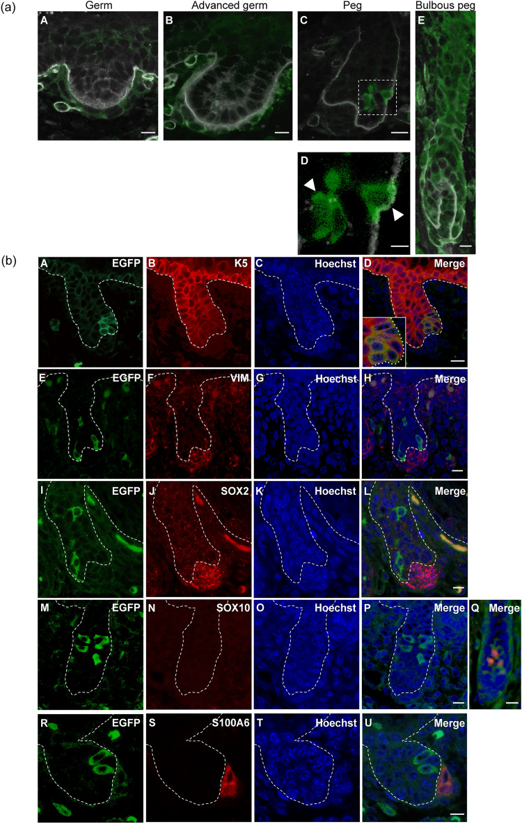
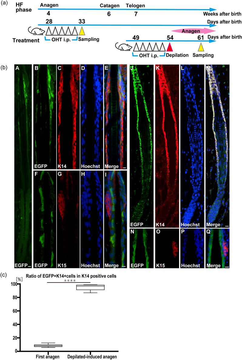
Nestin is an intermediate filament protein transiently expressed in neural stem/progenitor cells. We previously demonstrated that outer root sheath (ORS) keratinocytes of adult hair follicles (HFs) in mice descend from nestin-expressing cells, despite being an epithelial cell lineage. This study determined the exact stage when nestin-expressing ORS stem/precursor cells or their descendants appear during HF morphogenesis, and whether they are present in adult HFs. Using Nes-Cre/CAG-CAT-EGFP mice, in which enhanced green fluorescent protein (EGFP) is expressed following Cre-based recombination driven by the nestin promoter, we found that EGFP+ cells appeared in the epithelial layer of embryonic HFs as early as the peg stage. EGFP+ cells in hair pegs were positive for keratin 14 (K14) and K5, but not vimentin, SOX2, SOX10, or S100 alpha 6. Tracing of tamoxifen-induced EGFP+ cells in postnatal Nes-CreERT2/CAG-CAT-EGFP mice revealed labeling of some isthmus HF epithelial cells in the first anagen stage. EGFP+ cells in adult HFs were not immunolabeled for K15, an HF multipotent stem cell marker. However, when hairs were depilated in Nes-CreERT2/CAG-CAT-EGFP mice to induce the anagen stage after tamoxifen injection, the majority of ORS keratinocytes in depilation-induced anagen HFs were labeled for EGFP. Our findings indicate that nestin-expressing unipotent progenitor cells capable of differentiating into ORS keratinocytes are present in HF primordia and adult HFs.
© 2022. The Author(s).
Conflict of interest statement
The authors declare no competing interests.
Figures

Similar articles
-
Progenitor cells expressing nestin, a neural crest stem cell marker, differentiate into outer root sheath keratinocytes.Vet Dermatol. 2019 Oct;30(5):365-e107. doi: 10.1111/vde.12771. Epub 2019 Jul 12. Vet Dermatol. 2019. PMID: 31297916
-
Nestin expression in hair follicle sheath progenitor cells.Proc Natl Acad Sci U S A. 2003 Aug 19;100(17):9958-61. doi: 10.1073/pnas.1733025100. Epub 2003 Aug 6. Proc Natl Acad Sci U S A. 2003. PMID: 12904579 Free PMC article.
-
Comparison of nestin-expressing multipotent stem cells in the tongue fungiform papilla and vibrissa hair follicle.J Cell Biochem. 2014 Jun;115(6):1070-6. doi: 10.1002/jcb.24696. J Cell Biochem. 2014. PMID: 24142339
-
The potential of nestin-expressing hair follicle stem cells in regenerative medicine.Expert Opin Biol Ther. 2007 Mar;7(3):289-91. doi: 10.1517/14712598.7.3.289. Expert Opin Biol Ther. 2007. PMID: 17309321 Review.
-
Life of the B10 Mouse: A View from the Hair Follicles and Tissue Stem Cells.Cells Tissues Organs. 2024;213(3):213-222. doi: 10.1159/000533779. Epub 2023 Sep 13. Cells Tissues Organs. 2024. PMID: 37703854 Review.
Cited by
-
CRISPR/Cas9-mediated deletion of a GA-repeat in human GPM6B leads to disruption of neural cell differentiation from NT2 cells.Sci Rep. 2024 Jan 25;14(1):2136. doi: 10.1038/s41598-024-52675-3. Sci Rep. 2024. PMID: 38273037 Free PMC article.
-
Role of Long Non-Coding RNA X-Inactive-Specific Transcript (XIST) in Neuroinflammation and Myelination: Insights from Cerebral Organoids and Implications for Multiple Sclerosis.Noncoding RNA. 2025 Apr 29;11(3):31. doi: 10.3390/ncrna11030031. Noncoding RNA. 2025. PMID: 40407589 Free PMC article.
-
Hypoxic Preconditioning Prevents Oxidative Stress-Induced Cell Death in Human Hair Follicle Stem Cells.Iran J Biotechnol. 2024 Jul 1;22(3):e3888. doi: 10.30498/ijb.2024.447077.3888. eCollection 2024 Jul. Iran J Biotechnol. 2024. PMID: 39737203 Free PMC article.
-
From Adipose to Action: Reprogramming Stem Cells for Functional Neural Progenitors for Neural Regenerative Therapy.Int J Mol Sci. 2025 Jul 9;26(14):6599. doi: 10.3390/ijms26146599. Int J Mol Sci. 2025. PMID: 40724849 Free PMC article. Review.
References
-
- Ito M, Cotsarelis G, Kizawa K, Hamada K. Hair follicle stem cells in the lower bulge form the secondary germ, a biochemically distinct but functionally equivalent progenitor cell population, at the termination of catagen. Differentiation. 2004;72:548–557. doi: 10.1111/j.1432-0436.2004.07209008.x. - DOI - PubMed
Publication types
MeSH terms
Substances
LinkOut - more resources
Full Text Sources
Molecular Biology Databases
Research Materials
Miscellaneous
