. 2022 Oct 24;12(1):17825.
doi: 10.1038/s41598-022-22480-x.

Heterologously secreted MbxA from Moraxella bovis induces a membrane blebbing response of the human host cell

Affiliations

Heterologously secreted MbxA from Moraxella bovis induces a membrane blebbing response of the human host cell

Isabelle N Erenburg et al. Sci Rep. .

Abstract

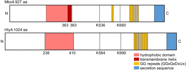
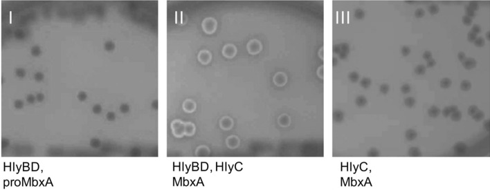
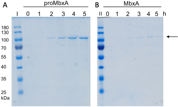
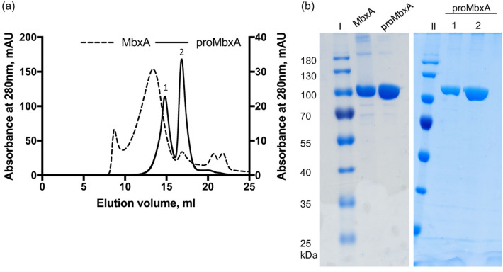
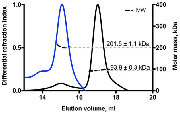
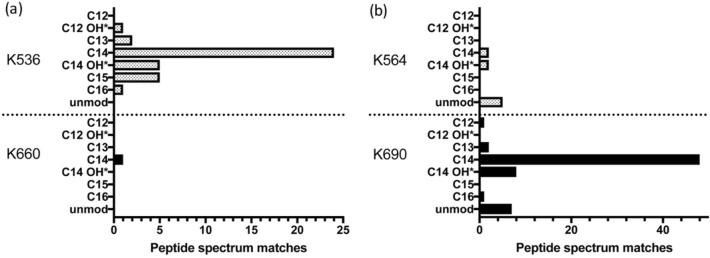
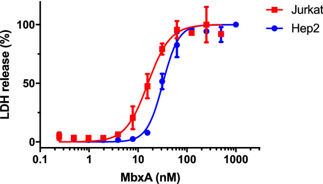
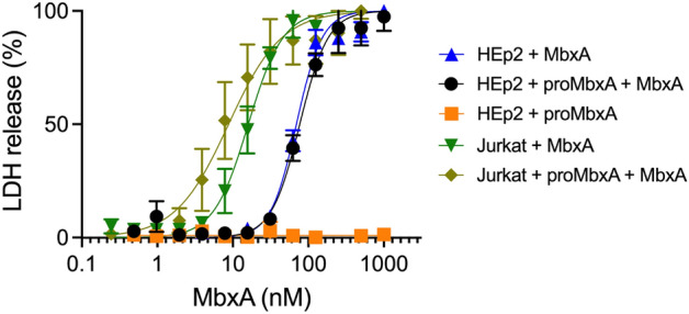
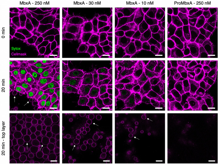
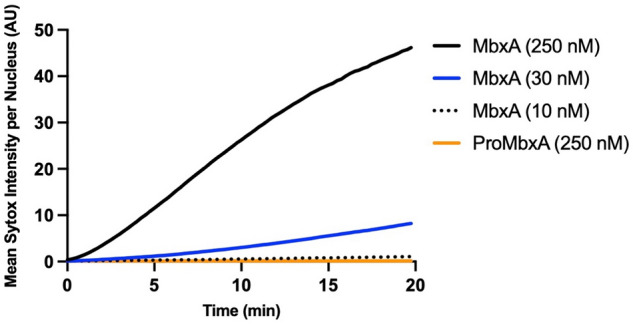
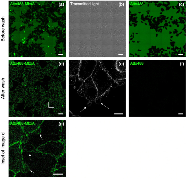
Many proteins of the Repeats in Toxins (RTX) protein family are toxins of Gram-negative pathogens including hemolysin A (HlyA) of uropathogenic E. coli. RTX proteins are secreted via Type I secretion systems (T1SS) and adopt their native conformation in the Ca2+-rich extracellular environment. Here we employed the E. coli HlyA T1SS as a heterologous surrogate system for the RTX toxin MbxA from the bovine pathogen Moraxella bovis. In E. coli the HlyA system successfully activates the heterologous MbxA substrate by acylation and secretes the precursor proMbxA and active MbxA allowing purification of both species in quantities sufficient for a variety of investigations. The activating E. coli acyltransferase HlyC recognizes the acylation sites in MbxA, but unexpectedly in a different acylation pattern as for its endogenous substrate HlyA. HlyC-activated MbxA shows host species-independent activity including a so-far unknown toxicity against human lymphocytes and epithelial cells. Using live-cell imaging, we show an immediate MbxA-mediated permeabilization and a rapidly developing blebbing of the plasma membrane in epithelial cells, which is associated with immediate cell death.


Conflict of interest statement

The authors declare no competing interests.

Figures

Figure 1
Figure 1
Schematic view of the primary structure of MbxA and HlyA. Five conserved GG repeats with the consensus sequence GGxGxDxUx are present in MbxA, while HlyA carries six conserved GG repeats (yellow boxes). They together form the Ca2+ binding RTX domain. As a RTX protein, HlyA characteristically possesses a C-terminal secretion signal of approximately 60 amino acids, which is shown in blue. For MbxA the length of the secretion sequence is unknown and drawn not to scale. In the N-terminal part of HlyA, a hydrophobic domain is involved in pore formation and marked in red. Depending on the prediction algorithm (see Materials and Methods) for MbxA either (1) a single membrane spanning helix is predicted from residue 363 to 383, (2) a set of transmembrane helices forming an ambiguous hydrophobic domain from residue 210 to 383, or (3) even from residue 120 to 383 (dashed lines) are predicted. The position of the acylated lysine residues K564 and K690 of HlyA and the homologous residues predicted on sequence comparisons to become acylated in MbxA, K536 and K660, are indicated and served as anker point for the scheme.
Figure 2
Figure 2
E. coli BL21(DE3) grown on Columbia agar with 5% sheep blood harboring the plasmids pK184-hlyBD with pSU-6H-mbxA (I), pK184-hlyBD with pSU-hlyC/6H-mbxA (II) or pSU-hlyC/6H-mbxA alone (III). Halo formation around colonies that expressed MbxA and HlyC together with the transporter components HlyBD (II) and absence of hemolysis around colonies that express only MbxA and HlyC (III) indicates functional secretion of active and acylated MbxA. Thus functional MbxA is not released from E. coli without additional expression of the HlyBD secretion system.
Figure 3
Figure 3
Heterologous secretion of proMbxA and MbxA via the HlyBD system. The Coomassie Brilliant Blue (CBB) SDS PAGE of culture supernatants of E. coli BL21(DE3) co-expressing HlyBD and MbxA (proMbxA, (A)) or HlyBD, HlyC and MbxA (MbxA, (B)) show accumulation of proMbxA or MbxA during a period of 0–5 h of expression (indicated by the numbers above the gel) after induction with IPTG. Samples of the supernatant were applied to the SDS gel without additional concentration by TCA precipitation. Molecular weight markers in kDa are shown in lanes I and II. An arrow indicates proMbxA and MbxA.
Figure 4
Figure 4
(a) SEC chromatographs of samples of active MbxA (dashed line) and proMbxA (solid line), respectively, applied to a Superose 6 Increase 6/30 column (GE Healthcare). The absorbance at 280 nm of the elution profile of proMbxA was plotted on the left Y axis, the elution profile of active MbxA on the right Y axis. proMbxA eluted in two separate main peaks, marked 1 and 2 in the chromatogram. (b) CBB stained SDS PAGE gels of pooled fractions of active MbxA and proMbxA obtained from IMAC purification (left panel) and used for SEC. The entire gels are shown in Figure S9. proMbxA was separated in two species, peak 1 and peak 2, during SEC and evaluated by SDS PAGE (right panel). Molecular weight markers in kDa are shown in lanes I and II.
Figure 5
Figure 5
MALS coupled to SEC analysis of the two proMbxA species separated via SEC (blue line peak 1 of Fig. 4, black line peak 2 of Fig. 4). The calculated molecular weight of 201.5 ± 1.1 kDa of the first peak (blue) corresponds to a dimer of proMbxA (theoretical MW (6H-MbxA)2 = 201.2 kDa). The molecular weight of 93.9 ± 0.3 kDa of the second peak (black) confirms a monomeric species of proMbxA (theoretical MW 6H-MbxA = 100.6 kDa).
Figure 6
Figure 6
MS analysis of MbxA acylation (a) and HlyA acylation (b). MbxA and HlyA were in vivo acylated by co-expressing HlyC. For both acylation sites in MbxA and HlyA, K536 and K660 or K564 and K690 respectively, all detected acyl modifications ranging from C12 to C16 fatty acids including hydroxy fatty acids are shown with the corresponding number of recorded PSM. Mass shifts contributing to hydroxy fatty acids could possibly originate from the oxidation of tyrosine residues in the peptide fragment and PSM of supposed hydroxy acylations are therefore marked (*). For proMbxA and proHlyA, both expressed in the absence of HlyC, only unmodified peptides were detected. Acyl modifications were detected only in peptides covering the predicted lysine residues K536 and K660 of MbxA. The PSM of C14 modified sequences suggest that the predominant modification is myristoylation.
Figure 7
Figure 7
MbxA induces LDH release in human epithelial cells (HEp-2, blue filled circle ) and human T cells (Jurkat, red filled square ), respectively. The cytotoxicity mediated by MbxA-induced membrane damage was measured using an LDH release assay. LDH release into the supernatant was measured after 1 h of incubation with MbxA. For the CD50 determination the MbxA cytotoxicity was plotted against the MbxA concentration and fitted with GraphPad Prism 7 according to Eq. (2) (see materials and methods). For each cell type, the highest LDH release was set to 100%. For simplicity, measurements with proMbxA were not included as no LDH release, even at concentrations of 1 µM, was detected.
Figure 8
Figure 8
proMbxA does not protect epithelial cells (HEp-2) from MbxA-induced membrane damage. Preincubation with 250 nM of proMbxA for 30 min at 37 °C resulted in a nearly identical dose–response curve to MbxA (black) compared to treatment with MbxA alone (37 °C for 30 min, blue). proMbxA was not cytotoxic (orange). A similar effect was observed for Jurkat cells. Preincubation with 250 nM of proMbxA for 30 min at 37 °C resulted in a not significant different dose–response curve to MbxA (light green) compared to treatment with MbxA alone (37 °C for 30 min, green). The cytotoxicity mediated by MbxA-induced membrane damage was measured using an LDH release assay. LDH release into the supernatant was measured after 1 h of incubation with MbxA. The relative LDH release was calculated from the maximal LDH release reached in each measurement, which was set to 100%. For the CD50 determination the MbxA cytotoxicity was plotted against the MbxA concentration and fitted with GraphPad Prism 7 according to Eq. (2) (see material and methods).
Figure 9
Figure 9
Confocal microscopy images of MbxA-induced membrane permeabilization and membrane blebbing in HEp-2 cells. HEp-2 cells were incubated with MbxA (250 nM, 30 nM and 10 nM at 37 °C) or proMbxA (250 nM) and analyzed at time point zero (first row) and after 20 min (second row). Plasma membrane was stained by CellMask Deep Red and membrane permeabilization was monitored using Sytox Green staining of the nuclear DNA. Incubation of HEp-2 cells with 250 nM, 30 nM and 10 nM MbxA resulted in DNA staining with Sytox Green cells in decreasing intensity corresponding to the decreasing concentration of MbxA and hence in membrane permeabilization. DNA staining was not observed when HEp-2 cells were incubated with 250 nM proMbxA. Membrane blebs were observed in the same focus layer as the HEp-2 cells (second row; white arrows) and above the HEp-2 cells (third row; white arrows). While incubation with 250 nM and 30 nM MbxA led to severe appearance of membrane blebbing, incubation with 10 nM of MbxA was less noticeable and 250 nM proMbxA incubation did not lead to any formation of blebs. Please see Fig. S4 for the entire images. Scale bar: 20 µm.
Figure 10
Figure 10
Quantification of MbxA-induced permeabilization of HEp-2 cells. Mean Sytox Green intensity per nucleus of HEp-2 cells (y-axis) was continuously measured over a period of 20 min at 37 °C (see mateirals and methods for details) and is plotted against time (x-axis) for different MbxA concentrations (250 nM, 30 nM and 10 nM) and 250 nM. Sytox Green staining of the HEp-2 cells´ DNA is faster and more intense when 250 nM MbxA were used and is reduced at lower MbxA concentrations (30 nM and 10 nM). In contrast to that, no significant Sytox Green staining of the nuclear DNA was observed with 250 nM proMbxA (number of nuclei used for quantification: 250 nM MbxA—71, 30 nM MbxA—74, 10 nM MbxA—79, 250 nM proMbxA—62). Nuclei have been analyzed from two independent experiments with two biological replicates each. The original images at different time points are provided in Fig. S7.
Figure 11
Figure 11
Incubation experiment of Atto488-MbxAS9C with HEp-2 cells. HEp-2 cells after 30 min of incubation with 250 nM Atto488-MbxAS9C, before (a and b) and after (d and e) removing excess unbound Atto488-MbxAS9C. Confocal overview images of fluorescent signal are shown in (a) and (d), transmitted light showing formation of blebbing process in (b) and super-resolution Airyscan micrograph in (e). Accumulations of Atto488-MbxAS9C tracing the outline of HEp-2 cells are clearly visible after removing the unbound Atto488-MbxAS9C (d). Control experiment with Atto488 (dye used to label MbxA) is shown in (c) and (f). HEp-2 cells after 30 min of incubation with Atto488, before (c) and after (f) washing off the unbound Atto488 from surrounding medium. The control experiment confirmed that accumulations tracing the outline of HEp-2 cells in (d) and (e) are Atto488-MbxAS9C, not the free dye. White arrows indicate the position of membrane blebs. (g) Zoom into white square in panel (d). Scale bars: a, b, c, d, f: 50 µm; e: 10 µm.

Similar articles

Cited by

References

    1. Rasko DA, Sperandio V. Anti-virulence strategies to combat bacteria-mediated disease. Nat. Rev. Drug. Discov. 2010;9:117–128. doi: 10.1038/nrd3013. - DOI - PubMed
    1. Krueger E, Brown AC. Inhibition of bacterial toxin recognition of membrane components as an anti-virulence strategy. J. Biol. Eng. 2019;13:4. doi: 10.1186/s13036-018-0138-z. - DOI - PMC - PubMed
    1. Linhartova I, et al. RTX proteins: A highly diverse family secreted by a common mechanism. FEMS Microbiol. Rev. 2010;34:1076–1112. doi: 10.1111/j.1574-6976.2010.00231.x. - DOI - PMC - PubMed
    1. Welch RA. Pore-forming cytolysins of gram-negative bacteria. Mol. Microbiol. 1991;5:521–528. doi: 10.1111/j.1365-2958.1991.tb00723.x. - DOI - PubMed
    1. Baumann U, Wu S, Flaherty KM, McKay DB. Three-dimensional structure of the alkaline protease of Pseudomonas aeruginosa: A two-domain protein with a calcium binding parallel beta roll motif. EMBO J. 1993;12:3357–3364. doi: 10.1002/j.1460-2075.1993.tb06009.x. - DOI - PMC - PubMed

Publication types

MeSH terms