. 2022 Oct 24;20(1):164.
doi: 10.1186/s12964-022-00963-8.

RBCK1 is an endogenous inhibitor for triple negative breast cancer via hippo/YAP axis

Affiliations

RBCK1 is an endogenous inhibitor for triple negative breast cancer via hippo/YAP axis

Zhongbo Li et al. Cell Commun Signal. .

Abstract

Background: Triple negative breast cancer (TNBC) is one of the most lethal breast cancer subtypes. Due to a lack of effective therapeutic targets, chemotherapy is still the main medical treatment for TNBC patients. Thus, it is important and necessary to find new therapeutic targets for TNBC. Recent genomic studies implicated the Hippo / Yap signal is over activated in TNBC, manifesting it plays a key role in TNBC carcinogenesis and cancer progression. RBCK1 was firstly identified as an important component for linear ubiquitin assembly complex (LUBAC) and facilitates NFKB signaling in immune response. Further studies showed RBCK1 also facilitated luminal type breast cancer growth and endocrine resistance via trans-activation estrogen receptor alpha.

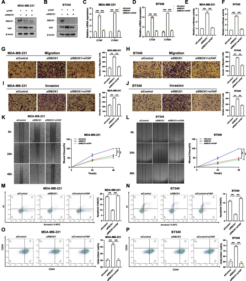
Methods: RBCK1 and YAP protein expression levels were measured by western blotting, while the mRNA levels of YAP target genes were measured by RT-PCR. RNA sequencing data were analyzed by Ingenuity Pathway Analysis. Identification of Hippo signaling activity was accomplished with luciferase assays, RT-PCR and western blotting. Protein stability assays and ubiquitin assays were used to detect YAP protein degradation. Ubiquitin-based immunoprecipitation assays were used to detect the specific ubiquitination modification on the YAP protein.

Results: In our current study, our data revealed an opposite function for RBCK1 in TNBC progression. RBCK1 over-expression inhibited TNBC cell progression in vitro and in vivo, while RBCK1 depletion promoted TNBC cell invasion. The whole genomic expression profiling showed that RBCK1 depletion activated Hippo/YAP axis. RBCK1 depletion increased YAP protein level and Hippo target gene expression in TNBC. The molecular biology studies confirmed that RBCK1 could bind to YAP protein and enhance the stability of YAP protein by promoting YAP K48-linked poly-ubiquitination at several YAP lysine sites (K76, K204 and K321).

Conclusion: Our study revealed the multi-faced RBCK1 function in different subtypes of breast cancer patients and a promising therapeutic target for TNBC treatment. Video abstract.

Keywords: Breast cancer; RBCK1; Ubiquitin; YAP.


Conflict of interest statement

All authors claim no conflict of interest.

Figures

Fig. 1
Fig. 1
RBCK1 depletion promotes cancer cell progression in TNBC AB RBCK1 deletion efficiency by two different siRNA oligonucleotides in breast cancer cell. *P < 0.05; **P < 0.01; ***P < 0.001 for RBCK1 mRNA level comparison. CD RBCK1 depletion has no effect on TNBC cancer cell proliferation. Each group was analyzed three times. *P < 0.05; **P < 0.01; ***P < 0.001 for cell growth comparisons. EF RBCK1 deletion promotes the migration of cell in TNBC.MDA-MB-231 and BT549 cells were transfected with siControl or siRBCK1. Calculate the number of cells and the number data showed as ± SD. *P < 0.05; **P < 0.01; ***P < 0.001 for cell comparisons. GH RBCK1 depletion promotes the invasion of cell in TNBC.MDA-MB-231 and BT549 cells were transfected with siControl or siRBCK1. Calculate the number of cells and the number data showed as ± SD. *P < 0.05; **P < 0.01; ***P < 0.001 for cell comparisons. IJ Wound healing experiment of TNBC cells was transfected with siRBCK1 or siControl. Wound closure was quantified at a specified point in time. *P < 0.05; ** P < 0.01; ***P < 0.001 for comparisons. KL RBCK1 depletion inhibited apoptosis in TNBC cells. MDA-MB-231 and BT549 cells were transfected with siControl or siRBCK1. Each group was analyzed three times. *P < 0.05; **P < 0.01; ***P < 0.001 for comparisons. MN RBCK1 depletion increased the expression of CD24-CD44 + in TNBC cells. MDA-MB-231 and BT549 cells were transfected with siControl or siRBCK1. Each group was analyzed in three times. *P < 0.05; ** P < 0.01; ***P < 0.001 for comparisons
Fig. 2
Fig. 2
RBCK1 overexpression inhibited TNBC cell progression in vitro and in vivo. A RBCK1 over-expression efficiency by plasmids. We overexpressed RBCK1 in TNBC cells via lenti-virus infect system. The level of Flag-RBCK1 protein was detected by western blot. β-Actin was used as the internal reference. BC Overexpression of RBCK1 inhibits the proliferation of TNBC cells. The metabolic activity of cells at the specified time points after transfection was determined by CCK-8 analysis. Each group was analyzed three times. *P < 0.05; **P < 0.01; ***P < 0.001 for cell growth comparisons. DE Overexpression of RBCK1 inhibited cell migration in TNBC cells. MDA-MB-231 and BT549 cells with stable expression of RBCK1 or empty vector. Calculate the number of cells and the number data showed as ± SD. *P < 0.05; **P < 0.01; ***P < 0.001 for cell comparisons. FG Overexpression of RBCK1 inhibited cell invasion in TNBC cells. MDA-MB-231 and BT549 cells with stable expression of RBCK1 or empty vector. Calculate the number of cells and the number data showed as ± SD. *P < 0.05; ** P < 0.01; ***P < 0.001 for cell comparisons. HI Wound healing experiment of MDA-MB-231 and BT549 cells with stable expression of RBCK1 or vector. Wound closure was quantified at a specified point in time. *P < 0.05; **P < 0.01; ***P < 0.001 for cell comparisons. JK Overexpression of RBCK1 increased apoptosis in TNBC cells. MDA-MB-231 and BT549 cells with stable expression of RBCK1 or empty vector were stained with PI and Annexin V. Each group was analyzed three times. *P < 0.05; **P < 0.01; ***P < 0.001 for comparisons. LM Overexpression of RBCK1 inhibited the expression of CD24-CD44 + in TNBC cells. MDA-MB-231 and BT549 cells with stable expression of RBCK1 or empty vector were stained with anti-CD44-FITC and anti-CD24-PE. Each group was analyzed in three times. *P < 0.05; **P < 0.01; ***P < 0.001 for comparisons. NP Overexpression of RBCK1 inhibited the tumor growth of MDA-MB-231 cells in a xenograft model. Tumor growth photographs, curves and weights are shown in panels N, O and P, respectively. The data are showed as ± SD. *P < 0.05; **P < 0.01; ***P < 0.001 for cell comparisons
Fig. 3
Fig. 3
Clinical data analysis reveals the correlation between RBCK1 and Hippo signaling in TNBC A Compared with breast tissue in TCGA database, RBCK1 mRNA levels in breast cancer data samples increased.. (https://tcga-data.nci.nih.gov). B Compared with normal breast tissue in TCGA database, RBCK1 mRNA levels in all subtypes of breast cancer are increased. (https://tcga-data.nci.nih.gov). C Kaplan–Meier map of progression free survival demonstrates that RBCK1 is associated with good prognosis in triple negative breast cancer patient. (http://kmplot.com/analysis/). D Kaplan–Meier map of progression free survival demonstrates that YAP is associated with poor prognosis in triple negative breast cancer patients. E Gene ontology analysis of the RNA sequencing data shows that RBCK1 depletion in TNBC cells activates the Hippo signaling pathway. MDA-MB-231 cells were transfected with siRBCK1 or siControl. After 48 h, the total mRNA was extracted for RNA sequencing analysis. The siControl and siRBCK1 groups were analyzed in three times. F Gene set enrichment analysis (GSEA) analysis showed that the gene sets of CORDENONSI YAP CONSERVED SIGNATURE was enriched in the RBCK1-low group. G The volcanic map analysis showed that CTGF and CYR61 genes downstream of YAP were significantly up-regulated in the RBCK1-low group. H Cases of tumor showed that RBCK1 protein is negatively correlated with YAP protein in IHC. I Statistical analysis of RBCK1 correlation between RBCK1 and YAP in 40 TNBC tumor samples (Additional file 2)
Fig. 4
Fig. 4
RBCK1 inhibits YAP protein level and Hippo target gene expression in TNBC AB RBCK1 depletion increases the level of YAP protein in TNBC cells. MDA-MB-231 and BT549 cells were transfected with siControl or siRBCK1. 48 h later, the cells were collected for western blot analysis. CD RBCK1 deletion increases expression of YAP target gene in TNBC cells. MDA-MB-231 and BT549 cells were transfected with siControl or siRBCK1. 48 h later, total RNA was collected for gene expression analysis. 36B4 was used as internal control. Each group was analyzed three times. *P < 0.05; **P < 0.01; ***P < 0.001 for target gene expression comparison. EF RBCK1 deletion increased the activity of TEAD luciferase in TNBC cells. MDA-MB-231 and BT549 cells were transfected with siControl, siRBCK1,YAP luciferase reporter plasmid and Renilla plasmid. After 24 h, the cells were harvested for the detection of luciferase activity. Each group was analyzed three times. *P < 0.05; **P < 0.01; ***P < 0.001 for gene expression comparisons. G Overexpression of RBCK1 decreased the levels of YAP protein in HEK293 cells. HEK293 cells were transfected with Flag-RBCK1 or Flag-tag plasmids. 48 h later, the cells were collected for western blot analysis. H Overexpression of RBCK1 reduced the expression of YAP target gene in HEK293cells. HEK293 cells were transfected with Flag-RBCK1 or Flag-tag plasmids. After 48 h, total RNA was extracted for the gene expression analysis. 36B4 was used as internal control. Each group was analyzed three times. IJ Overexpression of RBCK1 reduced the expression of YAP target gene. MDA-MB-231 and BT549 cells with stable expression of RBCK1 or empty vector, total RNA was extracted for the gene expression analysis. 36B4 was used as internal control. Each group was analyzed three times. KL Overexpression of RBCK1 reduced TEAD Luciferase activity in TNBC cells. MDA-MB-231 and BT549 cells with stable expression of RBCK1 or empty vector. After 24 h, the cells were transfected with the YAP luciferase reporter plasmid and Renilla plasmid. After 24 h, cells were harvested for the detection of luciferase activity. Each group was analyzed three times. *P < 0.05; **P < 0.01; ***P < 0.001 for gene expression comparisons
Fig. 5
Fig. 5
RBCK1 inhibits TNBC cancer progression via Hippo/YAP axis AB RBCK1 deletion boosted the level of YAP protein, and the function can be saved after YAP deletion in TNBC cell. MDA-MB-231 and BT459 cells were transfected with siControl, siRBCK1 or siRBCK1 + siYAP. 48 h later,the cells were collected for western blot analysis. CD RBCK1 deletion increased the expression of Hippo target gene, which can be reversed after YAP knocking-down in TNBC cell. MDA-MB-231 and BT459 cells were transfected with siControl, siRBCK1 or siRBCK1 + siYAP. 48 h later, total RNA was collected for gene expression analysis. 36B4 was used as internal control. Each group was analyzed three times. EF RBCK1 deletion increased the activity of TEAD luciferase, which can be reversed after YAP knocking-down in TNBC cell. MDA-MB-231 and BT459 cells were transfected with siControl, siRBCK1 or siRBCK1 + siYAP., YAP luciferase reporter plasmid and Renilla plasmid. 24 h later, the cells were collected for the detection of luciferase activity. Each group was analyzed three times. *P < 0.05; **P < 0.01; ***P < 0.001 for gene expression comparisons. G–H RBCK1 depletion promotes the migration of cell, which can be reversed after YAP knocking-down in TNBC cell. MDA-MB-231 and BT459 cells were transfected with siControl, siRBCK1 or siRBCK1 + siYAP. We used the trans-well assay analysis to examine the invasion capability of TNBC cell. Calculate the number of cells and the number data showed as ± SD. *P < 0.05; **P < 0.01; ***P < 0.001 for cell comparisons. IJ RBCK1 depletion promotes the invasion of cell, which can be reversed after YAP knocking-down in TNBC cell. MDA-MB-231 and BT459 cells were transfected with siControl, siRBCK1 or siRBCK1 + siYAP. We used the trans-well assay analysis to examine the invasion capability of TNBC cell. Calculate the number of cells and the number data showed as ± SD. *P < 0.05; **P < 0.01; ***P < 0.001 for cell comparisons. KL Wound-healing experiment confirmed that RBCK1 depletion increased the migration capacity of TNBC cells, the function could be saved after YAP deletion in TNBC cell. MDA-MB-231 and BT459 cells were transfected with siControl, siRBCK1 or siRBCK1 + siYAP. Wound closure was quantified at a specified point in time. Data are showed as ± SD. *P < 0.05; **P < 0.01; ***P < 0.001 for cell comparisons. MN RBCK1 depletion inhibited apoptosis, which can be reversed after YAP knocking-down in TNBC cell. MDA-MB-231 and BT459 cells were transfected with siControl, siRBCK1 or siRBCK1 + siYAP. 24 h later, the cells were stained with PI and Annexin V. Then, FACS analysis was performed on the cells to determine the proportion of apoptotic cells. Each group was analyzed three times. OP RBCK1 depletion increased the expression of CD24-CD44 + , in which can be reversed after YAP knocking-down in TNBC cell. MDA-MB-231 and BT459 cells were transfected with siControl, siRBCK1 or siRBCK1 + siYAP. After 24 h, cells were stained with anti-CD44-FITC and anti-CD24-PE. Then, FACS analysis was performed on the cells to determine the proportion of CD24-CD44 + cells. Each group was analyzed in three times
Fig. 6
Fig. 6
RBCK1 associates with YAP and modulates YAP stability in TNBC cells A The intracellular localization of RBCK1 and YAP was analyzed by immunofluorescence. MDA-MB-231 cells were cultured in normal medium before fixation. Intracellular localization of RBCK1 (red) and YAP (green) is shown. Nuclei (blue) were stained with 4’,6-diamidino-2-phenylindole (DAPI). B RBCK1 is mainly localized in the cytoplasm. YAP is mainly localized in the nuclear. We used a subcellular protein fractionation kit (Thermo Scientific, 78,840) for cytoplasm and nuclear separation experiment. CD Co-IP experiment revealed the association between endogenous RBCK1 and YAP protein in MDA-MB-231 cells. MDA-MB-231 cells were collected with RIPA lysis buffer. CO-IP was performed using an antibody as indicated EF RBCK1 full-length and deletion mutants are used in the study (Full-length, ΔUBL, ΔNZF, ΔRBR domains). YAP full-length and deletion mutants are used (Full length, ΔTA, ΔWW + ΔTA, ΔTBD, ΔTBD + ΔWW). G RBCK1 needs RBR domain to interact with YAP. HEK293 cells were transfected with 2 µg Myc-YAP and Flag-RBCK1 full-length or mutants (ΔUBL, ΔNZF, ΔRBR domains). 24 h later, the cells were collected with NP-40 lysis buffer. CO-IP was performed using an anti-Myc antibody. The interacting RBCK1 domains were detected by an anti-Flag antibody. H YAP needs TA domain to interact with RBCK1. HEK293 cells were transfected with 2 µg Flag-RBCK1 and Myc-YAP full-length or mutants (ΔTA, ΔWW + ΔTA, ΔTBD, ΔTBD + ΔWW). 24 h later, cells were harvested with NP-40 lysis buffer. The use of antibodies is shown in the figure. IJ RBCK1 did not further increase the level of YAP protein in TNBC cells in the presence of proteasome inhibitor MG132. MDA-MB-231 and BT549 cells were transfected with siControl or siRBCK1. 24 h later, the cells were treated with MG132 or vehicle for 6 h. The cell lysates were collected for western blot analysis. K The degradation effect of RBCK1 on YAP did not increase the levels of YAP protein any more in TNBC cell in the presence of the proteasome inhibitor MG132. HEK293 cells were transfected with 0.5 mg Flag-tag or Flag -RBCK1 plasmids. After 24 h, the cells were treated with 10 μM MG132/vehicle for 6 h. The cell lysates were prepared for western blot analysis. β-Actin was used as the internal reference. The results are representative of three independent experiments. LM RBCK1 depletion increases the level of YAP half-life in TNBC cells. MDA-MB-231 and BT549 cells were transfected with siControl or siRBCK1. 24 h later, the cells were treated with 100 µM cycloheximide/vehicle for the specified time. The cell lysates were prepared for western blot analysis. The relative YAP density was measured by ImageJ software. β-Actin was used as the internal reference. The results are representative of three independent experiments. N Overexpression of RBCK1 reduced YAP half-life in HEK293 cells. HEK293 cells were transfected with 0.5 mg Flag-tag or Flag -RBCK1 plasmids. 24 h later, the cells were treated with 100 µM cycloheximide/vehicle for the specified times. The cell were collected for western blot analysis. The relative YAP density was measured by ImageJ software. β-Actin was used as the internal reference. The results are representative of three independent experiments
Fig. 7
Fig. 7
RBCK1 functions as one ubiquitin ligase to promote YAP K48-linked poly-ubiquitination A RBCK1 deletion decreases the total polyubiquitination of YAP. siControl or siRBCK1 were transfected into MDA-MB-231 cells. 24 h later, 1 mg HA-Ub plasmid and 2 µg YAP plasmid were transfected into cells. 24 h later, the cells were immunoprecipitated with an anti-HA antibody. B RBCK1 boosted total polyubiquitination of YAP. 2 µg of YAP plasmid, 0.5 µg of HA-Ub plasmid and 0.5 µg of Flag-tag or Flag -RBCK1 plasmids were transfected into HEK293T cells. After 24 h, the cells were immunoprecipitated with an anti-HA antibody. C RBCK1 deletion decreases the K48-linked polyubiquitination of YAP. siControl or siRBCK1 were transfected into MDA-MB-231 cells. 24 h later, 1 mg HA-K48 Ubi plasmid and 2 µg of YAP plasmid were transfected into cells. 24 h later, the cell were immunoprecipitated with an anti-HA antibody. D RBCK1 boosted the K48-linked polyubiquitination of YAP. 2 µg of YAP plasmid, 0.5 µg of HA-K48 Ubi plasmid and 0.5 µg of Flag-tag or Flag -RBCK1 plasmids were transfected into HEK293T cells. 24 h later, the cells were immunoprecipitated with an anti-HA antibody. E RBCK1 deletion boosted the K63-linked polyubiquitination of YAP. siControl or siRBCK1 were transfected into MDA-MB-231 cells. 24 h later, 1 mg HA-K63 Ubi plasmid and 2 µg of YAP plasmid were transfected into cells. 24 h later, the cell were immunoprecipitated with an anti-HA antibody. F RBCK1 decreases K63-linked polyubiquitination of YAP. 2 µg of YAP plasmid, 0.5 µg of HA-K63 Ubi plasmid and 0.5 µg of Flag-tag or Flag -RBCK1 plasmids were transfected into HEK293T cells. After 24 h, the cell extracts were immunoprecipitated with an anti-HA antibody. G RBCK1 inhibits YAP degradation by RBR domain. 2 µg Flag-RBCK1 full length or mutant plasmids (ΔUBL, ΔNZF, ΔRBR domains) were transfected into HEK293T cells. After 24 h, the cells were harvested directly and used for the western blot analysis. β-Actin was used as the internal reference. H The UBL domain or RBR domain of RBCK1 is important to improve total ubiquitination YAP. 2 µg, Myc-YAP, 1 µg HA-UB plasmid and 1 µg Flag-RBCK1 full length or mutants (ΔUBL, ΔNZF, ΔRBR domains) were transfected into HEK293 cells. The polyubiquitinated YAP was detected through the western blot analysis. I The RBR domain of RBCK1 is important to improve K48-linked ubiquitination of YAP. 2 µg Myc-YAP, 1 µg HA-K48 Ubi plasmid and 1 µg Flag-RBCK1 full length or mutants (ΔUBL, ΔNZF, ΔRBR domains) were transfected into HEK293 cells. The K48-specific polyubiquitinated YAP was detected through the western blot analysis. J The ability of RBCK1 to degrade YAP protein was impaired by the mutations of RBCK1 in MDA-MB-231 cells. K The mutations of RBCK1 that impaired ubiquitination of RBCK1 activity and the ability to increase the total polyubiquitination of YAP. HEK293 cells were transfected with 2 µg of YAP plasmid, 0.5 µg of HA-Ub plasmid and 0.5 µg of Flag-tag or Flag -RBCK1 plasmids or Flag-RBCK1 C460A. After 24 h, the cells were immunoprecipitated with an anti-HA antibody. The total polyubiquitinated YAP was detected via western blot analysis. LM K76, K204, and K321 mutations (K76R, K204, and K321R) largely eliminated the ubiquitination effect of RBCK1 on YAP. The indicated vectors were transfected into HEK293 for ubiquitination analysis. Polyubiquitinated YAP was detected by Western blot analysis
Fig. 8
Fig. 8
The hypothetical model for RBCK1 regulating Hippo/ YAP signaling in TNBC progression RBCK1 protein relevant to YAP and improve YAP degradation by leading YAP K48-linked polyubiquitination, which reduced the Hippo/YAP axis activation and invasion and migration of TNBC cells

Similar articles

Cited by

References

    1. Naderi R, Gholizadeh-Ghaleh Aziz S, Haghigi-Asl AS. Evaluating the effect of Alantolactone on the expression of N-cadherin and Vimentin genes effective in epithelial-mesenchymal transition (EMT) in breast cancer cell line (MDA-MB-231) Ann Med Surg (Lond) 2022;73:103240. - PMC - PubMed
    1. Chan WL, Marinho J, Chavarri-Guerra Y, Hincapie-Echeverri J, Velasco RN, Jr, Akagunduz B, et al. Systemic treatment for triple negative breast cancer in older patients: A Young International Society of Geriatric Oncology Review Paper. J Geriatr Oncol. 2022;13:563–571. - PubMed
    1. Nagini S. Breast cancer: current molecular therapeutic targets and new players. Anticancer Agents Med Chem. 2017;17:152–163. - PubMed
    1. Yin L, Duan JJ, Bian XW, Yu SC. Triple-negative breast cancer molecular subtyping and treatment progress. Breast Cancer Res. 2020;22:61. - PMC - PubMed
    1. Liedtke C, Bernemann C, Kiesel L, Rody A. Genomic profiling in triple-negative breast cancer. Breast Care (Basel) 2013;8:408–413. - PMC - PubMed

Publication types