. 2022 Nov 22;7(22):e159436.
doi: 10.1172/jci.insight.159436.

IL-6-targeted therapies to block the cytokine or its receptor drive distinct alterations in T cell function

Affiliations

IL-6-targeted therapies to block the cytokine or its receptor drive distinct alterations in T cell function

Cate Speake et al. JCI Insight. .

Abstract

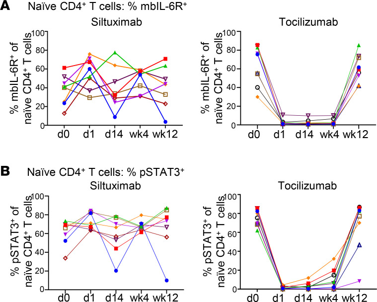
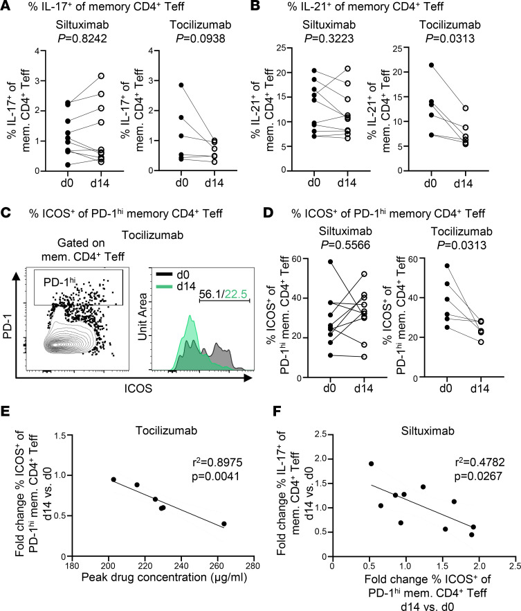
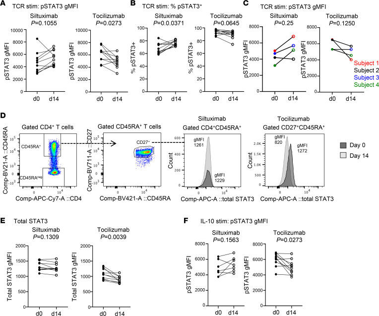
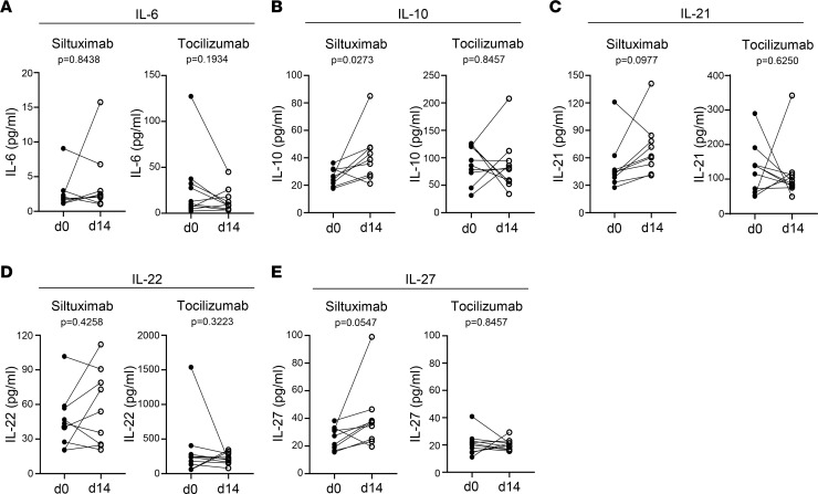
Therapeutics that inhibit IL-6 at different points in its signaling pathway are in clinical use, yet whether the immunological effects of these interventions differ based on their molecular target is unknown. We performed short-term interventions in individuals with type 1 diabetes using anti-IL-6 (siltuximab) or anti-IL-6 receptor (IL-6R; tocilizumab) therapies and investigated the impact of this in vivo blockade on T cell fate and function. Immune outcomes were influenced by the target of the therapeutic intervention (IL-6 versus IL-6R) and by peak drug concentration. Tocilizumab reduced ICOS expression on T follicular helper cell populations and T cell receptor-driven (TCR-driven) STAT3 phosphorylation. Siltuximab reversed resistance to Treg-mediated suppression and increased TCR-driven phosphorylated STAT3 and production of IL-10, IL-21, and IL-27 by T effectors. Together, these findings indicate that the context of IL-6 blockade in vivo drives distinct T cell-intrinsic changes that may influence therapeutic outcomes.

Keywords: Autoimmunity; Cytokines; Immunology; Immunotherapy; T cells.


Conflict of interest statement

Conflict of interest: The siltuximab study was funded by Janssen Pharmaceuticals under a contract research agreement with CJG and JHB. CS has consulted for Vertex Pharmaceuticals. She is a member of the Type 1 Diabetes TrialNet Study Group and of the Type 1 Diabetes in Acute Pancreatitis Consortium. CJG has received grant support and consulting fees from Novo Nordisk, consulting fees and travel support from Bristol Myers Squibb, grant support from Janssen Pharmaceuticals, and consulting fees from Eli Lilly. She is a member of the Type 1 Diabetes TrialNet Study Group and of the Type 1 Diabetes in Acute Pancreatitis Consortium. JHB is a scientific cofounder and scientific advisory board member of GentiBio; is a consultant for Bristol Myers Squibb and Hotspot Therapeutics; and has past and current research projects sponsored by Amgen, Bristol Myers Squibb, Janssen, Novo Nordisk, and Pfizer. She is a member of the Type 1 Diabetes TrialNet Study Group, a partner of the Allen Institute for Immunology, and a member of the scientific advisory boards for the La Jolla Institute for Allergy and Immunology and Bristol Myers Squibb Immunology.

Figures

Figure 1
Figure 1. Suppression of IL-6–induced p-STAT3 persists in tocilizumab-treated but not siltuximab-treated patient PBMCs.
Each line represents an individual patient; n = 10 for siltuximab and n = 9 for tocilizumab. (A) Frequency of mbIL-6R+ cells in naive CD4+ T cell compartment at baseline. (B) Frequency of IL-6–induced p-STAT3+ cells in the naive CD4+ T cell compartment. Thawed and rested PBMCs from siltuximab-treated or tocilizumab-treated patients with T1D were stimulated with recombinant IL-6 (2 ng/mL) for 10 minutes.
Figure 2
Figure 2. Tocilizumab but not siltuximab decreases ICOS expression of T follicular helper cells.
Thawed and rested PBMCs from siltuximab-treated or tocilizumab-treated patients with T1D were stimulated with PMA/ionomycin for 1 hour followed by an additional 3 hours in the presence of Brefeldin A. Each line represents an individual patient; n = 10 for siltuximab and n = 6 for tocilizumab. Solid circles represent d0 prior to drug infusion, and open circles represent d14 after drug infusion. (A) Frequency of IL-17+ cells in memory CD4+ Teffs. (B) Frequency of IL-21+ cells in memory CD4+ Teffs. (C) Representative histograms showing PD-1hi ICOS+ memory CD4+ Teffs at d0 and d14 after tocilizumab infusion from a single patient. (D) Frequency of ICOS+ cells in PD-1hi memory CD4+ Teffs. (E) Linear regression for tocilizumab cohort showing negative correlation between peak drug concentration on d1 and fold change d14 versus d0 for frequency of ICOS+ cells in PD-1hi memory CD4+ Teffs. (F) Linear regression for siltuximab cohort showing negative correlation between fold change d14 versus d0 for frequency of ICOS+ cells in PD-1hi memory CD4+ T cell compartment and fold change d14 versus d0 for frequency of IL-17+ cells in memory CD4+ T cell compartment. Statistical tests: (A, B, and D) Wilcoxon matched-pairs signed-rank test; and (E and F) linear regression.
Figure 3
Figure 3. Siltuximab but not tocilizumab reverses Teff resistance to Treg-mediated suppression.
Expanded allogeneic Tregs from a healthy control donor were cocultured at a ratio of 1:4 with Teffs from either siltuximab-treated or tocilizumab-treated patients with T1D in the presence of anti-CD3/anti-CD28–coated beads for 2 days. The percentage of suppression was determined by measuring the frequency of activated CD25+CD134+ Teffs. Each line represents an individual patient; n = 10 for siltuximab and n = 9 for tocilizumab. (A) Percentage suppression for siltuximab-treated patients and tocilizumab-treated patients. (B) Linear regression for siltuximab cohort showing positive correlation between peak drug concentration on d1 and percentage suppression at d14. (C) Percentage suppression for cohort that participated in both studies (n = 5); note these individuals are also included in A and B. Statistical tests: (A and C) Wilcoxon matched-pairs signed-rank test; (B) linear regression.
Figure 4
Figure 4. Tocilizumab and siltuximab have opposing effects on TCR-induced p-STAT3 signaling in naive CD4+ T cells.
Enriched pan T cells from siltuximab-treated or tocilizumab-treated patients with T1D were stimulated or not with anti-CD3/anti-CD28–coated beads for 4 hours. Cells were stained for p-STAT3 and total STAT3. Each line represents an individual patient; n = 10 for siltuximab (except for F where n = 7; gated CD4+CD45RA+ naive CD4+ T cells) and n = 10 for tocilizumab (gated CD4+CD27+CD45RA+ naive CD4+ T cells). Solid circles represent d0 prior to administration of the drug, and open circles represent d14 after drug administration. (A) p-STAT3 geometric MFI (gMFI). (B) Frequency of p-STAT3+ cells. (C) p-STAT3 MFI for cohort that participated in both studies (n = 4); note these individuals are also included in A, B, E, and F. (D) Gating strategy for total STAT3 gMFI of gated naive CD4+ T cells using unstimulated enriched pan T cells: histograms for representative siltuximab-treated patient and representative tocilizumab-treated patient. (E) Total STAT3 MFI of unstimulated cells. (F) p-STAT3 MFI after stimulation with IL-10 (20 ng/mL for 30 minutes). Statistical tests: Wilcoxon matched-pairs signed-rank test.
Figure 5
Figure 5. Siltuximab but not tocilizumab enhances T cell production of regulatory cytokines.
Meso Scale Discovery assays were used to measure cytokine production by enriched pan T cells from siltuximab-treated or tocilizumab-treated patients with T1D stimulated with anti-CD3/anti-CD28–coated beads for 4 hours. Each line represents an individual patient; n = 9 for siltuximab (except for A where n = 8) and n = 10 for tocilizumab. Solid circles represent d0 prior to administration, and open circles represent d14 after drug administration (d14). (A) IL-6, (B) IL-10, (C) IL-21, (D) IL-22, and (E) IL-27. Statistical test: Wilcoxon matched-pairs signed-rank test.

References

    1. Jones BE, et al. IL-6: a cytokine at the crossroads of autoimmunity. Curr Opin Immunol. 2018;55:9–14. doi: 10.1016/j.coi.2018.09.002. - DOI - PMC - PubMed
    1. Kang S, et al. Targeting interleukin-6 signaling in clinic. Immunity. 2019;50(4):1007–1023. doi: 10.1016/j.immuni.2019.03.026. - DOI - PubMed
    1. Johnson DE, et al. Targeting the IL-6/JAK/STAT3 signalling axis in cancer. Nat Rev Clin Oncol. 2018;15(4):234–248. doi: 10.1038/nrclinonc.2018.8. - DOI - PMC - PubMed
    1. Tocilizumab in patients admitted to hospital with COVID-19 (RECOVERY): a randomised, controlled, open-label, platform trial. Lancet. 2021;397(10285):1637–1645. doi: 10.1016/S0140-6736(21)00676-0. - DOI - PMC - PubMed
    1. Choy EH, et al. Translating IL-6 biology into effective treatments. Nat Rev Rheumatol. 2020;16(6):335–345. doi: 10.1038/s41584-020-0419-z. - DOI - PMC - PubMed

Publication types