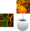
Live-Cell Imaging of Intact Ex Vivo Globes Using a Novel 3D Printed Holder
- PMID: 36282717
- PMCID: PMC10171086
- DOI: 10.3791/64510
Live-Cell Imaging of Intact Ex Vivo Globes Using a Novel 3D Printed Holder
Abstract
Corneal epithelial wound healing is a migratory process initiated by the activation of purinergic receptors expressed on epithelial cells. This activation results in calcium mobilization events that propagate from cell to cell, which are essential for initiating cellular motility into the wound bed, promoting efficient wound healing. The Trinkaus-Randall lab has developed a methodology for imaging the corneal wound healing response in ex vivo murine globes in real time. This approach involves enucleating an intact globe from a mouse that has been euthanized per established protocols and immediately incubating the globe with a calcium indicator dye. A counterstain that stains other features of the cell can be applied at this stage to assist with imaging and show cellular landmarks. The protocol worked well with several different live cell dyes used for counterstaining, including SiR actin to stain actin and deep red plasma membrane stain to stain the cell membrane. To examine the response to a wound, the corneal epithelium is injured using a 25 G needle, and the globes are placed in a 3D printed holder. The dimensions of the 3D printed holder are calibrated to ensure immobilization of the globe throughout the duration of the experiment and can be modified to accommodate eyes of different sizes. Live cell imaging of the wound response is performed continuously at various depths throughout the tissue over time using confocal microscopy. This protocol allows us to generate high-resolution, publication-quality images using a 20x air objective on a confocal microscope. Other objectives can also be used for this protocol. It represents a significant improvement in the quality of live cell imaging in ex vivo murine globes and permits the identification of nerves and epithelium.
Conflict of interest statement
Disclosures
We have no conflicts of interest to disclose.
Figures


References
-
- Klepeis VS, Cornell-Bell A, Trinkaus-Randal V Growth factors but not gap junctions play a role in injury-induced Ca2+ waves in epithelial cells. Journal of Cell Science. 114 (23), 4185–4195 (2001). - PubMed
Publication types
MeSH terms
Substances
Grants and funding
LinkOut - more resources
Full Text Sources
Miscellaneous
