. 2022 Oct 25;15(757):eabm0808.
doi: 10.1126/scisignal.abm0808. Epub 2022 Oct 25.

Host protein kinases required for SARS-CoV-2 nucleocapsid phosphorylation and viral replication

Affiliations

Host protein kinases required for SARS-CoV-2 nucleocapsid phosphorylation and viral replication

Tomer M Yaron et al. Sci Signal. .

Abstract

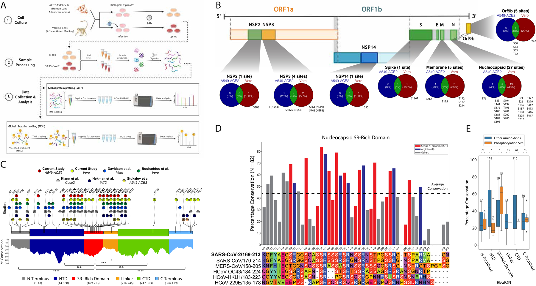
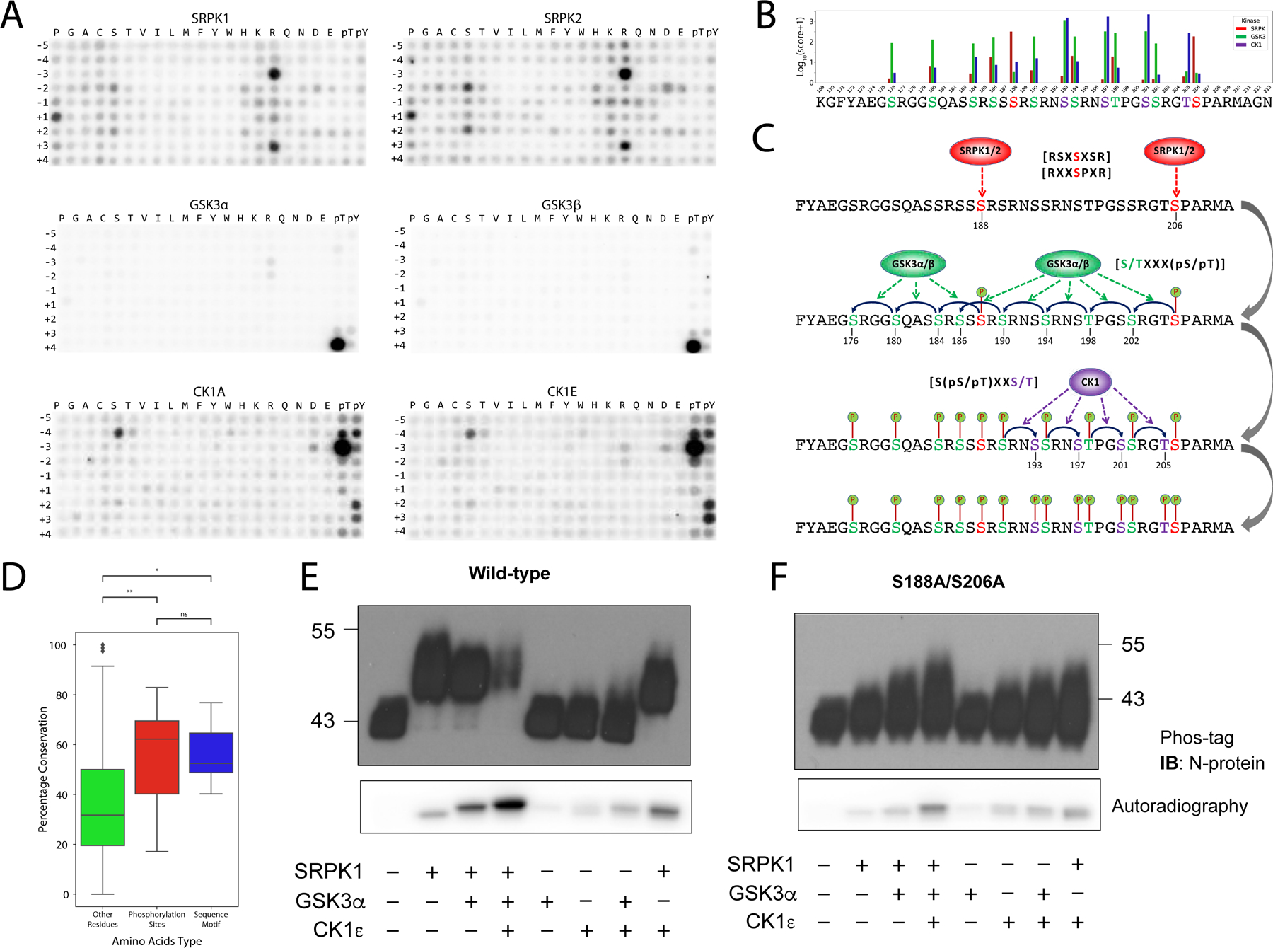
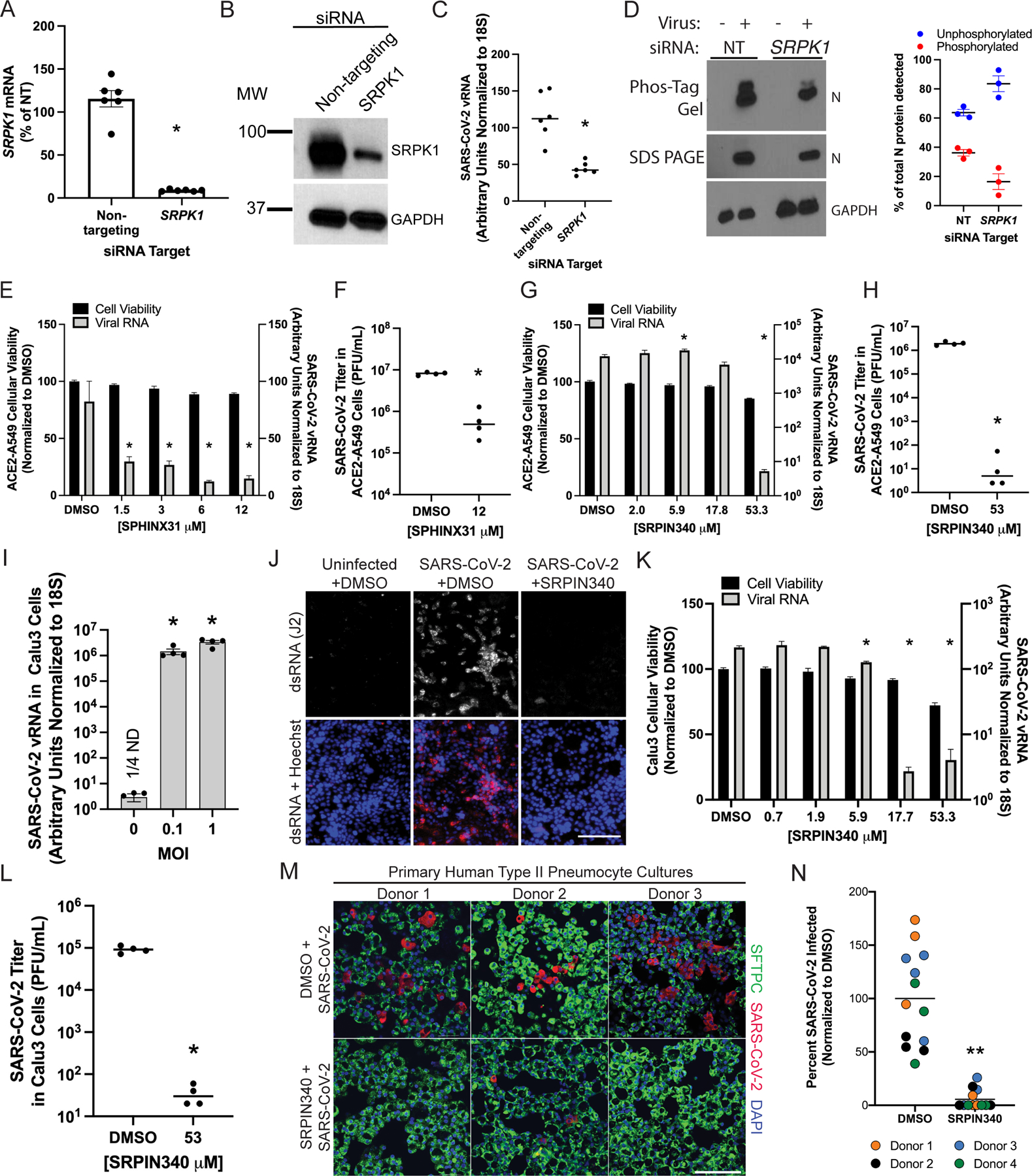
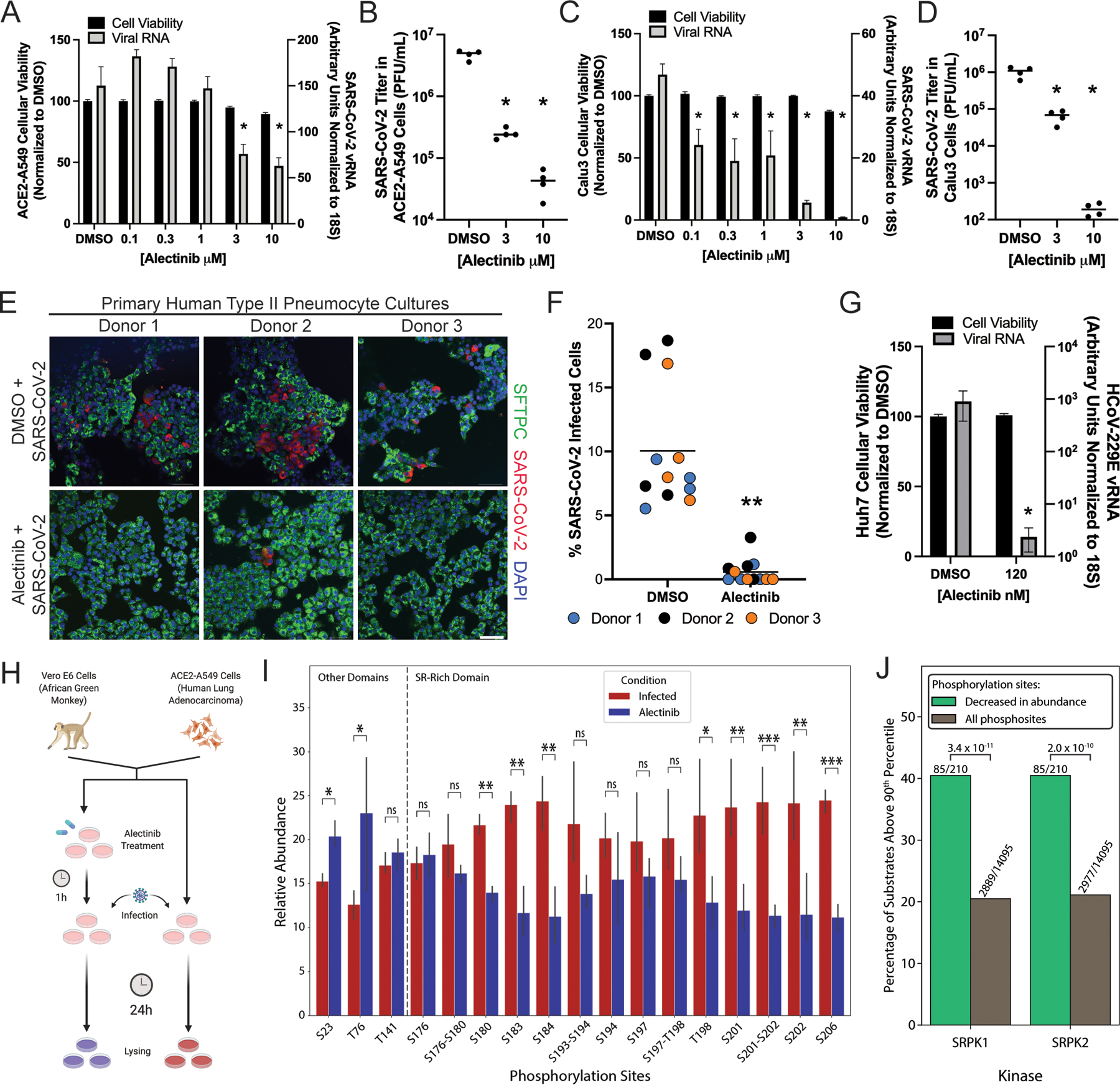
Multiple coronaviruses have emerged independently in the past 20 years that cause lethal human diseases. Although vaccine development targeting these viruses has been accelerated substantially, there remain patients requiring treatment who cannot be vaccinated or who experience breakthrough infections. Understanding the common host factors necessary for the life cycles of coronaviruses may reveal conserved therapeutic targets. Here, we used the known substrate specificities of mammalian protein kinases to deconvolute the sequence of phosphorylation events mediated by three host protein kinase families (SRPK, GSK-3, and CK1) that coordinately phosphorylate a cluster of serine and threonine residues in the viral N protein, which is required for viral replication. We also showed that loss or inhibition of SRPK1/2, which we propose initiates the N protein phosphorylation cascade, compromised the viral replication cycle. Because these phosphorylation sites are highly conserved across coronaviruses, inhibitors of these protein kinases not only may have therapeutic potential against COVID-19 but also may be broadly useful against coronavirus-mediated diseases.


Conflict of interest statement

Competing interests:

Duke University has filed for intellectual property protection regarding the use of SRPK inhibitors in the treatment of COVID-19. L.C.C. is a founder and member of the board of directors of Agios Pharmaceuticals and is a founder and receives research support from Petra Pharmaceuticals. L.C.C. is an inventor on patents (pending) for Combination Therapy for PI3K-associated Disease or Disorder, and The Identification of Therapeutic Interventions to Improve Response to PI3K Inhibitors for Cancer Treatment. L.C.C. is a co-founder and shareholder in Faeth Therapeutics. T.M.Y. is a stockholder and on the board of directors of DESTROKE, Inc., an early-stage start-up developing mobile technology for automated clinical stroke detection. O.E. is a founder and equity holder of Volastra Therapeutics and OneThree Biotech. O.E. is a member of the scientific advisory boards of Owkin, Freenome, Genetic Intelligence, Acuamark, and Champions Oncology. O.E. receives research support from Eli Lilly, Janssen, and Sanofi. R.E.S. is on the scientific advisory board of Miromatrix Inc. and is a consultant and speaker for Alnylam Inc. P.R.T serves as an acting CEO of Iolux Inc. P.R.T. serves as a consultant for Cellarity Inc. and Surrozen Inc. P.R.T. receives research support from United Therapeutics Inc. G.G. receives research funds from IBM and Pharmacyclics and is an inventor on patent applications related to MuTect, ABSOLUTE, MutSig, MSMuTect, MSMutSig, MSIdetect, POLYSOLVER, and TensorQTL. G.G. is a founder, consultant, and holds privately held equity in Scorpion Therapeutics. S.C. is a co-founder of OncoBeat LLC. The other authors declare that they have no competing interests.

Figures

Fig. 1.
Fig. 1.. The SARS-CoV-2 N protein is phosphorylated in the SR-rich domain.
(A) Diagram of the proteomics and phosphoproteomics workflow for cells infected with SARS-CoV-2. (B) Phosphorylation sites in SARS-CoV-2 proteins. S, serine; T, threonine, Y, tyrosine. (C) Top: Phosphorylation sites in the SARS-CoV-2 N protein identified in seven different phosphoproteomics analyses [two in the current study and five in previously published studies (–10)]. Bottom: Evolutionary conservation analysis of the different domains of the N protein across 82 different coronaviruses. *P < 0.05, **P < 0.01, ***P < 0.001, and ****P < 0.0001 by nonparametric Wilcoxon-Mann-Whitney U test. (D) Percentage identity of amino acids in the SR-rich domain of the N protein across 82 coronaviruses in multiple species (top) and their sequence alignment across six different human coronaviruses (bottom). (E) The conservation of the phosphorylation sites in each domain was compared to the conservation of the other amino acids in that domain. The numbers of amino acids compared in each domain are annotated above the boxes. *P < 0.05 by nonparametric Wilcoxon-Mann-Whitney U test.
Fig. 2.
Fig. 2.. Analysis of the phosphorylation of the SR-rich domain of the N protein by SRPK, GSK-3, and CK1.
(A) Biochemical substrate specificities of SRPK1/2 (top), GSK-3α/β (middle), and CK1A/ε (bottom). Data are representative of at least two replicate experiments. SRPK isoforms are selective for arginine at the −3 and +3 positions, serine at the −2 and +2 positions, and proline at the +1 position. GSK-3 isoforms are selective for phosphoserine or phosphothreonine at position +4. CK1 isoforms are selective for phosphoserine and phosphothreonine at position −3 and for serine at position −4. (See fig. S1 for the substrate specificities of additional kinases from these families). (B) Favorability scores for the different phosphorylation sites in the SR-rich domain according to the SRPK (SRPK1/2/3), GSK-3 (GSK-3α/β), and CK1 (CK1A/D/ε/G1) families. (C) Proposed scheme for the multisite phosphorylation of the SR-rich domain of the N protein. Simplified substrate consensus motifs are shown in parentheses, whereas detailed logos are provided in fig. S2. (D) Evolutionary conservation comparison of three types of amino acids in the SR-rich domain of the N protein across 82 different coronaviruses from multiple species. Sequence motif: amino acid residues predicted to be essential for the substrate specificity of the priming sites (Ser188: Arg185/Ser186/Ser190/Arg191; Ser206: Arg203/Pro207/Arg209); phosphorylation chains: phosphorylation sites described in the phosphorylation model in Fig. 2C; other amino acids: all other amino acids in the SR-rich domain. *P < 0.05 and **P < 0.01 by nonparametric Wilcoxon-Mann-Whitney U test. (E) Top: Western blotting analysis of recombinant N protein on Phos-tag gel after the indicated treatments with the recombinant kinases SRPK1, GSK-3α, and CK1ε. Bottom: SDS-PAGE/autoradiography of recombinant N protein after the indicated treatments with SRPK1, GSK-3α, and CK1ε in the presence of ATP[γ-32P]. Data are representative of three experiments. (F) Phos-tag gel analysis (top) and autoradiography (bottom) of recombinant N protein with the priming phosphorylation sites mutated (S188A and S206A), which was performed as described fir (E). For comparison, autoradiography of the WT and mutant N proteins was measured together. Data are representative of three experiments.
Fig. 3.
Fig. 3.. SRPK1/2 inhibitors suppress SARS-CoV-2 infection.
(A) qRT-PCR analysis of the relative abundance of SRPK1 mRNA in cells treated with nontargeting (NT) or SRPK1-specific siRNA ACE2-A549 cells. Data are means + SEM of six independent biological replicates. (B) Western blotting analysis of cells treated with nontargeting or SRPK1-specific siRNA. Blots are representative of three experiments. (C) SARS-CoV-2 viral RNA was measured by qRT-PCR assay 24 hours after the infection of ACE2-A549 cells that were treated with nontargeting or SRPK1-specific siRNA. Data are from six independent biological replicates. (D) Left: Western blotting analysis of cells treated with nontargeting or SRPK1-specific siRNA and then infected with SARS-CoV-2. Lysates were resolved on Phos-Tag gels or SDS PAGE gels. Blots are representative of three experiments. Right: Relative percentages of phosphorylated and unphosphorylated N protein quantified across replicate Western blots with ImageJ software. Data are from three experiments. (E) Cellular viability (left axis) was measured after ACE2-A549 cells were treated with the indicated concentrations of SPHINX31. Data are from four independent biological replicates. SARS-CoV-2 viral RNA abundance (right axis) was measured by qRT-PCR analysis 24 hours after infection of cells. Data are from four independent biological replicates. (F) Infectious viral titers were measured by plaque assay of the supernatants of cells that were treated with SPHINX31 and infected for 48 hours. Data are from four independent biological replicates. (G) Cellular viability (left axis) was measured after ACE2-A549 cells were treated with the indicated concentrations of SRPIN340 before infection. Data are from six independent replicates. SARS-CoV-2 viral RNA (right axis) was measured by qRT-PCR analysis 24 hours after infection. Data are from four independent biological replicates. (H) Infectious viral titers were measured by plaque assay of the supernatants of cells that were treated with SRPIN340 and infected for 48 hours. Data are from four independent biological replicates. (I) Calu-3 cells were infected with SARS-CoV-2 at the indicated MOI and SARS-CoV-2 viral RNA abundance was quantified by qRT-PCR after 24 hours of infection. Data are from four independent biological replicates; ND, not detected. (J) Calu-3 cells treated with DMSO as a control or SRPIN340 were infected with SARS-CoV-2 at an MOI of 2.5. Twenty-four hours after infection, the cells were fixed and stained for DNA with Hoechst and dsRNA with the J2 antibody. Scale bar, 150 μm. Images are representative of at least two images and two independent experiments. (K) Cellular viability (left axis) was measured after Calu-3 cells were treated with the indicated concentrations of SRPIN340, and SARS-CoV-2 viral RNA abundance (right axis) was measured by qRT-PCR analysis 24 hours after infection. Data are from four independent biological replicates. (L) Infectious viral titers were measured by plaque assay of the supernatants of Calu-3 cells treated with SRPIN340 and infected for 48 hours. Data are from four independent biological replicates. (M) Representative primary human type II pneumocyte culture images from samples that were pre-treated with SRPIN340 for 12 hours before they were infected with SARS-CoV-2. Twenty-four hours later, the cells were fixed and stained for SARS-CoV-2, DNA with DAPI, and surfactant protein C (SFTPC). Scale bar, 100 μm. (N) Quantification of the percentages of cells from the experiment described in (M) that were infected with SARS-CoV-2. At least three images each from the of four independent human donors are shown. For all panels, error bars represent SEM and statistical significance was calculated by nonparametric Wilcoxon-Mann-Whitney U test: *P < 0.05, **P < 0.001.
Fig. 4.
Fig. 4.. The FDA-approved kinase inhibitor Alectinib inhibits SARS-CoV-2 infection and reduces the extent of phosphorylation of the N protein.
(A) Cell viability (left axis) was measured after ACE2-A549 cells were treated with the indicated concentrations of Alectinib before being infected. SARS-CoV-2 viral RNA abundance (right axis) was measured by qRT-PCR assay 24 hours after infection. Data are from four independent biological replicates. (B) Infectious viral titers were measured by plaque assay of the supernatants of ACE2-A549 cells that were treated with Alectinib and infected for 48 hours. Data are from four independent biological replicates. (C) Cell viability (left axis) was measured after Calu-3 cells were treated with the indicated concentrations of Alectinib. SARS-CoV-2 viral RNA abundance (right axis) was measured by qRT-PCR assay 24 hours after infection. Data are from four independent biological replicates. (D) Infectious viral titers were measured by plaque assay of the supernatants of Calu-3 cells that were treated with Alectinib and infected for 48 hours. Data are from four independent biological replicates. (E) Representative images from primary human type II pneumocyte cultures treated with Alectinib for 12 hours before infection with SARS-CoV-2. Twenty-four hours later, the cells were fixed and stained for SARS-CoV-2 S protein, DNA (with DAPI), and SFTPC. Scale bar, 50 μm. (F) Quantification of the percentages of cells from the experiment described in (E) that were infected with SARS-CoV-2. Four independent images for each of the three different human donors are shown. (G) Cell viability (left axis) was measured after HuH7 cells were treated with Alectinib before infection. The relative abundance of 229E viral RNA was measured by qRT-PCR assay 24 hours after infection. Data are from four independent biological replicates. For all panels, statistical significance was calculated by nonparametric Wilcoxon-Mann-Whitney U test, unless otherwise stated, and measurements were taken from distinct samples; *P < 0.05, **P < 0.001. (H) Scheme for Alectinib treatment and infection for proteomics and phosphoproteomics analysis. Experiments were performed three times. (I) Relative abundance (normalized by total protein abundance) of the different phosphorylated sites in the N protein from ACE2-A549 cells treated with or without Alectinib before infection with SARS-CoV-2. Adjusted P values (FDR) were computed by moderated t test and adjusted by Benjamini-Hochberg correction. *FDR < 0.1, **FDR < 0.05, ***FDR < 0.01. (J) Comparison of the frequency of sites that scored high for SRPK1 and SRPK2 (>90th percentile) among phosphorylation sites that were decreased in abundance upon Alectinib treatment and their frequency among all the detected phosphorylation sites in ACE2-A549 cells. Denoted P values were computed by Fisher’s exact test.

Similar articles

Cited by

References

    1. Yuki K, Fujiogi M, Koutsogiannaki S, COVID-19 pathophysiology: A review. Clin Immunol 215, 108427 (2020). - PMC - PubMed
    1. Li Q et al., Early Transmission Dynamics in Wuhan, China, of Novel Coronavirus-Infected Pneumonia. N Engl J Med 382, 1199–1207 (2020). - PMC - PubMed
    1. Wu C-H et al., Glycogen synthase kinase-3 regulates the phosphorylation of severe acute respiratory syndrome coronavirus nucleocapsid protein and viral replication. Journal of Biological Chemistry 284, 5229–5239 (2009). - PMC - PubMed
    1. McBride R, Van Zyl M, Fielding BC, The coronavirus nucleocapsid is a multifunctional protein. Viruses 6, 2991–3018 (2014). - PMC - PubMed
    1. Fung TS, Liu DX, Post-translational modifications of coronavirus proteins: roles and function. Future Virology 13, 405–430 (2018). - PMC - PubMed

Publication types