

With childhood hemispherectomy, one hemisphere can support-but is suboptimal for-word and face recognition
- PMID: 36282918
- PMCID: PMC9636967
- DOI: 10.1073/pnas.2212936119
With childhood hemispherectomy, one hemisphere can support-but is suboptimal for-word and face recognition
Abstract
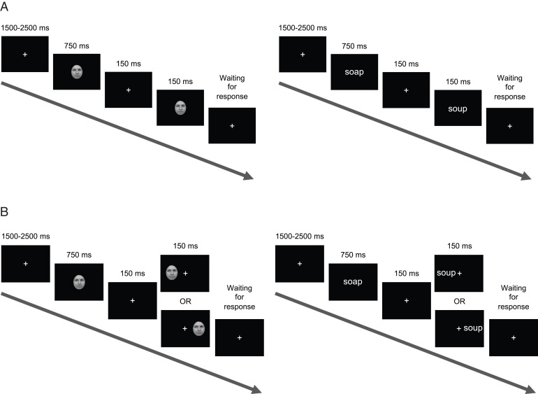
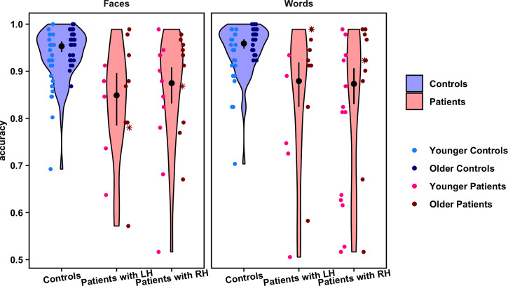
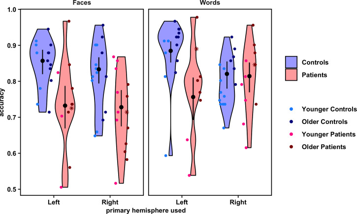
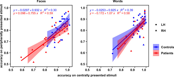
The right and left cerebral hemispheres are important for face and word recognition, respectively-a specialization that emerges over human development. The question is whether this bilateral distribution is necessary or whether a single hemisphere, be it left or right, can support both face and word recognition. Here, face and word recognition accuracy in patients (median age 16.7 y) with a single hemisphere following childhood hemispherectomy was compared against matched typical controls. In experiment 1, participants viewed stimuli in central vision. Across both face and word tasks, accuracy of both left and right hemispherectomy patients, while significantly lower than controls' accuracy, averaged above 80% and did not differ from each other. To compare patients' single hemisphere more directly to one hemisphere of controls, in experiment 2, participants viewed stimuli in one visual field to constrain initial processing chiefly to a single (contralateral) hemisphere. Whereas controls had higher word accuracy when words were presented to the right than to the left visual field, there was no field/hemispheric difference for faces. In contrast, left and right hemispherectomy patients, again, showed comparable performance to one another on both face and word recognition, albeit significantly lower than controls. Altogether, the findings indicate that a single developing hemisphere, either left or right, may be sufficiently plastic for comparable representation of faces and words. However, perhaps due to increased competition or "neural crowding," constraining cortical representations to one hemisphere may collectively hamper face and word recognition, relative to that observed in typical development with two hemispheres.
Keywords: development; face recognition; hemispherectomy; plasticity; word recognition.
Conflict of interest statement
Competing interest statement: Patent (provisional; final pending): Silence localization in brain using non-invasive recordings. P. Grover, A. Chamanzar, and M.B.
Figures
Similar articles
-
Neuromagnetic correlates of hemispheric specialization for face and word recognition.Neurosci Res. 2020 Jul;156:108-116. doi: 10.1016/j.neures.2019.11.006. Epub 2019 Nov 12. Neurosci Res. 2020. PMID: 31730780
-
An ERP investigation of the co-development of hemispheric lateralization of face and word recognition.Neuropsychologia. 2014 Aug;61:315-23. doi: 10.1016/j.neuropsychologia.2014.05.006. Epub 2014 Jun 13. Neuropsychologia. 2014. PMID: 24933662 Free PMC article.
-
The joint development of hemispheric lateralization for words and faces.J Exp Psychol Gen. 2013 May;142(2):348-58. doi: 10.1037/a0029503. Epub 2012 Aug 6. J Exp Psychol Gen. 2013. PMID: 22866684 Free PMC article.
-
A tale of two recognition systems: implications of the fusiform face area and the visual word form area for lateralized object recognition models.Neuropsychologia. 2009 Jan;47(1):1-16. doi: 10.1016/j.neuropsychologia.2008.08.024. Epub 2008 Sep 2. Neuropsychologia. 2009. PMID: 18805434 Review.
-
A vision of graded hemispheric specialization.Ann N Y Acad Sci. 2015 Nov;1359:30-46. doi: 10.1111/nyas.12833. Epub 2015 Jul 22. Ann N Y Acad Sci. 2015. PMID: 26199998 Review.
Cited by
-
Differential functional reorganization of ventral and dorsal visual pathways following childhood hemispherectomy.Dev Cogn Neurosci. 2023 Dec;64:101323. doi: 10.1016/j.dcn.2023.101323. Epub 2023 Nov 10. Dev Cogn Neurosci. 2023. PMID: 37976921 Free PMC article.
-
Holistic processing and face expertise after pediatric resection of occipitotemporal cortex.Neuropsychologia. 2024 Feb 15;194:108789. doi: 10.1016/j.neuropsychologia.2024.108789. Epub 2024 Jan 6. Neuropsychologia. 2024. PMID: 38191121 Free PMC article.
-
Functional Resilience of the Neural Visual Recognition System Post-Pediatric Occipitotemporal Resection.bioRxiv [Preprint]. 2024 May 8:2024.05.08.592792. doi: 10.1101/2024.05.08.592792. bioRxiv. 2024. Update in: iScience. 2024 Nov 22;27(12):111440. doi: 10.1016/j.isci.2024.111440. PMID: 38766137 Free PMC article. Updated. Preprint.
-
Hemispheric functional organization, as revealed by naturalistic neuroimaging, in pediatric epilepsy patients with cortical resections.Proc Natl Acad Sci U S A. 2024 Jul 9;121(28):e2317458121. doi: 10.1073/pnas.2317458121. Epub 2024 Jul 1. Proc Natl Acad Sci U S A. 2024. PMID: 38950362 Free PMC article.
-
Functional resilience of the neural visual recognition system post-pediatric occipitotemporal resection.iScience. 2024 Nov 22;27(12):111440. doi: 10.1016/j.isci.2024.111440. eCollection 2024 Dec 20. iScience. 2024. PMID: 39735436 Free PMC article.
References
-
- Güntürkün O., Ströckens F., Ocklenburg S., Brain lateralization: A comparative perspective. Physiol. Rev. 100, 1019–1063 (2020). - PubMed
-
- Barttfeld P., et al. , A lateral-to-mesial organization of human ventral visual cortex at birth. Brain Struct. Funct. 223, 3107–3119 (2018). - PubMed
-
- Grill-Spector K., Malach R., The human visual cortex. Annu. Rev. Neurosci. 27, 649–677 (2004). - PubMed
Publication types
MeSH terms
Substances
Grants and funding
LinkOut - more resources
Full Text Sources
