. 2022 Nov 16;13(22):3152-3167.
doi: 10.1021/acschemneuro.2c00444. Epub 2022 Oct 25.

Alzheimer's Disease Prevention through Natural Compounds: Cell-Free , In Vitro, and In Vivo Dissection of Hop (Humulus lupulus L.) Multitarget Activity

Affiliations

Alzheimer's Disease Prevention through Natural Compounds: Cell-Free , In Vitro, and In Vivo Dissection of Hop (Humulus lupulus L.) Multitarget Activity

Alessandro Palmioli et al. ACS Chem Neurosci. .

Abstract

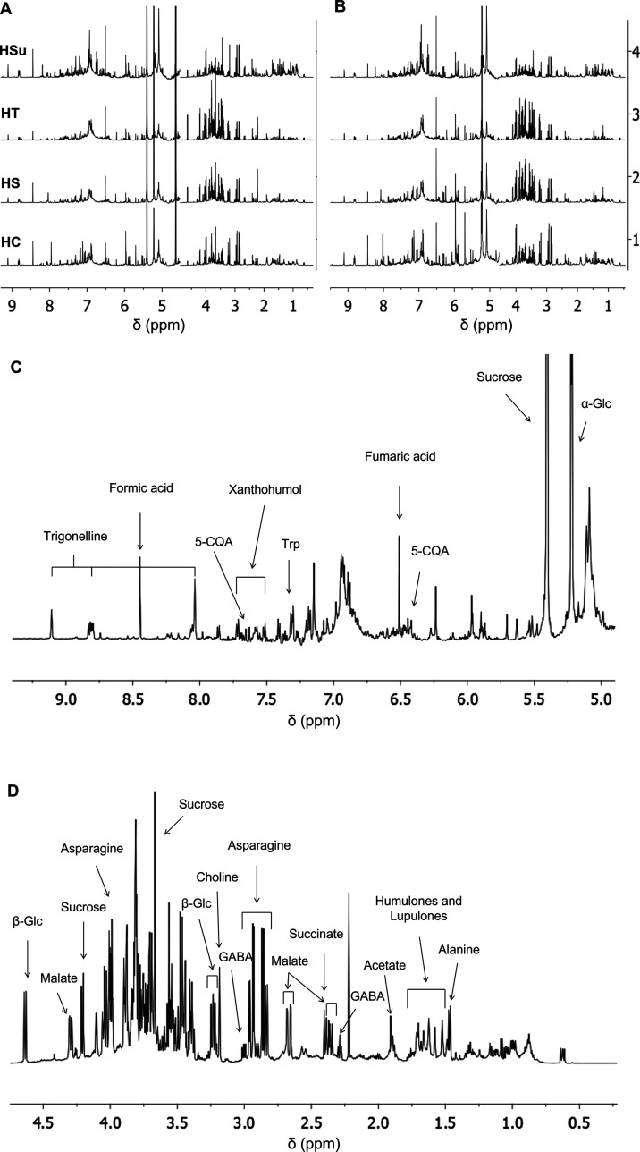
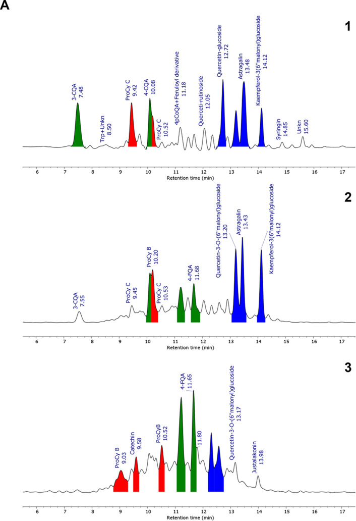
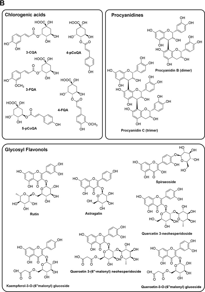
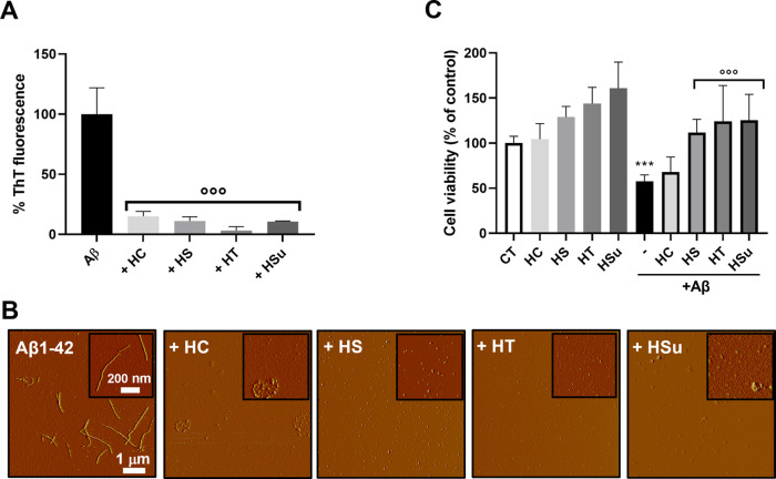
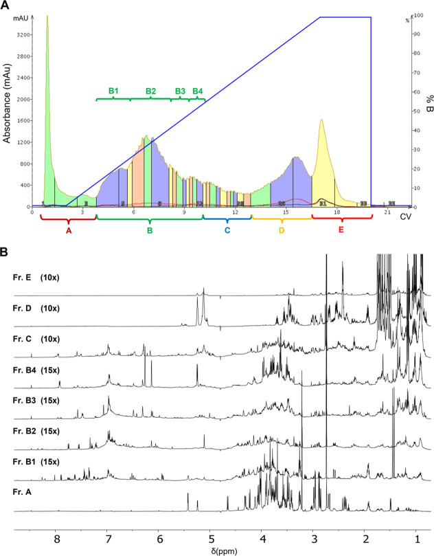
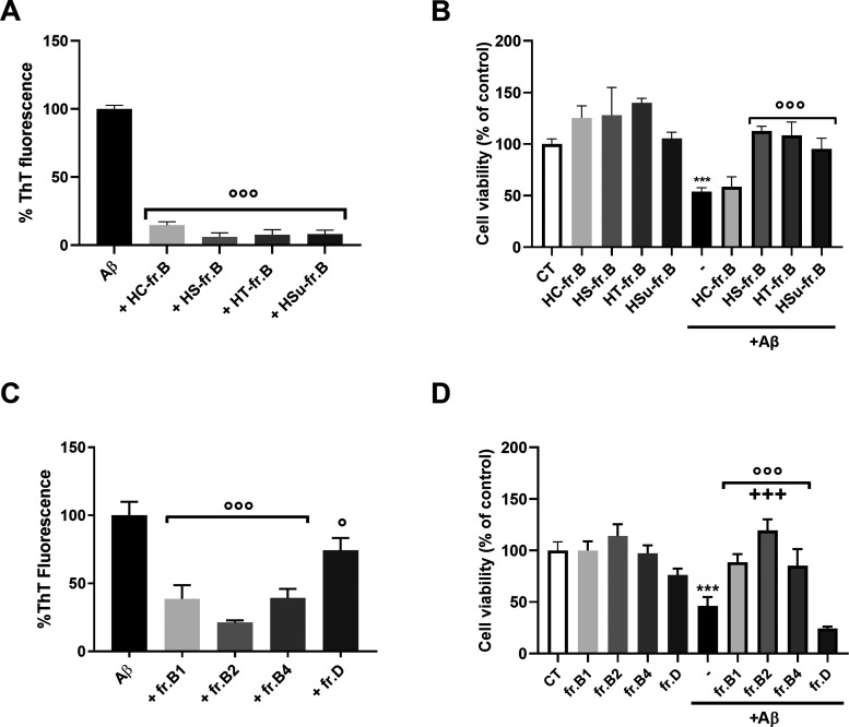
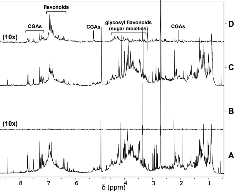
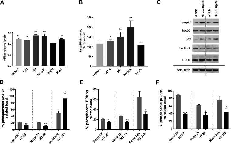
The relevant social and economic costs associated with aging and neurodegenerative diseases, particularly Alzheimer's disease (AD), entail considerable efforts to develop effective preventive and therapeutic strategies. The search for natural compounds, whose intake through diet can help prevent the main biochemical mechanisms responsible for AD onset, led us to screen hops, one of the main ingredients of beer. To explore the chemical variability of hops, we characterized four hop varieties, i.e., Cascade, Saaz, Tettnang, and Summit. We investigated the potential multitarget hop activity, in particular its ability to hinder Aβ1-42 peptide aggregation and cytotoxicity, its antioxidant properties, and its ability to enhance autophagy, promoting the clearance of misfolded and aggregated proteins in a human neuroblastoma SH-SY5Y cell line. Moreover, we provided evidence of in vivo hop efficacy using the transgenic CL2006Caenorhabditis elegans strain expressing the Aβ3-42 peptide. By combining cell-free and in vitro assays with nuclear magnetic resonance (NMR) and MS-based metabolomics, NMR molecular recognition studies, and atomic force microscopy, we identified feruloyl and p-coumaroylquinic acids flavan-3-ol glycosides and procyanidins as the main anti-Aβ components of hop.

Keywords: Alzheimer’s disease; Caenorhabditis elegans; NMR; UPLC-HR-MS; anti-Aβ compounds; hop.


Conflict of interest statement

The authors declare no competing financial interest.

Figures

Figure 1
Figure 1
NMR metabolic profiling of hop extracts. Comparison of NMR metabolic profiling of the hops [1, Cascade (HC); 2, Saaz (HS); 3, Tettnang (HT); and 4, Summit (Hsu)] extracted with (A) boiling water or (B) H2O/ethanol 9:1 solution, in 10 mM phosphate buffer (PB), pH 7.4 at 25 °C. (C, D) 1H NMR spectrum of HT obtained by boiling water extraction, 22 mg/mL, 10 mM PB, 25 °C, 1 mM sodium trimethylsilylpropanesulfonate (DSS). The expanded region with assigned peaks for aromatic compounds from 9.1 to 5.1 ppm (C), bitter acids, and sugars from 4.7 to 0.8 ppm (D).
Figure 2
Figure 2
UPLC-PDA-HR-MS analysis of hop extracts HT and polyphenol-enriched fractions. (A) Chromatographic trace extracted at 325 nm was obtained from (A1) total extract, (A2) fraction B, and (A3) fraction B2 (see Section 2.3 for details), and (B) structures of the main polyphenolic compounds identified in the hop extract. Main peaks (with relative area >5%) are color-filled on the basis of their family: chlorogenic acids (green), procyanidines (red), and glycosyl flavonoids (blue). HT, Tettnang. 3-CQA, 3-O-caffeoylquinic acid; 3-FQA, 3-O-feruloylquinic acid; 4-FQA, 4-O-feruloylquinic acid; 4-pCoQA, 4-p-coumaroylquinic acid; 5-pCoQA, 5-p-coumaroylquinic acid.
Figure 2
Figure 2
UPLC-PDA-HR-MS analysis of hop extracts HT and polyphenol-enriched fractions. (A) Chromatographic trace extracted at 325 nm was obtained from (A1) total extract, (A2) fraction B, and (A3) fraction B2 (see Section 2.3 for details), and (B) structures of the main polyphenolic compounds identified in the hop extract. Main peaks (with relative area >5%) are color-filled on the basis of their family: chlorogenic acids (green), procyanidines (red), and glycosyl flavonoids (blue). HT, Tettnang. 3-CQA, 3-O-caffeoylquinic acid; 3-FQA, 3-O-feruloylquinic acid; 4-FQA, 4-O-feruloylquinic acid; 4-pCoQA, 4-p-coumaroylquinic acid; 5-pCoQA, 5-p-coumaroylquinic acid.
Figure 3
Figure 3
Evaluation of the antioxidant activity of hop extracts. (A) Absorption spectra recorded on hop extract solution at 80 μg/mL concentration. (B) Comparison of the total reducing power (mg GAE/g) and radical scavenging activity (μmol TE/g) assessed on hop extracts by Folin Ciocalteu and 3-ethylbenzothiazoline-6-sulfonic acid (ABTS)-Trolox equivalent antioxidant capacity (TEAC)/2,2-diphenyl-1-picrylhydrazyl (DPPH) assays, respectively. (C) Values are reported as the mean (±SD) of a triplicate of three independent measurements. (D) Effect of HT extracts on hydrogen peroxide-induced cytotoxicity in human SH-SY5Y cells. Cell viability was assessed by the MTT assay after 1 h pretreatment with 0.25 or 0.1 mg/mL HT extracts followed by 24 h cotreatment with 100 μM hydrogen peroxide (H2O2). Values are expressed as % vs vehicle. Repeated-measures ANOVA test, followed by Dunnett’s post hoc test; **p < 0.01 vs HT alone and vs HT + H2O2.
Figure 4
Figure 4
Effects of hop extracts on Aβ1-42 aggregation and toxicity on the human neuroblastoma SH-SY5Y cell line. (A) The effect of incubation (24 h at 37 °C) of HC, HT, HS, or HSu extracts (0.25 mg/mL) on Aβ1-42 (2.5 μM) aggregation was determined by the ThT fluorescence assay. Data were expressed as the mean ± SD (N = 3), calculated by subtracting the relative control solutions (fraction alone) and were expressed as the percentage reduction of Aβ1-42 aggregation, °°°p < 0.001 vs Aβ alone, one-way ANOVA and Dunnett’s post hoc test. (B) AFM images were acquired after 24 h incubation at 37 °C of the Aβ 1-42 peptide (2.5 μM) with or without 0.25 mg/mL HC, HT, HS, or HSu extracts. (C) Human neuroblastoma SH-SY5Y cells were treated with 10 μM Aβ1-42 peptide in the absence or presence of 0.5 mg/mL HC, HT, HS, or HSu extracts for 24 h, and the toxicity was evaluated by the MTT assay. Control cells were treated with the vehicle (CT). Data are the mean ± SD of the percentage of viable cells (N = 6). ***p < 0.001 Aβ vs the respective control and °°°p < 0.001 Aβ + hop vs Aβ alone, according to one-way ANOVA and Dunnett’s post hoc test. HC, Cascade; HS, Saaz; HT, Tettnang; Hsu, Summit.
Figure 5
Figure 5
Hop extracts’ fractionation. (A) Chromatographic profile of the separation of the HT extract obtained by reverse-phase C18 chromatography (linear elution gradient from 2 to 100% MeOH in 15 CV). (B) 1H NMR spectra of the chromatographic fractions A–E. 1H NMR spectra were recorded on 2 mg/mL samples dissolved in D2O, 25 °C, at 600 MHz. The intensity ratios with respect to spectrum A, which has the highest signal-to-noise ratio, are shown in brackets. CV, column volume.
Figure 6
Figure 6
Effect of hop fractions on Aβ1-42 aggregation and in vitro toxicity. (A, C) Coincubation (24 h) of (A) fr.B from HC, HT, HS, or Hsu (0.03 mg/mL) or (C) HT fr. B1, B2, B4, or D (0.0125 mg/mL) with Aβ1-42 (2.5 μM) reduced the fibrillation determined by the ThT fluorescence assay. Data were expressed as mean ± SD (N = 3), calculated by subtracting the relative control solutions (fraction alone), and were expressed as the percentage reduction of Aβ1-42 aggregation, °°°p < 0.001 vs Aβ alone, one-way ANOVA and Dunnett’s post hoc test. (B, D) Human neuroblastoma SH-SY5Y cells were treated with the Aβ1-42 peptide (10 μM) in the absence or presence of (B) fr. B (0.03 mg/mL) from HC, HT, HS, or HSu or (D) HT fr. B1, B2, B4, or D (0.125 mg/mL). Control cells were treated with vehicle (CT). Cell viability was determined 24 h later by the MTT assay. Data are the mean ± SD of the percentage of viable cells (N = 3). ***p < 0.001 Aβ vs the respective control, °°°p < 0.001 Aβ + fr. B vs Aβ alone and +++p < 0.001 B2 + hop vs B1, B4, and D according to one-way ANOVA and Dunnett’s post hoc test. HC, Cascade; HS, Saaz; HT, Tettnang; Hsu, Summit.
Figure 7
Figure 7
NMR binding studies with Aβ1-42. (A) Off-resonance NMR spectrum of a solution containing HT extract fr. B2 (4 mg/mL). (B) STD NMR spectrum of the same sample of A. (C) Off-resonance NMR spectrum of a solution containing HT extract fr. B2 (4 mg/mL) and the Aβ1-42 peptide (120 μM). (D) STD NMR spectrum of the same sample of C. Samples were dissolved in deuterated PB, pH 7.4. STD spectra were acquired with 1024 scans and 2 s saturation time at 600 MHz, 25 °C. The intensity ratios with respect to spectrum A, which has the highest signal-to-noise ratio, are shown in brackets. HT, Tettnang.
Figure 8
Figure 8
Effect of HT extracts on autophagy markers and related kinase regulatory pathways (PI3K/AKT and ERK1/2) in human SH-SY5Y cells. (A) Relative quantification, calculated as the ratio to β-actin, of mRNA levels of macroautophagy (beclin-1, LC3, and p62) and CMA (lamp2A, hsc70) markers and BDNF after 24 h treatment with 0.1 mg/mL HT extract. Two-tailed paired t-test; *p < 0.05, **p < 0.01, ***p < 0.001 vs vehicle. (B) Protein expression of macroautophagy (beclin-1, LC3-II, and p62) and CMA (lamp2A, hsc70) markers after 24 h treatment with 0.1 mg/mL HT extract and (C) representative Western blot image showing immunoreactivity for the target proteins and the corresponding β-actin, used as the internal standard. Two-tailed paired t-test; * p < 0.05, ** p < 0.01 vs vehicle. Time course of the phosphorylation status of AKT (D), ERK1/2 (E), and p70S6K (F) kinases after 30 min, 2 h, and 24 h treatment with 0.1 mg/mL HT extract. Values represent the percentage of the ratio between the phosphorylated and total kinase levels. Student’s t-test *p < 0.05, **p < 0.01, ***p < 0.001 vs related basal.
Figure 9
Figure 9
HT protected CL2006 worms from paralysis caused by Aβ expression. (A) Dose-response effect of HT on the paralysis of CL2006 worms. Synchronized CL2006 worms were treated in the L3 larval stage with different concentrations (10–250 μg/mL) of HT dissolved in water. Control worms were treated with the same volume of water only (Vehicle). Paralysis was scored 120 h after treatment. (B) Synchronized CL2006 worms were treated in the L3 larval stage with 50 μg/mL HT dissolved in water. CL2006 worms were treated in the same experimental conditions with 100 μM doxycycline (Doxy) dissolved in water as a positive control. CL802 and CL2006 worms were treated with the same volume of water only (CL802 and Vehicle, respectively) as negative controls. Paralysis was scored 120 h after treatment. ****p < 0.0001 and **p < 0.1 vs CL2006 treated with Vehicle according to one-way ANOVA and Bonferroni’s post hoc test. HT, Tettnang.

References

    1. Scheltens P.; De Strooper B.; Kivipelto M.; Holstege H.; Chételat G.; Teunissen C. E.; Cummings J.; van der Flier W. M. Alzheimer’s disease. Lancet 2021, 397, 1577–1590. 10.1016/S0140-6736(20)32205-4. - DOI - PMC - PubMed
    1. McDade E.; Bateman R. J. Stop Alzheimer’s before it starts. Nature 2017, 547, 153–155. 10.1038/547153a. - DOI - PubMed
    1. Long J. M.; Holtzman D. M. Alzheimer Disease: An Update on Pathobiology and Treatment Strategies. Cell 2019, 179, 312–339. 10.1016/j.cell.2019.09.001. - DOI - PMC - PubMed
    1. Knopman D. S.; Amieva H.; Petersen R. C.; Chételat G.; Holtzman D. M.; Hyman B. T.; Nixon R. A.; Jones D. T. Alzheimer disease. Nat. Rev. Dis. Primers 2021, 7, 33 10.1038/s41572-021-00269-y. - DOI - PMC - PubMed
    1. Bolton J. L.; Dunlap T. L.; Hajirahimkhan A.; Mbachu O.; Chen S. N.; Chadwick L.; Nikolic D.; van Breemen R. B.; Pauli G. F.; Dietz B. M. The Multiple Biological Targets of Hops and Bioactive Compounds. Chem. Res. Toxicol. 2019, 32, 222–233. 10.1021/acs.chemrestox.8b00345. - DOI - PMC - PubMed

Publication types