. 2022 Nov 3;109(11):2068-2079.
doi: 10.1016/j.ajhg.2022.09.012. Epub 2022 Oct 24.

Bi-allelic CAMSAP1 variants cause a clinically recognizable neuronal migration disorder

Affiliations

Bi-allelic CAMSAP1 variants cause a clinically recognizable neuronal migration disorder

Reham Khalaf-Nazzal et al. Am J Hum Genet. .

Abstract

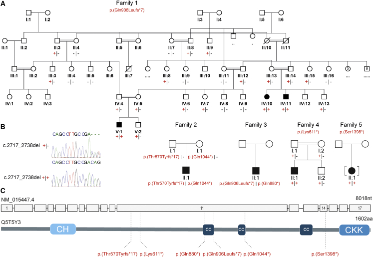
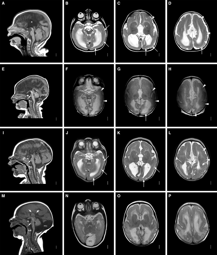
Non-centrosomal microtubules are essential cytoskeletal filaments that are important for neurite formation, axonal transport, and neuronal migration. They require stabilization by microtubule minus-end-targeting proteins including the CAMSAP family of molecules. Using exome sequencing on samples from five unrelated families, we show that bi-allelic CAMSAP1 loss-of-function variants cause a clinically recognizable, syndromic neuronal migration disorder. The cardinal clinical features of the syndrome include a characteristic craniofacial appearance, primary microcephaly, severe neurodevelopmental delay, cortical visual impairment, and seizures. The neuroradiological phenotype comprises a highly recognizable combination of classic lissencephaly with a posterior more severe than anterior gradient similar to PAFAH1B1(LIS1)-related lissencephaly and severe hypoplasia or absence of the corpus callosum; dysplasia of the basal ganglia, hippocampus, and midbrain; and cerebellar hypodysplasia, similar to the tubulinopathies, a group of monogenic tubulin-associated disorders of cortical dysgenesis. Neural cell rosette lineages derived from affected individuals displayed findings consistent with these phenotypes, including abnormal morphology, decreased cell proliferation, and neuronal differentiation. Camsap1-null mice displayed increased perinatal mortality, and RNAScope studies identified high expression levels in the brain throughout neurogenesis and in facial structures, consistent with the mouse and human neurodevelopmental and craniofacial phenotypes. Together our findings confirm a fundamental role of CAMSAP1 in neuronal migration and brain development and define bi-allelic variants as a cause of a clinically distinct neurodevelopmental disorder in humans and mice.

Keywords: MARK2; agyria; autosomal recessive; lissencephaly; neurodevelopmental disorder; pachygyria; patronin; tubulinopathy.


Conflict of interest statement

Declaration of interests The authors declare no competing interests.

Figures

Figure 1
Figure 1
Family pedigrees and bi-allelic CAMSAP1 variants associated with a syndromic neuronal migration disorder (A) Simplified pedigrees of families in the study showing cosegregation of the variants identified (“–” wild-type allele; “+” familial variant), with reference to transcript GenBank: NM_015447.4. (B) Chromatogram for the c.2717_2738 deletion is shown with heterozygous (top) and homozygous variant (bottom) individuals shown. (C) Intron/exon genomic organization of CAMSAP1 (top) and protein domain architecture of CAMSAP1 (bottom) illustrating the calponin homology (CH), coiled coil (CC), and calmodulin-regulated spectrin-associated (CKK [CAMSAP1, KIAA1078/CAMSAP2, KIAA1543/CAMSAP3] domain) domains alongside the location of each of the identified pathogenic variants (dotted line).
Figure 2
Figure 2
Neuroradiology of affected individuals Neuroimaging in four individuals with the CAMSAP1-related neuronal migration disorder (for further imaging see Figure S2): row 1 (A-D) is family 1, V:1 aged 7 months; row 2 (E-H) is family 2, II:1 aged 2 days; row 3 (I-L) is family 3, II:1 aged 3 months; row 4 (M-P) is family 4, II:1 aged 3 months. T1-weighted midline sagittal images show absent (∗ in A, also E and I) or short and thin (short white arrows in M) corpus callosum and small base of the pons (thin white arrow in E, I, and M). Enlarged posterior fossa or “mega cisterna magna” (PFC in E and M) was seen in 2/4 subjects. T2-weighted axial images show posterior-more-severe-than-anterior gradient with areas of agyria or severe pachygyria with prominent cell sparse zones and reduced thickness of the cerebral mantle/wall in posterior regions (white arrows in B–D, G, J, and K) and areas of less severe pachygyria with thicker cerebral mantle/wall in anterior regions (white arrowheads in C, D, F–H, and L). The gradient in family 4, II:1 (N–P) was less clear with lower resolution images. The boundaries of the basal ganglia and thalami were difficult to see, and the internal capsules are not seen ( in C, also in G, K, and O). The third ventricle was enlarged in all and dramatically enlarged into a midline interhemispheric cyst in family 1, V:1 (3V in D).
Figure 3
Figure 3
iPSCs from affected individuals display decreased proliferation and differentiation and increased apoptosis of neural progenitor cells (A and B) Brightfield images showing abnormal clustering of cells in rosettes from an affected individual at 11 days in vitro. Control cell rosettes have a clearly visible interior region of reduced cell density as compared to more dense cells from the affected individual. Scale bar, 500 μm. (C–L) Immunohistochemistry (IHC) analysis highlights molecular features of affected-individual-derived rosettes. Scale bars, 50 μm. (C–H) IHC for PAX6 (green, a marker of neuronal progenitors), and TUJ1/TUBB3 (red, marks differentiated neurons) demonstrated significantly fewer differentiated neurons at 11 days in rosettes derived from affected individual iPSCs compared to control iPSCs. (E–H) PAX6 (E and F) and TUJ1 (G and H) shown independently. (I and J) IHC for phosphohistone H3 (pHH3) shows reduced staining suggesting a proliferation defect in rosettes derived from iPSCs obtained from an affected individual. (K and L) IHC for cleaved caspase-3 (CC3) demonstrating increased apoptosis in rosettes derived from iPSCs obtained from an affected individual. (M and N) Quantification of counts for PHH3+ cells (n = 3 images × 3 replicates) and CC3+ cells (n = 3 images × 3 replicates). iPSCs from the affected individual were derived from family 2, II:1, one clone of which was received from the Genome Engineering and Stem Cell Center, Department of Genetics, School of Medicine, Washington University in Saint Louis. Control cells are from iPSC line iPSC72.3.
Figure 4
Figure 4
Expression of Camsap1 in the CNS and developing facial primordia RNAScope probe for Camsap1 demonstrate robust mRNA expression at E10.5 (A and B) and E14.5 (C and D) in the developing head (A and C), and neural tube (B and D) with a clear enrichment in the neural tissues (A–J) in addition to the developing pharyngeal arches (A). Expression at E18.5 (E and F), P7 (G and H), and P27 (I and J) remains high in the brain with slightly enriched expression in upper layers (F, H, and J are higher magnification views of E, G, and I, respectively). All scale bars, 500 μm.

References

    1. Oegema R., Barakat T.S., Wilke M., Stouffs K., Amrom D., Aronica E., Bahi-Buisson N., Conti V., Fry A.E., Geis T., et al. International consensus recommendations on the diagnostic work-up for malformations of cortical development. Nat. Rev. Neurol. 2020;16:618–635. - PMC - PubMed
    1. Severino M., Geraldo A.F., Utz N., Tortora D., Pogledic I., Klonowski W., Triulzi F., Arrigoni F., Mankad K., Leventer R.J., et al. Definitions and classification of malformations of cortical development: practical guidelines. Brain. 2020;143:2874–2894. - PMC - PubMed
    1. Di Donato N., Chiari S., Mirzaa G.M., Aldinger K., Parrini E., Olds C., Barkovich A.J., Guerrini R., Dobyns W.B. Lissencephaly: Expanded imaging and clinical classification. Am. J. Med. Genet. 2017;173:1473–1488. - PMC - PubMed
    1. Fry A.E., Cushion T.D., Pilz D.T. The genetics of lissencephaly. Am. J. Med. Genet. C Semin. Med. Genet. 2014;166C:198–210. - PubMed
    1. Smith D.S., Niethammer M., Ayala R., Zhou Y., Gambello M.J., Wynshaw-Boris A., Tsai L.H. Regulation of cytoplasmic dynein behaviour and microtubule organization by mammalian Lis1. Nat. Cell Biol. 2000;2:767–775. - PubMed

Publication types