. 2022 Oct 25;6(1):e202201585.
doi: 10.26508/lsa.202201585. Print 2023 Jan.

A high-content endogenous GLUT4 trafficking assay reveals new aspects of adipocyte biology

Affiliations

A high-content endogenous GLUT4 trafficking assay reveals new aspects of adipocyte biology

Alexis Diaz-Vegas et al. Life Sci Alliance. .

Abstract

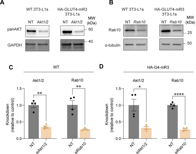
Insulin-induced GLUT4 translocation to the plasma membrane in muscle and adipocytes is crucial for whole-body glucose homeostasis. Currently, GLUT4 trafficking assays rely on overexpression of tagged GLUT4. Here we describe a high-content imaging platform for studying endogenous GLUT4 translocation in intact adipocytes. This method enables high fidelity analysis of GLUT4 responses to specific perturbations, multiplexing of other trafficking proteins and other features including lipid droplet morphology. Using this multiplexed approach we showed that Vps45 and Rab14 are selective regulators of GLUT4, but Trarg1, Stx6, Stx16, Tbc1d4 and Rab10 knockdown affected both GLUT4 and TfR translocation. Thus, GLUT4 and TfR translocation machinery likely have some overlap upon insulin-stimulation. In addition, we identified Kif13A, a Rab10 binding molecular motor, as a novel regulator of GLUT4 traffic. Finally, comparison of endogenous to overexpressed GLUT4 highlights that the endogenous GLUT4 methodology has an enhanced sensitivity to genetic perturbations and emphasises the advantage of studying endogenous protein trafficking for drug discovery and genetic analysis of insulin action in relevant cell types.


Conflict of interest statement

The authors declare that they have no conflict of interest.

Figures

Figure 1.
Figure 1.. The anti-GLUT4 exofacial antibody LM048 is ideal for studying the insulin stimulated trafficking of endogenous GLUT4 to the plasma membrane.
(A) Quantification of plasma membrane GLUT4 staining (PM GLUT4) of non-permeabilized mouse 3T3-L1 adipocytes stimulated with or without 100 nM insulin for 20 min and stained with the antibodies LM048, LM052, and LM059 imaged by confocal microscopy (see Fig S1A for representative images). Data are presented as fold-increase in fluorescent signal relative to unstimulated (basal) adipocytes for each antibody. FOB: Fold over basal. Error bars = SD, n = 2. *P < 0.05, ****P < 0.0001 by two-way ANOVA with Dunnett’s multiple comparisons test. (B) To demonstrate the specificity of the anti-GLUT4 antibody LM048, cells were treated with a non-targeting siRNA (NT) or siRNA targeting Slc2a4/Glut4, stimulated with 100 nM insulin (20 min), stained with LM048 followed by permeabilization and further staining with an antibody against total GLUT4. Representative confocal images of surface and total staining (scale bars = 40 µm). LDs = lipid droplets. (C) Quantification of surface and total GLUT4 labelling in adipocytes treated with non-targeting (NT) siRNA or siRNA targeting GLUT4 (siG4). Error bars = SD, n = 3. *P < 0.05, ***P < 0.001 by unpaired t test with Welch’s correction. (D) Representative bright-field and confocal images of 3T3-L1 adipocytes treated with indicated doses of insulin and stained with the anti-GLUT4 antibody LM048, and DAPI. Images at the bottom surface and midplane (Rims) are presented (scale bars = 100 µm, scale bar of crop = 40 µm). (E, F) Quantification of PM GLUT4 content in response to specified insulin doses in either bicarbonate (E) or HEPES (F) buffered media. Data points from individual experiments were fitted to a 3-parameter Hill equation. The EC50 for cells in bicarbonate buffer is indicated on the graph. For HEPES buffered media, a single curve did not adequate fit all datasets using Akaike’s Information Criterion. (G) Quantification of PM GLUT4 using the anti-GLUT4 antibody LM048 in adipocytes treated with the indicated doses of insulin for specified times. Data are presented as fluorescence intensity of pmGLUT4 signals a percentage of the maximum response n = 4. (H, I) Population (H) and single cell (I) plasma membrane endogenous GLUT4 abundance data from wild-type 3T3-L1 adipocytes treated with or without 100 nM insulin in combination with DMSO (control), the PI3K inhibitor GDC0941, Akt inhibitors GDC0068 and MK2206, or PDK1 inhibitor GSK23344. Cells were stained with the LM048 antibody against endogenous GLUT4 in non-permeabilized cells and expressed as a proportion of DMSO-treated controls n = 6.
Figure S1.
Figure S1.. Efficacy of antibodies targeting distinct exofacial GLUT4 epitopes for labelling cell surface GLUT4 in mouse and human adipocytes.
(A, B) Confocal images of non-permeabilised cells stained for surface GLUT4 using integral antibodies LM048, LM052, or LM059 in mouse 3T3-L1 (A) and SGBS (B) adipocytes. Cells were either unstimulated (basal) or stimulated with 100 nM insulin for 20 min as indicated. (C) Quantification of data surface staining in SGBS adipocytes. Data are presented as a fold-increase in fluorescent signal relative to the signal from unstimulated (basal) adipocytes for each antibody. FOB = Fold over basal. Error bars = SD, n = 2, *P < 0.05 by two-way ANOVA with Dunnet’s multiple comparisons test.
Figure 2.
Figure 2.. Studying insulin-stimulated translocation of endogenous GLUT4 in insulin resistant adipocytes.
(A) PM GLUT4 in 3T3-L1 adipocytes stimulated with 1 or 100 nM insulin after treatment with either medium, dexamethasone (DEX; 20 nM for 8 d), TNF-α (TNF; 2 ng/ml for 4 d) or chronic insulin (CI; 10 nM every 4 h for 24 h). Data are presented as mean ± SD. ****P < 0.0001, *P < 0.05 versus basal by two-way ANOVA with Šídák’s multiple comparisons test. (B) Total cellular GLUT4 was assessed by Western blot (upper panel). Each track represents an independent biological replicate, from left to right control, Dexamethasone, TNF and CI. Quantitation (lower panel) of GLUT4 normalized to control cells (set to 100%). Data are mean ± SD, n = 3 with *P < 0.05 compared with control by 1-way ANOVA with Dunnet’s test for multiple comparisons. (C) Dose response with CI over 24 h. Data are min:max normalized to the control basal and 100 nM response and are presented as the population mean ± SD of n = 6 experiments. *P < 0.05 by mixed effect model with Geisser-Greenhouse Correction and Dunnet’s multiple comparisons test. (D, E, F) Single cell analysis of surface GLUT4 staining using LM048 in 3T3-L1 cells after CI or TNF treatment for 24 h, ±1 or 100 nM insulin. (C, D) Histogram displaying single cell responses from an average of 25,611 cells per indicated condition (C). Dotted lines represent the peak of the control basal, 0.5 and 100 nM responses. (E) Dose response with TNF over 24 h. Data are min:max normalized to the control basal and 100 nM response and are presented as the population mean ± SD of n = 6 experiments. *P < 0.05, **P < 0.01, ***P < 0.001, ****P < 0.0001 by mixed effect model with Geisser–Greenhouse Correction and Dunnet’s multiple comparisons test. (D) and (F) were run simultaneously and they share the control cells, the control cells have been plotted twice for clarity. (E, F) Histogram displaying single cell responses from an average of 21,456 cells per indicated condition (E). Dotted lines represent the peak of the control basal, 0.5 and 100 nM responses. (G) Total GLUT4 was assessed after CI (left) or TNFα (right treatment 14-3-3 was used as loading control [bottom panel]). (H, I) Densitometric analysis of GLUT4 abundance from (I), ± SD of n = 3 experiments. P-values as indicated by one-way ANOVA with Dunnet’s test for multiple comparisons. (I) Representative image of 3T3-L1 adipocytes in basal (left panel) and after 0.5 nM insulin stimulation (right panel) in control and insulin resistance cells. Green = PM-GLUT4, orange = nucleus, cyan = lipid droplets.
Figure S2.
Figure S2.. 2-Deoxy-D-glucose uptake in 3T3-L1 adipocytes subjected to models of insulin resistance for 24 h at the indicated doses, ±1 nM insulin.
Figure 3.
Figure 3.. siRNA-based workflow to identify regulators of GLUT4 trafficking.
(A) siRNA-mediated knockdown of known GLUT4 trafficking mediators in fully differentiated 3T3-L1 adipocytes and the densitometry of the blots n = 2–4. (B) Quantification of PM GLUT4 abundance (using LM048) in cells treated with control siRNA or siRNA targeting Rab10 in response to varying doses of insulin n = 3. EC50s of each curve are expressed on the graph. (C) Quantification of PM GLUT4 abundance (using LM048) in cells treated with control siRNA or siRNA targeting IRS1/2, Rab10 or Kif13A in response to 0.5 and 100 nM insulin (data presented as mean ± SD of n = 4 experiments). ***P < 0.001, ****P < 0.0001 by mixed effect model with Geisser–Greenhouse Correction and Dunnet’s multiple comparisons test.
Figure 4.
Figure 4.. Assessment of insulin-stimulated TfR translocation in 3T3-L1 adipocytes.
(A) Surface labelling of TfR in non-permeabilized cells, treated with either non-targeting siRNA (Ctrl) or siRNA targeting Transferrin Receptor (siTfR), in the presence and absence of 100 nM insulin. (A, B) Quantification of GLUT4 (upper) and TfR trafficking responses (lower) of cells in (A). (C) Western blot of total cellular TfR in lysates from cells treated with non-targeting siRNA (NT) or siRNA targeting TfR (siTfR). Membranes were blotted with antibodies recognizing TfR or α-Tubulin as a loading control. (D) Time-course of surface Transferrin Receptor (TfR) and GLUT4, using LM048, in 3T3-L1 adipocytes in response to 100 nM insulin (data presented as mean ± SD of n = 3 experiments).
Figure S3.
Figure S3.. Sensitivity of TFR and GLUT4 to PI3K/Akt inhibition and the identification of TRARG1 interactors.
(A) Insulin stimulated GLUT4 (upper) and TfR (lower) responses of cells expressing HA-GLUT4-mRuby3 in the presence of insulin signalling pathway inhibitors. The surface labelling with HA was quantified and expressed as a proportion of the total GLUT4 (mRuby3) signal in response to 0.5 and 100 nM insulin. Cells were treated with the inhibitors torin, wortmannin, MK2206, or GDC0068 and GLUT4 responses were expressed relative to the DMSO control. Data are presented as mean ± SEMb. n = 7. (B) Representative Western blotting of TRARG1 immunoprecipitation in control and insulin stimulated cells. (C) Volcano plot of TRARG1 interactome under basal (Bas) or insulin-stimulated (INS) conditions (left)—TRARG1 interactors regulated by insulin treatment (highlighted in blue) as identified by median absolute deviation analysis (4.22.2.); and correlation plot of TRARG1 interactor enrichment under basal (Bas) and insulin (Ins) treated conditions (right). (D) Bcl9l expression in 3T3-L1 adipocytes after treatment with non-targeting siRNA (NT) or siRNA targeting Bcl9l. Data expressed as mean ± SD, n = 2–4. (D, E) Western blotting of cell lysates for Bcl9l and α-tubulin under the same conditions as in (D).
Figure 5.
Figure 5.. Validation of digital phase contrast (DPC) imaging to measure lipid droplet content in 3T3-L1 adipocytes.
(A) Bright-field and DPC images of 3T3-L1 cells throughout differentiation from preadipocytes (day 0) to adipocytes (day 8), and the resultant lipid droplet mask used to quantify lipid accumulation. (A, B, C) Number of lipid droplets (B) and average lipid droplet volume (C) observed throughout the differentiation time-course in (A) (Data are presented as mean + SD, n = 2, 40 wells per experiment ****P < 0.001, by one-way ANOVA with Dunnet’s test for multiple comparisons). (D) The effect of siRNA-mediated knockdown of Cidec and Plin1 on lipid droplets in differentiated 3T3-L1 adipocytes, as measured by brightfield and DPC in comparison with a non-targeting siRNA control. Hoechst 33342 was used as a control for cell number/density. (D, E, F) Number of lipid droplets (E) and average lipid droplet volume (F) observed for the cells in (D) (Data is presented as mean + SD, n = 5, ****P < 0.001, by one-way ANOVA with Dunnet’s test for multiple comparisons).
Figure 6.
Figure 6.. High-content screen for regulators of insulin-stimulated membrane traffic and lipid droplet content.
(A, B) Quantification of fluorescently labelled surface GLUT4 (using LM048) (A) and TfR (B) in response to 0.5 and 100 nM insulin after knockdown of a panel of regulators of GLUT4 trafficking, in non-permeabilized 3T3-L1 adipocytes. (A, B, C) Number of nuclei per field of view in the cells in (A, B). (A, B, D) Average lipid droplet area for cells in (A, B). (A, B, E) Number of lipid droplets for the cells in (A, B). Legend on the right indicates the siRNA conditions in each graph. Data presented as mean ± SD, n = 4–8, *P < 0.05, by mixed effect model with Geisser–Greenhouse Correction and Dunnet’s multiple comparisons test.
Figure 7.
Figure 7.. Trafficking of overexpressed HA-GLUT4-mR3 protects cells from genetic perturbations in comparison with endogenous GLUT4.
(A, C, E) Correlations between the endogenous GLUT4 or HA-GLUT4-mR3 trafficking responses in wild-type and HA-GLUT4-mR3 expressing cells, respectively, in response to siRNA knockdown of several regulators of GLUT4 trafficking. (B, D, F) Correlations between the TfR trafficking responses in wild-type and HA-GLUT4-mRuby3-expressing cells, in response to siRNA knockdown of regulators of GLUT4 trafficking (as indicated). (A, B, C, D, E, F) Plasma membrane GLUT4 (pmG4) and TfR were determined under basal (A, B), and 0.5 nM (C, D) or 100 nM insulin-stimulated conditions (E, F). FOC = Fold over control. Correlations are indicated by the respective r2 and P-values as determined by simple linear regression.
Figure S4.
Figure S4.. Akt1/2 and Rab10 knockdown in wild type and HA-G4-mRuby3 overexpressing adipocytes.
(A) Pan AKT expression in 3T3-L1 adipocytes following treatment with non-targeting siRNA (NT) or siRNA targeting a combination of Akt1 and Akt2 (Akt1/2). (B) Rab10 expression in 3T3-L1 adipocytes following treatment with non-targeting siRNA (NT) or siRNA targeting Rab10. (C, D) Densitometric analysis of Akt1/2 and Rab10 protein abundance in wild type (C) and HA-G4-mR3 (D) adipocytes. Data expressed as mean ± SEM, n = 4. *P < 0.05, **P < 0.01, ****P < 0.0001. Two-tailed Welch’s t test.
Figure S5.
Figure S5.. Assessment of GLUT4 overexpression in HA-GLUT4-mR cells and sensitivity of insulin-stimulated endogenous GLUT4 or HA-GLUT4-mR translocation to the Akt inhibitor MK2206.
(A) Western blotting of cell lysates for GLUT4 and Caveolin1 (CAV1) after subcellular fractionation in the presence and absence of 100 nM insulin for 20 min. WCH, whole cell homogenate; M/N, mitochondrial/nuclear; HDM, high-density microsomes; LDM, low-density microsomes; PM, plasma membrane. n = 1. (A, B) PM GLUT4 levels in response to 100 nM insulin in wild type and HA-GLUT4-mR3 overexpressing cells n = 2 (B). (C, D) PM GLUT4 abundance in wild-type or HA-G4 overexpressing 3T3-L1 adipocytes treated with 1 nM insulin (C) or 100 nM insulin (D) in combination with DMSO (control) or the Akt inhibitor MK2206 for 10 min prior insulin addition. (E) Sensitivity to MK2206 in WT cells in response to 1 or 100 nM insulin for GLUT4 (E) is shown. Data were min:max normalized for each insulin concentration. n = 4. Non-linear regression comparing independent fits with a global fit shared parameter (IC50) was used to test statistical significance.

References

    1. Al-Hasani H, Hinck CS, Cushman SW (1998) Endocytosis of the glucose transporter GLUT4 is mediated by the GTPase dynamin. J Biol Chem 273: 17504–17510. 10.1074/jbc.273.28.17504 - DOI - PubMed
    1. Al-Hasani H, Yver DR, Cushman SW (1999) Overexpression of the glucose transporter GLUT4 in adipose cells interferes with insulin-stimulated translocation. FEBS Lett 460: 338–342. 10.1016/s0014-5793(99)01369-1 - DOI - PubMed
    1. Atkinson BJ, Griesel BA, King CD, Josey MA, Olson AL (2013) Moderate GLUT4 overexpression improves insulin sensitivity and fasting triglyceridemia in high-fat diet-fed transgenic mice. Diabetes 62: 2249–2258. 10.2337/db12-1146 - DOI - PMC - PubMed
    1. Babbey CM, Ahktar N, Wang E, Chen CC-H, Grant BD, Dunn KW (2006) Rab10 regulates membrane transport through early endosomes of polarized Madin-Darby canine kidney cells. Mol Biol Cell 17: 3156–3175. 10.1091/mbc.e05-08-0799 - DOI - PMC - PubMed
    1. Beaton N, Rudigier C, Moest H, Müller S, Mrosek N, Röder E, Rudofsky G, Rülicke T, Ukropec J, Ukropcova B, et al. (2015) TUSC5 regulates insulin-mediated adipose tissue glucose uptake by modulation of GLUT4 recycling. Mol Metab 4: 795–810. 10.1016/j.molmet.2015.08.003 - DOI - PMC - PubMed

Publication types

LinkOut - more resources