. 2023 Feb 1;34(2):220-240.
doi: 10.1681/ASN.2022060644. Epub 2022 Oct 31.

Translation Rescue by Targeting Ppp1r15a through Its Upstream Open Reading Frame in Sepsis-Induced Acute Kidney Injury in a Murine Model

Affiliations

Translation Rescue by Targeting Ppp1r15a through Its Upstream Open Reading Frame in Sepsis-Induced Acute Kidney Injury in a Murine Model

Ashley Kidwell et al. J Am Soc Nephrol. .

Abstract

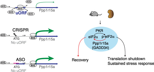
Background: Translation shutdown is a hallmark of late-phase, sepsis-induced kidney injury. Methods for controlling protein synthesis in the kidney are limited. Reversing translation shutdown requires dephosphorylation of the eukaryotic initiation factor 2 (eIF2) subunit eIF2 α ; this is mediated by a key regulatory molecule, protein phosphatase 1 regulatory subunit 15A (Ppp1r15a), also known as GADD34.

Methods: To study protein synthesis in the kidney in a murine endotoxemia model and investigate the feasibility of translation control in vivo by boosting the protein expression of Ppp1r15a, we combined multiple tools, including ribosome profiling (Ribo-seq), proteomics, polyribosome profiling, and antisense oligonucleotides, and a newly generated Ppp1r15a knock-in mouse model and multiple mutant cell lines.

Results: We report that translation shutdown in established sepsis-induced kidney injury is brought about by excessive eIF2 α phosphorylation and sustained by blunted expression of the counter-regulatory phosphatase Ppp1r15a. We determined the blunted Ppp1r15a expression persists because of the presence of an upstream open reading frame (uORF). Overcoming this barrier with genetic and antisense oligonucleotide approaches enabled the overexpression of Ppp1r15a, which salvaged translation and improved kidney function in an endotoxemia model. Loss of this uORF also had broad effects on the composition and phosphorylation status of the immunopeptidome-peptides associated with the MHC-that extended beyond the eIF2 α axis.

Conclusions: We found Ppp1r15a is translationally repressed during late-phase sepsis because of the existence of an uORF, which is a prime therapeutic candidate for this strategic rescue of translation in late-phase sepsis. The ability to accurately control translation dynamics during sepsis may offer new paths for the development of therapies at codon-level precision.

Podcast: This article contains a podcast at.


Conflict of interest statement

E.H. Doud reports receiving honoraria from PEAKS Bioinformatics Solutions. A. Halim reports having ownership interest in OVIBIO Corporation. All remaining authors have nothing to disclose.

Figures

None
Graphical abstract
Figure 1
Figure 1
Ppp1r15a uORF inhibits translation of Ppp1r15a CDS. (A) Polyribosome profiling of kidney extracts from mice treated with LPS 5 mg/kg intravenously for indicated durations. Ribosomal subunit 40S, 60S, monoribosome (80S), and polyribosomes were separated using sucrose density gradient (10%–50%). The increased polyribosome signal peaked at 4 hours, indicating increased translation. Conversely, at later time points, an increase in monosomal signal and decrease in polyribosomal signal indicate decreased global translation, with a nadir observed at 16 hours after endotoxin challenge. Data are representative of two independent experiments. Polysome/monosome ratios: 8.3, 6.3, 3.8, and 4.7 at 0, 4, 16, and 28 hours, respectively. (B) Time-course analysis of eIF2α serine-51 phosphorylation and Ppp1r15a protein expression by Western blot are shown (mouse kidney extracts). Signal intensity was normalized to 0-hour samples and scaled (hence values range from −2 to +2; right panel). (C) Reanalysis of published mouse kidney Ribo-seq and RNA-seq data. Combined Ribo-seq (red, blue, green) and RNA-seq (gray) analysis enables the determination of translation efficiency. P-site offset was computed from ribosome-protected mRNA fragments mapped to transcriptome genome wide. P-site read coverage is shown for Ppp1r15a and Ppp1r15b, and these reads are color coded on the basis of their codon frame use (red, blue, green). The codon-frame periodicity was calculated from the transcription start site (TSS) in the genome coordinate. Thus, codon frame colors can be different before and after an intronic region for a given coding sequence (CDS). Note the low translation efficiency of Ppp1r15a (but not Ppp1r15b), as determined by the low Ribo-seq/RNA-seq ratio over the CDS. Arrow in the left upper panel points to distinct three-nucleotide periodicity throughout the 26 amino acid codons of the third Ppp1r15a uORF (blue). (D) Scatterplot of translation efficiency changes (TE, Ribo-seq/RNA-seq; x axis) and mRNA abundance changes (y axis) from 0-hour baseline to 16 hours after LPS for all protein coding genes with counts value >100. (E) Reporter constructs used in HEK293T cell lines and the Sanger sequencing chromatograms. Full-length mouse Ppp1r15a 5′ UTR was fused to firefly luciferase in frame on the pCDH backbone vector. In the mutant construct, the third uORF (uORF3) was abolished by introducing a single nucleotide mutation in the start codon. (F) Luciferase activity levels are shown for HEK293T cells transfected with Ppp1r15a 5′ UTR wild-type (WT) and mutant plasmids. Chr, chromosome; H3, histone; Ile, isoleucine; Met, methionine; p-eIF2α, phosphorylated eIF2α; PuroR, puromycin resistance; Ser51-p, phosphorylated serine 51; T2A, thosea asigna virus 2A-like self-cleaving peptide.
Figure 2
Figure 2
Modulation of Ppp1r15a uORF sequence differentially affects Ppp1r15a translation. (A) Schematic of mutant HEK293T cell lines generated with CRISPR/Cas9. No-uORF cell line lacks the uORF methionine (Met, ATG) start codon (instead leucine [CTC, Leu]). Short uORF harbors a truncated 11-mer uORF instead of 26 amino acids in wild type (WT). Long uORF encodes 99-mer uORF due to a single nucleotide deletion and frameshift. Wild-type uORF ends with polyprolines (PP; proline-proline-glycine-stop). Polyproline(+) cell line retains this polyproline sequence followed by a linker and single Strep II Tag (proline-proline-glycine-linker-StrepTag-stop). Polyproline(−) cell line lacks this polyproline sequence, and instead glycine is inserted (glycine-glycine-glycine-linker-StrepTag-stop). (B) Sanger sequencing chromatograms for the indicated cell lines. Chromatograms for the rest of cell lines are shown in Supplemental Figure 2. (C) Western blot and traditional PCR gel electrophoresis for PPP1R15A of indicated cell lines at baseline and after 1 µg/ml poly(I:C) transfection. The shift of PCR products in PP(+) and PP(−) is due to the StrepTag insert. (D) Quantitation of PPP1R15A protein levels, as determined by Western blot under indicated conditions. n=4, independent replicates. (E) Quantitation of PPP1R15A mRNA levels as determined by real-time quantitative PCR (qPCR) under indicated conditions. n=3, independent replicates. (F) Polyribosome profiling of wild-type versus no-uORF cell line at baseline (upper panel) and 16 hours after poly(I:C) transfection (lower panel). Polysome/monosome ratios: 1.3 for both cell lines at baseline, and 1.8 and 0.5 at 16 hours for wild-type and no-uORF cell lines, respectively. Ratios indicate higher translation in no-uORF cell line after poly(I:C). Data are representative of two independent experiments. (G) Western blot for PPP1R15A and eIF2α under indicated conditions. Poly(I:C) concentrations used were 0.1, 0.5, and 1.0 µg/ml for 4 hours. Data are representative of three independent experiments. (H) RNA-seq analysis. Representative antiviral genes upregulated after 16 hours of poly(I:C) treatment are shown (all within top 40 differentially expressed genes, as shown in Supplemental Figure 3A). aa, amino acids; ctrl, control; GAPDH, glyceraldehyde-3-phosphate dehydrogenase; H3, histone; p-eIF2α, phosphorylated eIF2α.
Figure 3
Figure 3
Ppp1r15a uORF-deficient mice show less kidney injury compared with wild type upon endotoxin challenge. Phenotype characterization of Ppp1r15a uORF-deficient mice. (A) Sanger sequencing chromatograms of Ppp1r15a uORF mutant and wild-type (WT) mice. (B) Representative immunohistochemistry staining of Ppp1r15a (brown) is shown for Ppp1r15a uORF−/− and WT mouse kidneys under indicated time points. Ppp1r15a signal is most pronounced in S1 proximal tubule, thick ascending limb (TA), and collecting duct (CD). Quantitation is on the basis of percentage area over threshold staining positive for Ppp1r15a (nine representative areas from n=3 mice per condition). *P<0.05 versus WT. (C) Ribo-seq analysis of Ppp1r15a mouse kidney tissues under indicated conditions. RNA-seq reads are shown in gray and Ribo-seq reads in blue (all three codon frames combined). Data are representative of n=3 mice per condition. (D) Western blot analysis of liver, lung, and brain of Ppp1r15a uORF mutant and WT mice at baseline and 26 hours after 5 mg/kg LPS tail vein injection. Lower panel: Ppp1r15a signal intensity was normalized to 0 hours and scaled for each organ. *P<0.05 versus WT. (E) Polyribosome profiling of kidney extracts from mice treated with LPS intravenously for indicated durations. For clarity, traces are overlayed within (upper four panels) or between (lower five panels) Ppp1r15a uORF−/− and WT mice for select time points. Data are representative of two independent experiments. (F) Heatmap of select antiviral genes in Ppp1r15a uORF mutant and wild-type mouse kidneys under indicated conditions, as determined by RNA-seq. (G) Serum creatinine levels at baseline and 24 hours after 5 mg/kg LPS intravenously for Ppp1r15a uORF−/− and WT male mice. (H) Survival curves after CLP are shown for homozygous (Homo), heterozygous (Hetero) Ppp1r15a uORF mutant, and WT mice (n=7–9 per condition). *P<0.05 versus heterozygous mice. ATA(Ile), isoleucine; ATG(Met), methionine; chr, chromosome; H3, histone.
Figure 4
Figure 4
Antisense oligonucleotide targeting Ppp1r15a uORF increases Ppp1r15a protein expression and mitigates endotoxin-induced kidney injury. In vivo therapeutic effects of uORF-targeted oligonucleotides in sepsis. (A) ASO-based strategy to increase translation efficiency is shown. (B) Luciferase activity levels are shown for HEK293T cells transfected with wild-type PPP1R15A 5′ UTR luciferase reporter (Figure 1E) and indicated ASOs. Control consists of scramble ASO mix. (C) In vivo time-course experiment demonstrating the upregulation of Ppp1r15a protein levels in the kidneys of ASO2-treated wild-type mice, as determined by Western blot. Mice were injected with 10 mg/kg ASO via tail vein at indicated time points after 5 mg/kg LPS intravenously and tissues were harvested 24 hours after LPS. n=3 per condition. (D) Serum creatinine levels 24 hours after 5 mg/kg LPS intravenously under indicated conditions. ASO2, scramble or saline vehicle were administered one time approximately 8 hours after LPS. n=5–7 per condition. (E) Representative polyribosome profiling of kidney extracts from wild-type mice treated with indicated conditions. Polysome/monosome ratios: 8.4 (control), 4.4 (LPS), 7.4 (LPS plus 20 mg/kg ASO 5 hours after LPS), 5.2 (LPS plus 10 mg/kg ASO 5 hours after LPS), and 4.7 (LPS plus 10 mg/kg ASO 12 hours after LPS). H3, histone.
Figure 5
Figure 5
Modulation of Ppp1r15a enables accelerated renal recovery in a model of endotoxemia. Ppp1r15a uORF−/− mouse kidney exhibits distinct MHC-I immunopeptidome. (A) Nonmetric multidimensional scaling (NMDS) plots of MHC-I associated peptides detected from wild-type (WT) and Ppp1r15a uORF−/− mouse kidneys. H2-Kb haplotype, nine-mers are shown. Each dot represents a unique peptide. Peptide distance was defined on the basis of amino acid sequence similarity. Overlay of WT and Ppp1r15a uORF−/− NMDS plots are shown in Supplemental Figure 6. (B) Jaccard similarity index analysis performed on H2-Kb nine-mers per sample. (C) Predicted immunogenicity of peptide/MHC-I complex (T-cell recognition score) is shown for the indicated conditions (nine-mers from n=3 samples are combined for each condition in the scatterplot; no significant differences). (D) Summary of noncanonic peptides (light blue) supported by both Ribo-seq and MHC-I immunopeptidomics under indicated conditions. Liquid chromatography with tandem mass spectrometry spectrum of Polr1a uORF is shown on the right. (E) Ratios of phosphorylated peptides adjusted for total peptide counts are shown under indicated conditions. dORF, downstream ORF; iORF, internal out-of-frame ORF; uoORF, upstream overlapping ORF.
Figure 6
Figure 6
Modulation of Ppp1r15a enables accelerated renal recovery in a murine model of endotoxemia. Scheme depicting overall approaches used and findings of the study. Overexpression of Ppp1r15a protein was achieved by genetic and antisense oligonucleotide approaches targeting Pppr15a uORF. ATG, methionine; p-eIF2α, phosphorylated eIF2α.

Comment in

References

    1. Deutschman CS, Tracey KJ: Sepsis: Current dogma and new perspectives. Immunity 40: 463–475, 2014 - PubMed
    1. van der Poll T, Shankar-Hari M, Wiersinga WJ: The immunology of sepsis. Immunity 54: 2450–2464, 2021 - PubMed
    1. Casanova JL, Abel L: Mechanisms of viral inflammation and disease in humans. Science 374: 1080–1086, 2021 - PMC - PubMed
    1. Hato T, Dagher PC: How the innate immune system senses trouble and causes trouble. Clin J Am Soc Nephrol 10: 1459–1469, 2015 - PMC - PubMed
    1. Hato T, Maier B, Syed F, Myslinski J, Zollman A, Plotkin Z, et al. : Bacterial sepsis triggers an antiviral response that causes translation shutdown. J Clin Invest 129: 296–309, 2019 - PMC - PubMed

Publication types

Substances