. 2022 Oct 25;8(1):426.
doi: 10.1038/s41420-022-01141-y.

Ferrostatin-1 alleviates tissue and cell damage in diabetic retinopathy by improving the antioxidant capacity of the Xc--GPX4 system

Affiliations

Ferrostatin-1 alleviates tissue and cell damage in diabetic retinopathy by improving the antioxidant capacity of the Xc--GPX4 system

Jingzhi Shao et al. Cell Death Discov. .

Abstract

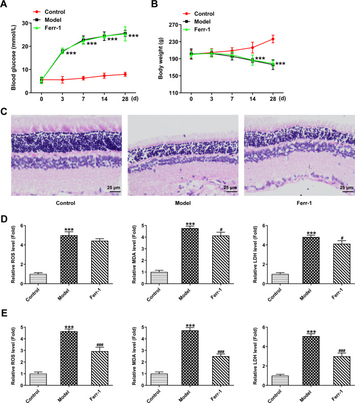
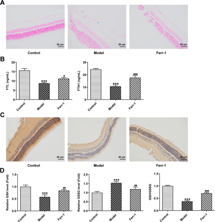
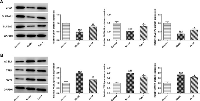
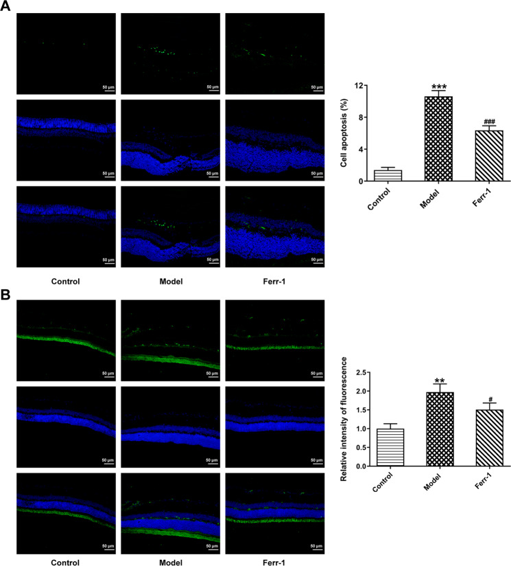
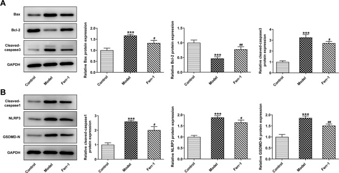
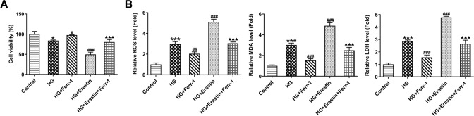
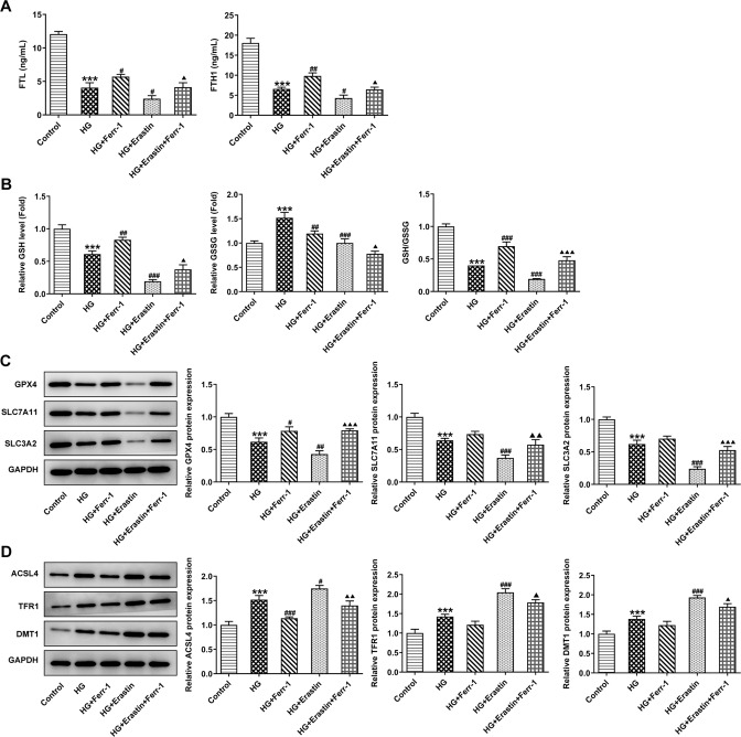
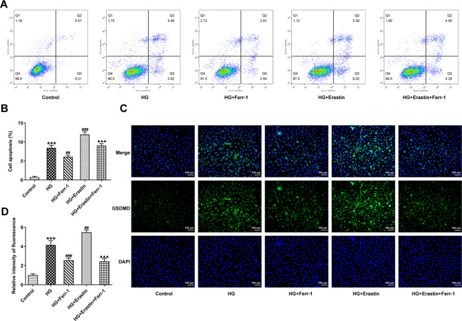
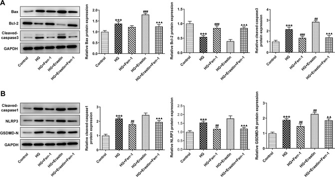
Diabetic retinopathy (DR) is a common microvascular complication leading to a high blindness rate among patients with diabetes. Ferroptosis is a type of cell death caused by the accumulation of iron-dependent lipid peroxides. Studies have shown that ferroptosis plays an important role in DR. The rat model of DR was constructed and treated with Ferrostatin-1 (Ferr-1). Haematoxylin and eosin (HE) were used to detect the degree of retinopathy. Oxidative stress levels were detected by ELISA. Perl's staining was used to detect iron deposition in retinal tissues. Ferritin levels were measured by ELISA. The expression of GPX4 was detected by immunohistochemistry (IHC). GSH/GSSG kit was used to detect the content and proportion of reduced/oxidized glutathione. Western blot was used to detect the expression of ferroptosis-related proteins. TUNEL assay was used to detect cell apoptosis. The expression of GSDMD was detected by fluorescence in situ hybridization (FISH). Western blot was used to detect the expression of apoptosis and pyroptosis-related proteins. Then, high glucose (HG)-induced retinal epithelial cell line ARPE-19 was treated by Erastin (ferroptosis activator) and Ferr-1. CCK-8, ELISA, western blot, flow cytometry, and immunofluorescence (IF) staining were used to detect oxidative stress levels, ferroptosis and cell damage. The mechanism was further explored by adding ferroptosis agonist Erastin. In vitro and in vivo results showed that oxidative stress was increased in DR model, resulting in ferroptosis and tissue or cell damage. After administration of Ferr-1, the antioxidant capacity was improved, ferroptosis levels were reduced and tissue or cell damage was alleviated. In vitro results showed that Ferr-1 reversed the impacts of Erastin on oxidative stress, ferroptosis, and cell damage in HG-induced ARPE-19 cells. Ferr-1 alleviated tissue and cell damage by improving the antioxidant capacity of the Xc--GPX4 system.


Conflict of interest statement

The authors declare no competing interests.

Figures

Fig. 1
Fig. 1. Ferr-1 ameliorates retinal tissue damage in DR rats.
A Changes in blood glucose in rats. B Changes in the body weight of rats. C HE detected the degree of retinopathy. The levels of oxidative stress in peripheral blood (D) and retinal tissues (E) were detected by corresponding kits. ***P < 0.001 vs control. #P < 0.05, ###P < 0.001 vs Model.
Fig. 2
Fig. 2. Ferr-1 inhibits ferroptosis in retinal tissues of DR rats.
A Perl’s staining detected iron deposition in retinal tissues. B Ferritin levels in retinal samples were detected by ELISA. C IHC detected GPX4 expression level in retinal tissues. D GSH/GSSG kit was used to detect the content and proportion of reduced/oxidized glutathione in tissues. ***P < 0.001 vs control. #P < 0.05, ##P < 0.01, ###P < 0.001 vs Model.
Fig. 3
Fig. 3. Ferr-1 regulates the expression of ferroptosis associated proteins in retinal tissues of DR rats.
A, B Western blot detected the expression of GPX4, SLC7A11, and SLC3A2. ***P < 0.001 vs control. #P < 0.05, ##P < 0.01, ###P < 0.001 vs Model.
Fig. 4
Fig. 4. Ferr-1 attenuates cell damage in retinal tissues of DR rats.
A TUNEL assay detected the cell apoptosis. B FISH assay detected the expression of GSDMD in tissues. **P < 0.01, ***P < 0.001 vs control. #P < 0.05, ###P < 0.001 vs Model.
Fig. 5
Fig. 5. Ferr-1 regulates the expression of apoptosis and pyroptosis-related proteins in retinal tissues of DR rats.
A Western blot detected the expression of apoptosis-related proteins Bax, Bcl-2, and the cleavage of caspase-3. B Western blot detected the expression of pyroptosis-related proteins the cleavage of caspase-1, NLRP3, and GSDMD-N. ***P < 0.001 vs control. #P < 0.05, ##P < 0.01 vs Model.
Fig. 6
Fig. 6. Ferr-1 regulates the Xc--GPX4 system to regulate cell activity and oxidative stress levels in retinal tissues of DR rats.
A CCK-8 kit was used to detect cell viability. B The levels of oxidative stress were detected by corresponding kits. *P < 0.05, ***P < 0.001 vs control. #P < 0.05, ##P < 0.01, ###P < 0.001 vs HG. △△△P < 0.001 vs HG + Erastin.
Fig. 7
Fig. 7. Ferr-1 regulates the redox capacity of the Xc--GPX4 system to regulate ferroptosis in DR cells.
A Ferritin levels in DR cells were detected by ELISA. B GSH/GSSG kit was used to detect the content and proportion of reduced/oxidized glutathione in DR cells. C, D Western blot detected the expression of ferroptosis-related proteins. ***P < 0.001 vs control. #P < 0.05, ##P < 0.01, ###P < 0.001 vs HG. P < 0.05, △△P < 0.01, △△△P < 0.001 vs HG + Erastin.
Fig. 8
Fig. 8. Ferr-1 regulates the redox capacity of Xc--GPX4 system to inhibit DR cell damage.
A Cell apoptosis was detected by flow cytometry. B Statistical diagram of apoptotic cells. C IF detected the expression of GSDMD in cells. D GSDMD expression statistics. ***P < 0.001 vs control. ##P < 0.01, ###P < 0.001 vs HG. △△△P < 0.001 vs HG + Erastin.
Fig. 9
Fig. 9. Ferr-1 regulates the expression of apoptosis and pyroptosis-related proteins to inhibit DR cell damage.
A Western blot detected the expression of apoptosis-related proteins Bax, Bcl-2, and the cleavage of caspase-3. B Western blot detected the expression of pyroptosis-related proteins and the cleavage of caspase-1, NLRP3, and GSDMD-N. ***P < 0.001 vs control. ##P < 0.01, ###P < 0.001 vs HG. △△P < 0.01, △△△P < 0.001 vs HG + Erastin.

References

    1. Wang W, Lo ACY. Diabetic retinopathy: pathophysiology and treatments. Int J Mol Sci. 2018;19. 10.3390/ijms19061816. - PMC - PubMed
    1. Cho NH, Shaw JE, Karuranga S, Huang Y, da Rocha Fernandes JD, Ohlrogge AW, et al. IDF Diabetes Atlas: global estimates of diabetes prevalence for 2017 and projections for 2045. Diabetes Res Clin Pr. 2018;138:271–81. doi: 10.1016/j.diabres.2018.02.023. - DOI - PubMed
    1. Dixon SJ, Lemberg KM, Lamprecht MR, Skouta R, Zaitsev EM, Gleason CE, et al. Ferroptosis: an iron-dependent form of nonapoptotic cell death. Cell. 2012;149:1060–72. doi: 10.1016/j.cell.2012.03.042. - DOI - PMC - PubMed
    1. Shen Z, Liu T, Li Y, Lau J, Yang Z, Fan W, et al. Fenton-reaction-acceleratable magnetic nanoparticles for ferroptosis therapy of orthotopic brain tumors. ACS Nano. 2018;12:11355–65. doi: 10.1021/acsnano.8b06201. - DOI - PubMed
    1. Shu W, Baumann BH, Song Y, Liu Y, Wu X, Dunaief JL. Ferrous but not ferric iron sulfate kills photoreceptors and induces photoreceptor-dependent RPE autofluorescence. Redox Biol. 2020;34:101469. doi: 10.1016/j.redox.2020.101469. - DOI - PMC - PubMed