Review
. 2023 Apr;24(4):273-287.
doi: 10.1038/s41580-022-00543-1. Epub 2022 Oct 25.

An expanded lexicon for the ubiquitin code

Affiliations
Review

An expanded lexicon for the ubiquitin code

Ivan Dikic et al. Nat Rev Mol Cell Biol. 2023 Apr.

Abstract

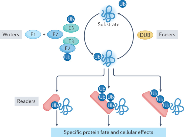
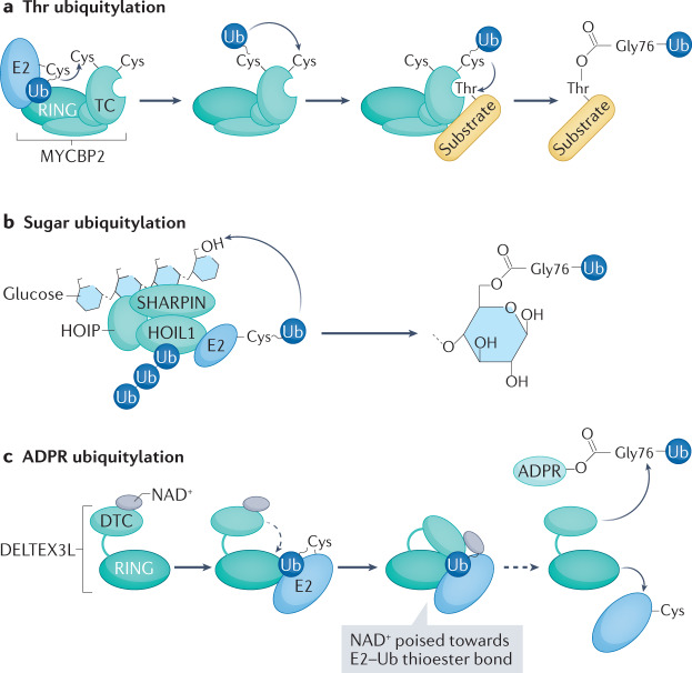
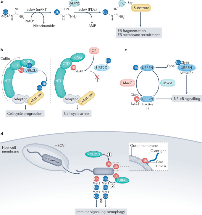
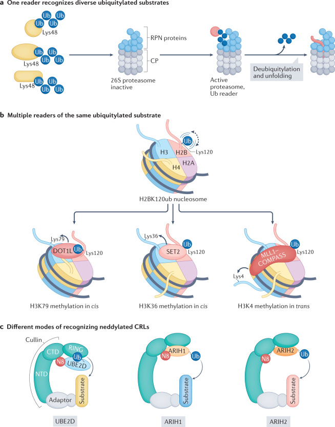
Our understanding of the ubiquitin code has greatly evolved from conventional E1, E2 and E3 enzymes that modify Lys residues on specific substrates with a single type of ubiquitin chain to more complex processes that regulate and mediate ubiquitylation. In this Review, we discuss recently discovered endogenous mechanisms and unprecedented pathways by which pathogens rewrite the ubiquitin code to promote infection. These processes include unconventional ubiquitin modifications involving ester linkages with proteins, lipids and sugars, or ubiquitylation through a phosphoribosyl bridge involving Arg42 of ubiquitin. We also introduce the enzymatic pathways that write and reverse these modifications, such as the papain-like proteases of severe acute respiratory syndrome coronavirus (SARS-CoV) and SARS-CoV-2. Furthermore, structural studies have revealed that the ultimate functions of ubiquitin are mediated not simply by straightforward recognition by ubiquitin-binding domains. Instead, elaborate multivalent interactions between ubiquitylated targets or ubiquitin chains and their readers (for example, the proteasome, the MLL1 complex or DOT1L) can elicit conformational changes that regulate protein degradation or transcription. The newly discovered mechanisms provide opportunities for innovative therapeutic interventions for diseases such as cancer and infectious diseases.


Conflict of interest statement

The authors declare no competing interests.

Figures

Fig. 1
Fig. 1. Elements of the ubiquitin code.
E3 enzymes (in combination with E2 and E1 enzymes) function as writers of the ubiquitin code. E3 enzymes are endowed with substrate specificity and — in a multistep mechanism that also engages E1 and E2 enzymes — attach ubiquitin to one or more residues of the substrate. Ubiquitin was primarily thought to modify Lys residues until recent studies discovered modification of many moieties on proteins and other macromolecules. This reaction can be repeated using a Lys of ubiquitin for attachment of the next ubiquitin molecule, giving rise to a ubiquitin chain. Depending on the E2–E3 pair that catalyses the last step of the ubiquitylation reaction, different Lys residues of ubiquitin are used, resulting in chains with different linkage types. Besides the seven Lys residues of ubiquitin, linkage can also occur with the amino-terminal Met. While ubiquitylation can be reversed by deubiquitylating enzymes (DUBs) that function as erasers of the ubiquitin code, it can be expanded by post-translational modifications of ubiquitin itself (Box 1). Distinct ubiquitin chains (and distinctly modified ubiquitin chains) have been shown to encode different cellular functions. They are decoded by readers equipped with ubiquitin-binding domains that are able to distinguish ubiquitin modifications and link the ubiquitylated substrate to downstream events, such as protein degradation, relocation, formation of multiprotein complexes and activation of enzymatic pathways. Ub, ubiquitin.
Fig. 2
Fig. 2. Unconventional ubiquitylation.
a | Thr ubiquitylation by the E3 MYCBP2. The RING–Cys–relay (RCR) mechanism depends on the RING domain, which binds E2–ubiquitin, and on the tandem Cys (TC) domain, which contains two catalytic Cys residues. Upon E2–E3 transthiolation and E2 dissociation, ubiquitin is relayed from Cys4520 to Cys4572 within the TC domain by intramolecular transthiolation. The TC domain also positions the substrate to allow Thr esterification. b | Linear ubiquitin assembly complex (LUBAC)-mediated ubiquitylation of unbranched glucosaccharides. The LUBAC components haem-oxidized IRP2 ubiquitin ligase 1 (HOIL1)-interacting protein (HOIP) and SHANK-associated RH domain-interacting protein (SHARPIN) bind glucosaccharides, whereas HOIL1 ubiquitylates the C6 hydroxyl moiety of glucose in one catalytic step. The reaction is accelerated by non-covalent binding of unconjugated Met1-linked or Lys63-linked ubiquitin oligomers to the RING-between-RING (RBR) domain of HOIL1. c | Ubiquitylation of ADP-ribose (ADPR) by DELTEX3L. E2–ubiquitin recruitment by the RING domain of DELTEX3L promotes a conformational change that positions the NAD+ bound to the carboxy-terminal domain (DTC) in close proximity to E2–ubiquitin, which facilitates ubiquitylation of ADPR at Gly76. Ub, ubiquitin.
Fig. 3
Fig. 3. Pathogen-induced ubiquitin modifications.
a | Ser ubiquitylation catalysed by the Legionella pneumophila effector SdeA. Using its mono-ADP-ribosyltransferase (mART) activity, SdeA first generates ADP-ribose (ADPR)–ubiquitin by transferring ADPR from NAD+ to Arg42 of ubiquitin. Subsequently, the phosphodiesterase (PDE) domain conjugates ADPR–ubiquitin to a Ser residue on substrates, thereby generating a ubiquitin-phosphoribosylated (PR) protein. The substrates modified in this way lead to pleiotropic changes in the host cell, including enhanced endoplasmic reticulum (ER) fragmentation and recruitment of ER membrane to L. pneumophila-containing vesicles. b | The carboxy-terminal domain (CTD) of cullin is modified by neural precursor cell-expressed developmentally downregulated protein 8 (NEDD8; N8), which leads to conformational changes that allow interactions between N8 and ubiquitin-conjugating enzyme E2 D (UBE2D) family members, the RING domain and ubiquitin. Deamidation of N8 at Gln40 (resulting in conversion to Glu40) by the enteropathogenic Escherichia coli (EPEC) virulence factor cycle-inhibiting factor (Cif) causes disruption of these mutual allosteric regulations of cullin–RING ligases (CRLs) and N8 that are required for CRL enzymatic activity. CRL substrates accumulate and cause cell cycle arrest. c | In normal conditions, ubiquitin is transferred to the catalytic Cys87 of UBE2N through a thioester bond involving Gly76 of ubiquitin. In this state, E2 is active and can participate in ubiquitylation reactions that trigger NF-κB signalling. The L. pneumophila effector MavC acts as a transglutaminase that links Ub via Gln40 to Lys92 or Lys94 of UBE2N, thereby inactivating E2 and preventing it from stimulating NF-κB signalling. The highly homologous effector MvcA reverses the reaction by hydrolysing the isopeptide bond created by MavC. d | Ubiquitylation of lipids by E3 ubiquitin ligase RING finger protein 213 (RNF213). Following escape from a damaged Salmonella enterica-containing vacuole (SCV), RNF213 is the first E3 ligase to attack cytosolic S. enterica, targeting a lipopolysaccharide composed of lipid A, core sugars and O antigen, in the outer bacterial membrane. Using an atypical zinc-finger domain, RNF213 attaches ubiquitin to a hydroxyl group of lipid A very close to the outer bacterial membrane (1). This first ubiquitin molecule then serves as the docking site for linear ubiquitin assembly complex (LUBAC), which builds linear ubiquitin chains (linked through Met1) on pre-existing ubiquitin molecules (2). The assembled ubiquitin coat is recognized by autophagy receptors such as p62, NDP52 and the optineurin effector protein NF-κB essential modulator (NEMO), which activate xenophagy (p62 and NDP52) and NF-κB-dependent immune signalling (optineurin), respectively (3). NTD, amino-terminal domain; Ub, ubiquitin. In part c, blue Ub, Ub linked via Gly76; red Ub, Ub linked via Gln40. In part d, blue Ub, ubiquitin molecule attached by LUBAC; red Ub, ubiquitin molecule attached by RNF213.
Fig. 4
Fig. 4. Different readers of the ubiquitin code — three examples.
a | Recognition of multiple diverse ubiquitylated substrates by one reader, the 26S proteasome. Lys48-linked polyubiquitin chain-tagged proteins are recognized by ubiquitin receptors (RPN1, RPN10 and RPN13) that are part of the base subcomplex of the proteasome. Interactions between ubiquitin and the ubiquitin-binding domains of the ubiquitin receptors regulate the proteasome conformations that activate unfolding, deubiquitylation and degradation of the substrate. b | Multiple downstream machineries recognize the ubiquitylated substrate, histone H2B. Ubiquitin attached to Lys120 of H2B can flexibly adopt different positions to bind various chromatin-modifying methyltransferases. The positioning of ubiquitin in combination with additional binding sites of the methyltransferase on the nucleosome activates methylation of specific Lys residues. DOT1L and SET2 target two different Lys residues of histone H3, Lys 79 (H3K79) and H3K36, respectively, in cis, whereas MLL1-including complex of proteins associated with Set1 (COMPASS) methylates H3K4 in trans. c | Different modes through which various ubiquitin-carrying enzymes recognize neddylated cullin–RING ligases (CRLs). Neural precursor cell-expressed developmentally downregulated protein 8 (NEDD8; N8) can allosterically regulate the conformation of the cullin it modifies, and vice versa, the cullin affects the positioning of N8 within the complex so that different readers, even homologous readers (ARIH1 and ARIH2) are accommodated distinctly on different CRLs. CP, core particle; CTD, carboxy-terminal domain; H2BK120ub, Lys120-ubiquitinylated histone H2B; H4, histone H4; NTD, amino-terminal domain; Ub, ubiquitin; UBE2D, ubiquitin-conjugating enzyme E2 D.

References

    1. Seet BT, Dikic I, Zhou MM, Pawson T. Reading protein modifications with interaction domains. Nat. Rev. Mol. Cell Biol. 2006;7:473–483. doi: 10.1038/nrm1960. - DOI - PubMed
    1. Zheng N, Shabek N. Ubiquitin ligases: structure, function, and regulation. Annu. Rev. Biochem. 2017;86:129–157. doi: 10.1146/annurev-biochem-060815-014922. - DOI - PubMed
    1. Harper JW, Schulman BA. Cullin-RING ubiquitin ligase regulatory circuits: a quarter century beyond the F-Box hypothesis. Annu. Rev. Biochem. 2021;90:403–429. doi: 10.1146/annurev-biochem-090120-013613. - DOI - PMC - PubMed
    1. Wang Y, Argiles-Castillo D, Kane EI, Zhou A, Spratt DE. HECT E3 ubiquitin ligases-emerging insights into their biological roles and disease relevance. J. Cell Sci. 2020;133:jcs258087. doi: 10.1242/jcs.258087. - DOI - PMC - PubMed
    1. van Wijk SJL, Fiškin E, Dikic I. Selective monitoring of ubiquitin signals with genetically encoded ubiquitin chain-specific sensors. Nat. Protoc. 2013;8:1449–1458. doi: 10.1038/nprot.2013.089. - DOI - PubMed

Publication types