. 2022 Oct 25;20(1):165.
doi: 10.1186/s12964-022-00966-5.

Metformin alleviates bone loss in ovariectomized mice through inhibition of autophagy of osteoclast precursors mediated by E2F1

Affiliations

Metformin alleviates bone loss in ovariectomized mice through inhibition of autophagy of osteoclast precursors mediated by E2F1

Xudong Xie et al. Cell Commun Signal. .

Abstract

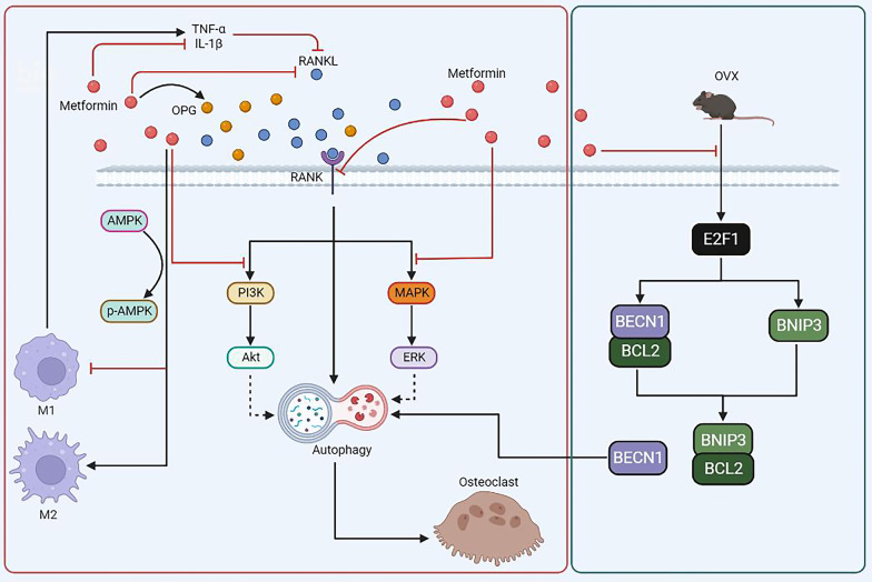
Background: Postmenopausal bone loss, mainly caused by excessive bone resorption mediated by osteoclasts, has become a global public health burden. Metformin, a hypoglycemic drug, has been reported to have beneficial effects on maintaining bone health. However, the role and underlying mechanism of metformin in ovariectomized (OVX)-induced bone loss is still vague.

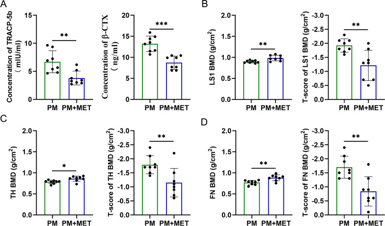
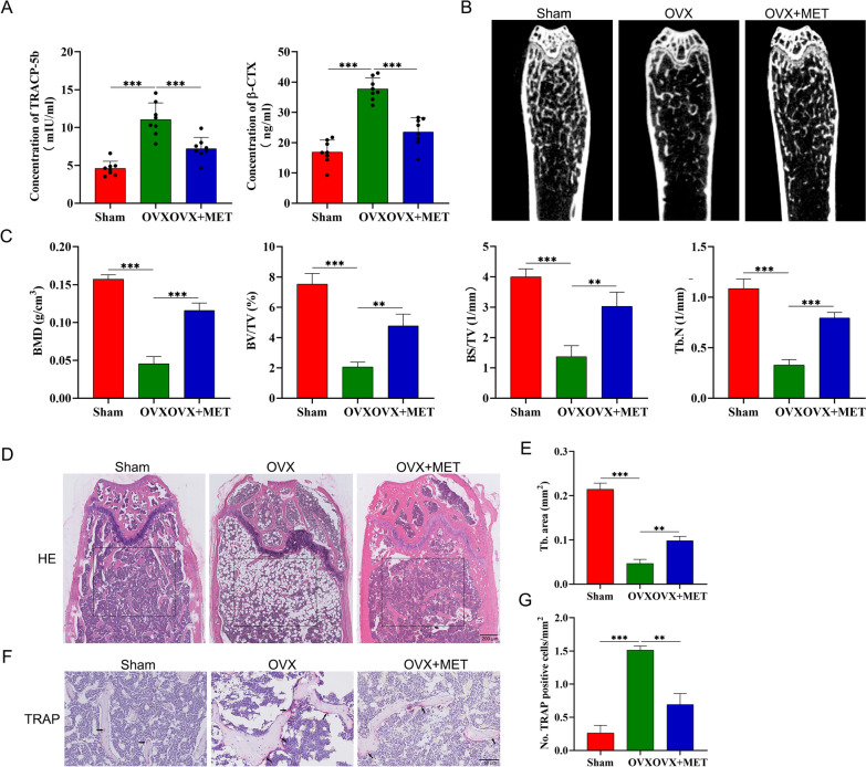
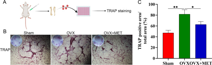
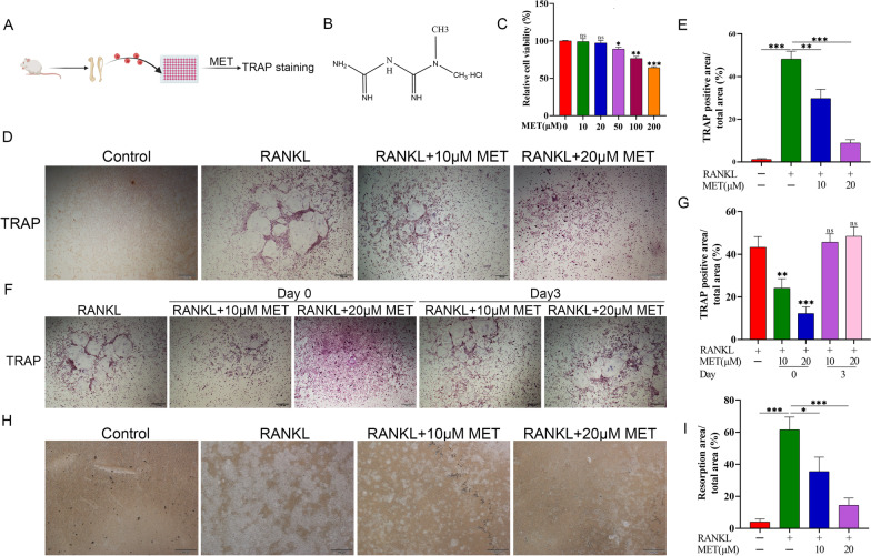
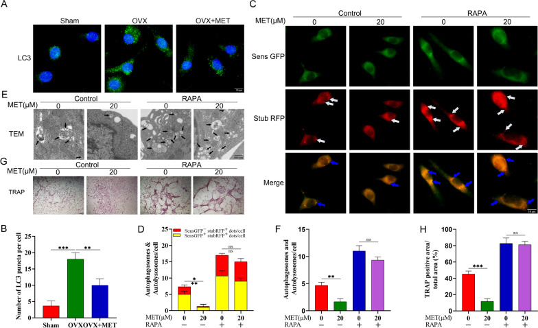
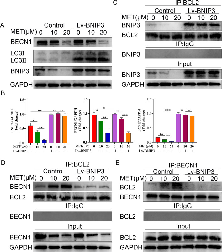
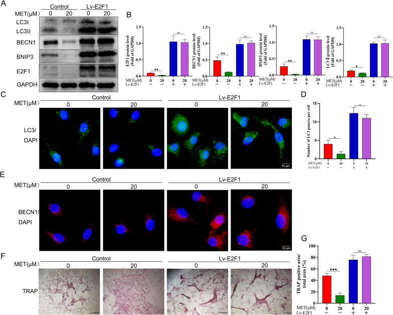
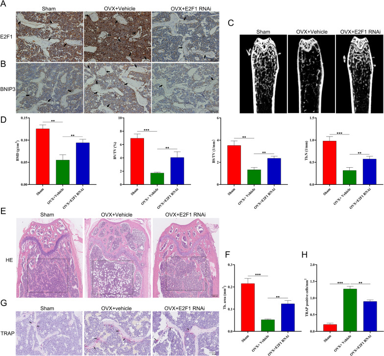
Results: In this study, we demonstrated for the first time that metformin administration alleviated bone loss in postmenopausal women and ovariectomized mice, based on reduced bone resorption markers, increased bone mineral density (BMD) and improvement of bone microstructure. Then, osteoclast precursors administered metformin in vitro and in vivo were collected to examine the differentiation potential and autophagical level. The mechanism was investigated by infection with lentivirus-mediated BNIP3 or E2F1 overexpression. We observed a dramatical inhibition of autophagosome synthesis and osteoclast formation and activity. Treatment with RAPA, an autophagy activator, abrogated the metformin-mediated autophagy downregulation and inhibition of osteoclastogenesis. Additionally, overexpression of E2F1 demonstrated that reduction of OVX-upregulated autophagy mediated by metformin was E2F1 dependent. Mechanistically, metformin-mediated downregulation of E2F1 in ovariectomized mice could downregulate BECN1 and BNIP3 levels, which subsequently perturbed the binding of BECN1 to BCL2. Furthermore, the disconnect between BECN1 and BCL2 was shown by BNIP3 overexpression.

Conclusion: In summary, we demonstrated the effect and underlying mechanism of metformin on OVX-induced bone loss, which could be, at least in part, ascribed to its role in downregulating autophagy during osteoclastogenesis via E2F1-dependent BECN1 and BCL2 downregulation, suggesting that metformin or E2F1 inhibitor is a potential agent against postmenopausal bone loss. Video abstract.

Keywords: Autophagy; BECN1; BNIP3; Bone loss; E2F1; Metformin; Osteoclast precursors.


Conflict of interest statement

All authors declare that they have no competing financial interests.

Figures

Fig. 1
Fig. 1
Change in bone resorption and BMD with metformin in postmenopausal women with T2DM. A The β-CTX and TRACP-5b levels in the serum of postmenopausal women taking or not taking metformin for T2DM. B BMD and BMD T-score at LS1; C BMD and BMD T-score at TH. D BMD and BMD T-score at FN. PM, Postmenopausal women (n = 8); PM + MET, metformin-treated postmenopausal women(n = 8); LS1, Lumbar Spine; TH, Total Hip; FN, Femoral Neck. Data are presented as means ± SD of 8 independent experiments (*p < 0.05; **p < 0.01; ***p < 0.001)
Fig. 2
Fig. 2
Metformin attenuates OVX-induced bone loss in vivo. A The β-CTX and TRACP-5b levels in the serum from sham group (n = 8), ovariectomy (OVX; n = 8) group and OVX + MET group (n = 8). B Representative micro-CT images of the distal femur in sham group, OVX group, and OVX + MET group. C Quantitative analysis of bone mineral density (BMD), trabecular bone surface area/total value (BS/TV), volume per tissue volume (BV/TV) and trabecular number (Tb. N). D Representative images of Hematoxylin and Eosin staining of distal femur sections. E Quantitative analysis of trabecular (Tb) bone area in (D). F Representative images of femur stained with TRAP. G Histomorphometric analysis of the osteoclast number. sham, sham operated mice group; OVX, ovariectomized mice group, OVX+MET, ovariectomized mice with metformin treatment group. (*p < 0.05; **p < 0.01; ***p < 0.001)
Fig. 3
Fig. 3
Metformin in vivo administration inhibits the formation of osteoclasts. A schematic diagram of the experiment. B, C TRAP-staining and quantitation of TRAP-positive cells. Data are presented as means ± SD of 3 independent experiments (*p < 0.05; **p < 0.01; ***p < 0.001)
Fig. 4
Fig. 4
Metformin suppresses RANKL-induced osteoclast differentiation in vitro. A schematic diagram of the experiment. B Chemical structural formula of metformin. C Osteoclast precursors were incubated with different concentrations of metformin for 5 d in the presence of 30 ng/ml M-CSF and 50 ng/mL RANKL. Subsequently, cell viability was evaluated with Cell Counting Kit 8 (CCK8) reagent. D Representative images of TRAP-positive cells after treatment with metformin. E Quantitative analysis of the area of TRAP-positive osteoclasts. F, G Effect of metformin on RANKL-induced osteoclast precursors differentiation at different stages. H, I Pit formation assay of osteoclasts and quantification of the pits area. Data are presented as means ± SD of 3 independent experiments (*p < 0.05; **p < 0.01; ***p < 0.001)
Fig. 5
Fig. 5
Metformin inhibits osteoclast differentiation via decreasing autophagy. A Immunofluorescence staining for LC3 in osteoclast precursors isolated from the femur and tibia of sham group mice, OVX group mice, and OVX + MET group mice. B Quantification of the LC3 puncta per cell in (A). C, D After infection with tandem GFP-red fluorescent protein (RFP)-LC3 lentivirus, osteoclast precursors were treated with metformin and/or rapamycin (10 nM) for 24 h in presence of 30 ng/mL M-CSF and 50 ng/mL RANKL, and then the fluorescence was observed by fluorescence microscopy and quantitative analysis was performed. E, F metformin and/or rapamycin were used to treat osteoclast precursors for 24 h, and then the formation of autophagosomes and/or autolysosomes in osteoclast precursors were observed under transmission electron microscopy. G After metformin or/and rapamycin, TRAP-stained multinucleated osteoclasts. H Quantitative analysis of the area of TRAP-positive osteoclasts. Data are presented as means ± SD of 3 independent experiments (*p < 0.05; **p < 0.01; ***p < 0.001, ns, non-significant)
Fig. 6
Fig. 6
Metformin regulates autophagy through a mechanism involving BECN1 and BNIP3. A Cell lysates of osteoclast precursors were used to examine the expression of BECN1, BNIP3 and LC3 proteins by western bolt. After overexpression of BNIP3 (Lv-BNIP3), osteoclast precursors were treated with metformin (0, 10, 20 μM) for 5 d in the presence of 30 ng/mL M-CSF and 50 ng/mL RANKL. B The quantitative analysis of immunoblots relative to GAPDH protein level in (A). The results were from pairwise comparisons within- and between-group. C Osteoclast precursors were treated as in (A), and the cell lysates were preformed to co-immunoprecipitation with BCL2 antibody, followed by immunoblotting with the indicated antibodies. Among them, IgG group was considered as negative control. D, E Osteoclast precursors were treated as in (A), and the cell lysates were subjected to co-immunoprecipitation with either BCL2 or BECN1 antibody, followed by immunoblotting with the indicated antibodies. Among them, IgG group was considered as negative control. Values displayed are means ± SD of 3 independent experiments (*p < 0.05; **p < 0.01; ***p < 0.001, ns, non-significant)
Fig. 7
Fig. 7
Metformin-mediated autophagy downregulation via BECN1 and BNIP3 is E2F1 dependent. A Cell lysates of osteoclast precursors were used to examine the expression of E2F1, BNIP3 and LC3 proteins by western bolt. After overexpression of E2F1 (Lv-E2F1), osteoclast precursors were treated with metformin (0, 20 μM) for 5 d in the presence of 30 ng/mL M-CSF and 50 ng/mL RANKL. B The quantitative analysis of immunoblots relative to GAPDH protein level in (A). C After overexpression of E2F1 (Lv-E2F1), osteoclast precursors were treated with metformin (0, 20 μM) for 24 h, and immunofluorescence staining for LC3 was performed. D Quantification of the LC3 puncta per cell in (C). E After overexpression of E2F1 (Lv-E2F1), osteoclast precursors were treated with metformin (0, 20 μM) for 24 h, immunofluorescent staining (IF) analyses of osteoclast precursors using anti-BECN1 antibody. (F, G) After osteoclast precursors were treated as in (A), TRAP staining was performed and TRAP-positive multinucleated osteoclasts (≥ 3 nuclei) were counted. Values displayed are means ± SD of 3 independent experiments (*p < 0.05; **p < 0.01; ***p < 0.001, ns, non-significant)
Fig. 8
Fig. 8
E2F1-siRNA administration rescues OVX-related bone loss in vivo. Mice were treated with Vehicle (Saline) or E2F1-RNAi (250 nmol/kg) through tail vein twice per week for 4 weeks after four weeks following ovariectomy or sham surgery. A Representative immunohistochemical staining for E2F1 in distal femur sections from sham (n = 3), OVX + vehicle (n = 3) and OVX + E2F1 RANi (E2F1 siRNA; n = 3). B Representative immunohistochemical staining for BNIP3 in distal femur sections from sham (n = 3), OVX + Vehicle (n = 3) and OVX + E2F1 RANi (n = 3). C Micro CT analysis of the distal femur from sham, OVX + Vehicle and OVX + E2F1 RNAi group. D Calculations of bone mineral density (BMD), bone value/total value (BV/TV), bone surface area/total value (BS/TV) and trabecular number (Tb.N). E Histological H&E staining of distal femur sections. (F) Quantitative histomorphometric assessment of trabecular number (Tb. N) based on H&E-stained femur sections. G Histological TRAP staining of femur sections. H Quantitative assessment of osteoclasts number based on TRAP-stained tibial sections. Values displayed are means ± SD of 3 independent experiments (*p < 0.05; **p < 0.01; ***p < 0.001)
Fig. 9
Fig. 9
Schematic showing that the molecular mechanism underlying the inhibition of osteoclastogenesis by metformin in our study (Green box), and the possible relationship between autophagy and other signaling pathways involving in osteoclast differentiation and function upon metformin stimulation in previous studies (red box). A Metformin treatment inhibits OVX-induced E2F1 upregulation. Then, metformin downregulates OVX-induced BECN1 and BNIP3 overexpression, followed by reduction of the connection between BNIP3 and BCL2, which subsequently promotes the association of BECN1 and BCL2, resulting in reduction of the free BECN1 level. In this way, OVX-triggered autophagy upregulation is suppressed by metformin, leading to the alleviation of OVX-induced bone loss. B Increased level of autophagy was observed during the RANKL-induced osteoclastogenesis. (a, b, c) Metformin was demonstrated to downregulate the expression level of RANK (a), RANKL (b) and OPG (c). (d) Inflammation factors, such as TNF-α and IL-1β, could reduce RANKL expression. (e) Metformin was shown to downregulate the level of TNF-α and IL-1β. (f, g) Metformin could promote the polarization of anti-inflammatory M2 macrophages and inhibit the polarization of proinflammatory M1 macrophages (f), followed by reduction of TNF-α and IL-1β level (g). (h, i) Metformin could inhibit the PI3K/Akt (h) and MAPK/ERK (i) signaling pathways during osteoclastogenesis

Similar articles

Cited by

References

    1. Boyle WJ, Simonet WS, Lacey DL. Osteoclast differentiation and activation. Nature. 2003;423:337–342. - PubMed
    1. Compston JE, McClung MR, Leslie WD. Osteoporosis. Lancet. 2019;393:364–376. - PubMed
    1. Cosman F, de Beur SJ, LeBoff MS, Lewiecki EM, Tanner B, Randall S, et al. Clinician's guide to prevention and treatment of osteoporosis. Osteoporos Int. 2014;25:2359–2381. - PMC - PubMed
    1. Tella SH, Gallagher JC. Prevention and treatment of postmenopausal osteoporosis. J Steroid Biochem Mol Biol. 2014;142:155–170. - PMC - PubMed
    1. Reid IR. Short-term and long-term effects of osteoporosis therapies. Nat Rev Endocrinol. 2015;11:418–428. - PubMed

Publication types

LinkOut - more resources