. 2023 Jan;29(1):306-316.
doi: 10.1111/cns.14004. Epub 2022 Oct 25.

Inhibition of the expression of rgs-3 alleviates propofol-induced decline in learning and memory in Caenorhabditis elegans

Affiliations

Inhibition of the expression of rgs-3 alleviates propofol-induced decline in learning and memory in Caenorhabditis elegans

Ayang Zhao et al. CNS Neurosci Ther. 2023 Jan.

Abstract

Background: Exposure to anesthesia leads to extensive neurodegeneration and long-term cognitive deficits in the developing brain. Caenorhabditis elegans also shows persistent behavioral changes during development after exposure to anesthetics. Clinical and rodent studies have confirmed that altered expression of the regulators of G protein signaling (RGS) in the nervous system is a factor contributing to neurodegenerative and psychological diseases. Evidence from preclinical studies has suggested that RGS controls drug-induced plasticity, including morphine tolerance and addiction. This study aimed to observe the effect of propofol exposure in the neurodevelopmental stage on learning and memory in the L4 stage and to study whether this effect is related to changes in rgs-3 expression.

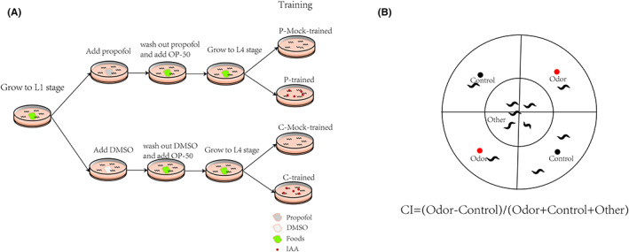
Methods: Caenorhabditis elegans were exposed to propofol at the L1 stage, and learning and memory abilities were observed at the L4 stage. The expression of rgs-3 and the nuclear distribution of EGL-4 were determined to study the relevant mechanisms. Finally, RNA interference was performed on rgs-3-expressing cells after propofol exposure. Then, we observed their learning and memory abilities.

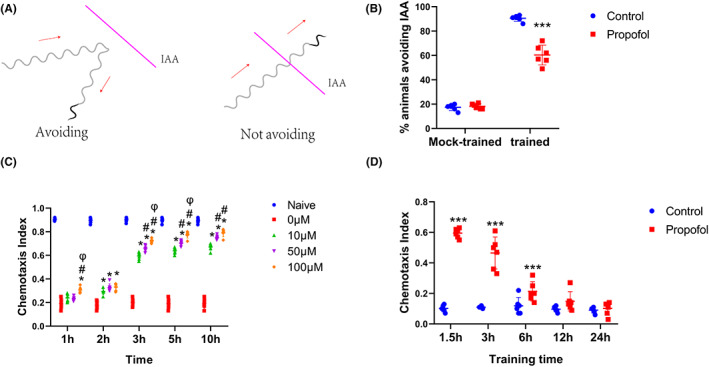
Results: Propofol time- and dose-dependently impaired the learning capacity. Propofol induced a decline in non-associative and associative long-term memory, rgs-3 upregulation, and a failure of nuclear accumulation of EGL-4/PKG in AWC neurons. Inhibition of rgs-3 could alleviate the propofol-induced changes.

Conclusion: Inhibition of the expression of rgs-3 alleviated propofol-induced learning and memory deficits in Caenorhabditis elegans.

Keywords: learning; memory; neurodevelopment; propofol; regulators of G protein signaling.


Conflict of interest statement

The authors declare no conflicting financial interest.

Figures

FIGURE 1
FIGURE 1
Experiment flow chart. (A) Schematic of aversive olfactory learning assay. (B) Schematic diagram of chemotaxis plate.
FIGURE 2
FIGURE 2
Propofol induces a decline in learning in a time‐ and dose‐dependent manner. (A) Travel paths corresponding to two different behaviors observed upon exposure to IAA. (B) Percentage of animals avoiding IAA. (C) C. elegans was exposed to propofol at concentrations of 0, 10, 50, and 100 μM for 1, 2, 3, 5, and 10 h. Naive represents untrained wild‐type nematodes. (D) Chemotaxis index of nematodes exposed to IAA calculated for different training intervals. p values were generated by one‐way analysis of variance (ANOVA). n = 40–60 animals per condition. The experiments were repeated six times. *p < 0 0.05 versus 0 μM, # p < 0.05 versus 10 μM group; φ p < 0 0.05 versus 50 μM group; ***p < 0 0.001 versus Control.
FIGURE 3
FIGURE 3
The HPLC profile of propofol. (A) The HPLC profile and retention time of propofol. (B) Comprehensive chromatogram of pending text sample and standard. The Chinese character in the picture means propofol). n = 3 independent experiments.
FIGURE 4
FIGURE 4
Propofol induces a decline in non‐associative and associative long‐term memory. (A) Chemotaxis index of nematodes exposed to IAA calculated at different recovery times. n = 40–60 animals per condition and the experiment was repeated six times. A one‐way analysis of variance (ANOVA) test was used. (B) Odor‐induced memory retrieval resulted in the rapid (within 20 min) translocation of DAF‐16 / FOXO to the nucleus (red circle) of gonadal sheath cells. To visualize protein spatial dynamics, we used a strain expressing translation fusion DAF‐16:: DAF‐16:: GFP (TJ356). Each of the four typical worms is imaged separately, cut along its edge, and stacked on top of the other. (C–D) Density plots of the number of cells with nuclear DAF‐16/FOXO localization per worm in different groups. n = ~100 animals per condition. A nonparametric Kruskal‐Wallis test was used. (E–H) Cumulative distribution of the number of cells with nuclear DAF‐16/FOXO localization per worm before or after the challenge. A nonparametric Kolmogorov–Smirnov test was used. (I) Expression of genes downstream of the DAF‐16/FOXO transcription factor was upregulated following odor‐induced memory reactivation. n = 3 independent experiments. One‐way analysis of variance (ANOVA) was used. (J) Each line represents a single independent experimental repeat (total n = 5), each with ~100 animals scored for each of the groups. Paired t‐test was used (*p < 0.05 versus other groups; ***p < 0.001 versus other groups). Before: before challenging; After: after challenging for 20 min.
FIGURE 5
FIGURE 5
Propofol induces upregulation of the expression of rgs‐3 and a decline in the ability of nuclear accumulation of EGL‐4/PKG in AWC neurons. (A) Fluorescent confocal images of AWC neurons in propofol and control groups. Integrality of the cilia of AWC neurons was observed in the propofol and control groups. Each of the two typical worms is imaged separately, cut along its edge, and stacked on top of the other. (B) The expression of rgs‐3 was up‐regulated after exposure to propofol at the L4 stage. n = 3 independent experiments. The t‐test was used. (C) Cumulative distribution (AWC neuronal nuclear index) for EGL‐4::GFP in different groups of animals after conditioning. An increase in AWC neuronal nuclear index was observed after conditioning in the C‐trained group, while a decrease was observed after conditioning in the P‐trained group. n = 40–60 animals per condition. A nonparametric Kruskal‐Wallis test was used. (D)Nuclear enrichment of EGL‐4 in AWC neurons. Representative images of EGL‐4::GFP fluorescence in the AWC neurons. F nucleus, F cytoplasm = fluorescence measured in AWC neuron (nucleus or cytoplasm of the same neuron). (E) Percentage of animals with nuclear EGL‐4 at different training intervals. n = 40–60 animals per condition, and the experiments were repeated six times. One‐way analysis of variance (ANOVA) was used (***p < 0.001; **p < 0.01).
FIGURE 6
FIGURE 6
Inhibition of the expression of rgs‐3 could alleviate the propofol‐induced decline in learning and memory. (A) The expression of rgs‐3 was down‐regulated after feeding RNAi. n = 3 independent experiments. One‐way analysis of variance (ANOVA) was used. (B) Fluorescent confocal images of AWC neurons in rgs‐3 siRNA and L4440. The integrality of the cilia of AWC neurons was observed. Each of the two typical worms is imaged separately, cut along its edge, and stacked on top of the other. (C) AWC neuronal nuclear index for EGL‐4::GFP in different groups of animals after conditioning. The bar represents the median and interquartile range. n = 40–60 animals per condition. A nonparametric Kruskal–Wallis test was used. (D) Percentage of animals avoiding IAA after feeding RNAi. One‐way analysis of variance (ANOVA) was used. (E) Chemotaxis index of nematodes exposed to IAA at 1.5 h of training interval. One‐way analysis of variance (ANOVA) was used. (F) Chemotaxis index of nematodes exposed to IAA at different recovery times. n = 40–60 animals per condition and repeated thrice. A one‐way analysis of variance (ANOVA) test was used. (G‐H) Density plots of the number of cells with nuclear DAF‐16/FOXO localization per worm in different groups. n = ~100 animals per condition. A nonparametric Kruskal–Wallis test was used. (I–L) Cumulative distribution of the number of cells with nuclear DAF‐16/FOXO localization per worm before or after the test in P‐trained worms. A nonparametric Kolmogorov–Smirnov test was used (***p < 0.001; **p < 0.01).

References

    1. Bosnjak ZJ, Logan S, Liu Y, Bai X. Recent insights into molecular mechanisms of propofol‐induced developmental neurotoxicity: implications for the protective strategies. Anesth Analg. 2016;123(5):1286‐1296. - PMC - PubMed
    1. Chidambaran V, Costandi A, D'Mello A. Propofol: a review of its role in pediatric anesthesia and sedation. CNS Drugs. 2015;29(7):543‐563. - PMC - PubMed
    1. Gundacker C, Forsthuber M, Szigeti T, et al. Lead (Pb) and neurodevelopment: A review on exposure and biomarkers of effect (BDNF, HDL) and susceptibility. Int J Hyg Environ Health. 2021;238:113855. - PubMed
    1. Nakaoka H, Hisada A, Matsuzawa D, et al. Associations between prenatal exposure to volatile organic compounds and neurodevelopment in 12‐month‐old children: the Japan Environment and Children's Study (JECS). Sci Total Environ. 2021;794:148643. - PubMed
    1. Bansal E, Hsu HH, de Water E, et al. Prenatal PM2.5 exposure in the second and third trimesters predicts neurocognitive performance at age 9‐10 years: a cohort study of Mexico City children. Environ Res. 2021;202:111651. - PMC - PubMed

Publication types

MeSH terms

Substances