. 2022 Oct 15:2022:3590326.
doi: 10.1155/2022/3590326. eCollection 2022.

Pancreatic Cancer-Derived Exosomes Promote the Proliferation, Invasion, and Metastasis of Pancreatic Cancer by the miR-3960/TFAP2A Axis

Affiliations

Pancreatic Cancer-Derived Exosomes Promote the Proliferation, Invasion, and Metastasis of Pancreatic Cancer by the miR-3960/TFAP2A Axis

Jinhong Wu. J Oncol. .

Abstract

Background: The microRNAs (miRNAs) in cancer-derived exosomes have the ability to change tumor microenvironment. This study aims to investigate the role of miRNA in cancer-derived exosomes in pancreatic cancer (PC).

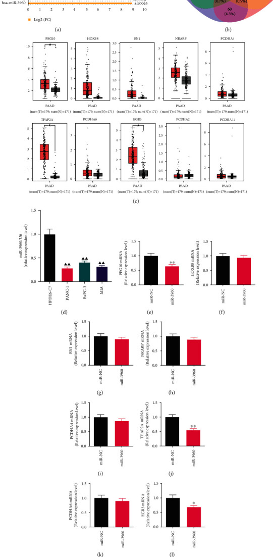
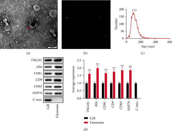
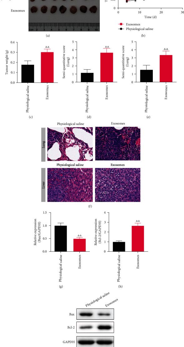
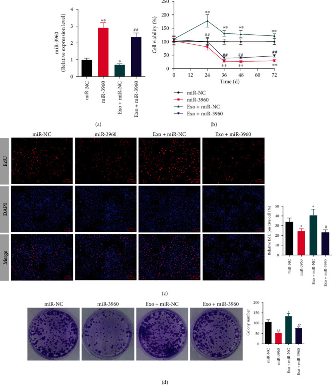
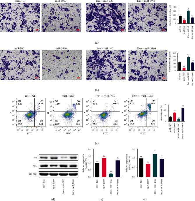
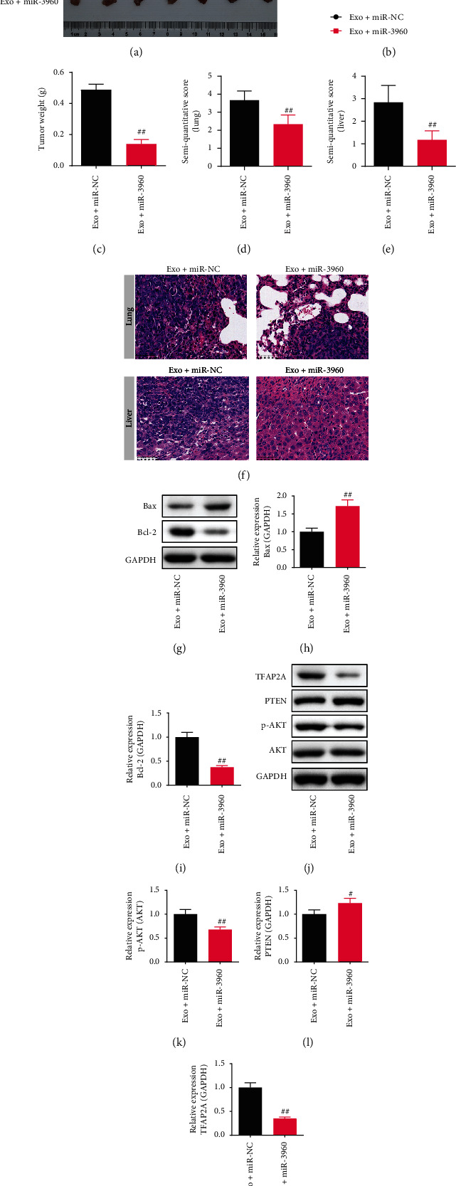
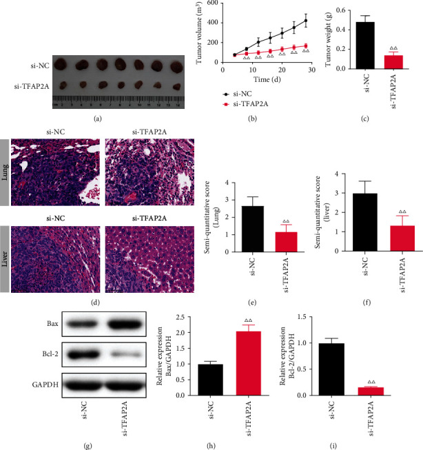
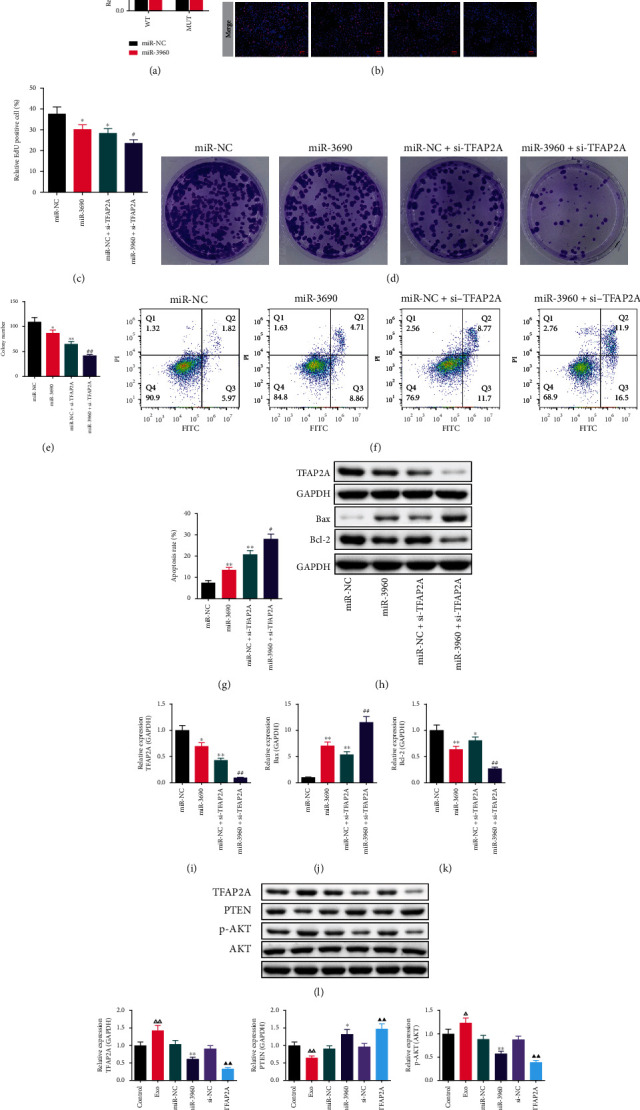
Methods: Based on the analysis of PC-derived and healthy exosomes by bioinformatics analysis and quantitative real-time PCR validation, the miR-3960 was identified to be the most significantly different miRNA, and TFAP2A proved as its potential target gene. Besides, the exosomes were isolated from PANC-1 cells and identified. After that, PANC-1 cells were treated with the isolated exosomes or transfected with miR-3960 mimics or si-TFAP2A, the effect of PC-derived exosomes, as well as the miR-3960/TFAP2A axis in PC cells, were assessed by the CCK-8, EDU staining, Transwell, cell colony formation, and flow cytometry assays. Furthermore, the effects of exosomes and the miR-3960/TFAP2A axis on PC tumor growth were observed in tumor-bearing mice by the measurement of tumor weight and volume, and hematoxylin-eosin staining. Moreover, the expressions of TFAP2A/PTEN/AKT signaling proteins were detected by Western blot.

Results: PC-derived exosomes were isolated successfully and proved to have promotion effects on the proliferation, metastasis, and invasion of PC cells both in vitro and tumor growth in vivo. Also, the PC-derived exosomes upregulated the TFAP2A, Bcl-2, and p-AKT/AKT protein levels, and inhibited PTEN and Bax levels and PANC-1 cell apoptosis. Overexpression of miR-3960 antagonized the promotion effect of exosomes on PC cells and the TFAP2A/PTEN/AKT signaling pathway, inhibiting the growth of tumors. Besides, si-TFAP2A enhanced the inhibitory effect of miR-3960 in PC.

Conclusion: MiR-3960 antagonizes the promotion effect of tumor-derived exosomes on the proliferation, invasion, and metastasis of PC via suppressing TFAP2A.


Conflict of interest statement

The authors declare that the research was conducted in the absence of any commercial or financial relationships that could be construed as a potential conflict of interest.

Figures

Figure 1
Figure 1
Screening of microRNAs of cancer-derived exosomes in pancreatic cancer based on bioinformatics and quantitative real-time PCR (qRT-PCR) analysis. (x¯±s) (a) The bar graph shows the top 30 differentially expressed genes of GSE50632 on the GPL16294 platform using GEO2R for analysis. The Log2 (FC) of miR-3960 is 8.90065. (b) The Venn diagram presents the intersection of mRNAs retrieved by different databases for miR-3960. (c) The expression levels of PEG10, HOXB8, EN1, NRARP, PCDHA4, TFAP2A, PCDHA6, EGR3, PCDHA2, and PCDHA11 mRNA in PC tumor and normal groups were analyzed by the TCGA normal and GTEx data on the website of GEPIA. Red indicates the tumor group; grey indicates the normal group. (d) The miR-3960 level in HPDE6-C7, PANC-1, BxPC-3 and MIA cell lines by qRT-PCR analysis (n =3). (e-n) The effect of miR-3960 on the levels of PEG10, HOXB8, EN1, NRARP, PCDHA4, TFAP2A, PCDHA6, EGR3, PCDHA2, and PCDHA11 mRNA by qRT-PCR (n =3). P <0.01 compared to the normal group; ▲▲P <0.01 compared to the HPDE6-C7 group; P <0.05, ∗∗P <0.01 compared to the miR-NC group.
Figure 2
Figure 2
The identification of PANC-1-derived exosomes. (x¯±s, n =3). (a) Transmission electron microscopy. (Scale bar =200 nm) (b and c) Nanoparticle Tracking analysis. (d) The average levels of TSG101, Alix, CD81, CD9, CD63, HSP70 and C-myc. ∗∗P <0.01 compared to the cell group.
Figure 3
Figure 3
The effect of tumor-derived exosomes on pancreatic cancer (PC) on the xenograft tumor model. (x¯±s, n =7) (a) The photographs of tumors that were dissected from PC tumor bearing mice every fourth day. (b) The tumor volume. (c) The tumor weight. (d-f) The semi-quantitative score and typical picture of Hematoxylin-eosin (HE) staining in the lung and liver. (×400) (g-i) The Bax and Bcl-2 protein levels in the lung of PC mice (n =3). ΔΔP<0.01 compared to the cell group.
Figure 4
Figure 4
The effect of tumor-derived exosomes on the proliferation in miR-3960-overexpressed PANC-1 cells. (x¯±s) (a) The quantitative real-time PCR result of miR-3960 (n =3). (b) The cell viability analyzed by Cell Counting Kit-8 (n =5). (c) The typical pictures and relative positive cells of 5-Ethynyl-2'-deoxyuridine (EdU) assay (n =3). Red indicates EdU positive cells and blue indicates DAPI positive cells. (×400) (d) The typical pictures and colonies number of cell colony formation assay (n =3). P <0.05, ∗∗P <0.01 compared to the miR-NC group, #P <0.05, ##P <0.01 compared to the Exo + miR-NC group.
Figure 5
Figure 5
The effect of tumor-derived exosomes on the migration, invasion and apoptosis in miR-3960-overexpressed PANC-1 cells. (x¯±s, n =3) (a) The migration of PANC-1 cells tested by Transwell assay. (×200) (b) The invasion of PANC-1 cells tested by Transwell assay. (c) The apoptosis of PANC-1 cells tested by flow cytometry. (d-f) The Bax and Bcl-2 protein levels measured by Western blot. P <0.05, ∗∗P <0.01 compared to the miR-NC group, #P <0.05, ##P <0.01 compared to the Exo + miR-NC group.
Figure 6
Figure 6
The effect of tumor-derived exosomes on miR-3960-overexpressed pancreatic cancer (PC) on the xenograft tumor model. (x¯±s, n =7) (a) The photographs of tumors dissected from PC mice. (b) The tumor volume. (c) The tumor weight. (d-f) The semi-quantitative score and typical picture of Hematoxylin-eosin staining in the lungs and livers. (×400) (g-i) The Bax and Bcl-2 protein levels in the lungs of PC mice. (j-m) The p-AKT/AKT, PTEN, TFAP2A levels in the lungs of PC mice (n =3). ##P <0.01 compared to the Exo + miR-NC group.
Figure 7
Figure 7
The effect of TFAP2A-silencing on pancreatic cancer. (x¯±s) (a) The photographs of tumors dissected from PC mice. (n =7) (b) The tumor volume. (n =7) (c) The tumor weight. (n =7) (d-f) The semi-quantitative score and typical picture of Hematoxylin-eosin staining in the lung and liver. (×400, n =7) (g-i) The Bax and Bcl-2 protein levels in the lung of PC mice (n =3). ΔΔP<0.01 compared to the si-TFAP2A group.
Figure 8
Figure 8
The effect of TFAP2A-knockdown on miR-3960-overexpressed PANC-1 cells and the expression of TFAP2A and PTEN/Akt signaling proteins in PANC-1 cells. (x¯±s, n =3) (a) The dual-luciferase reporter assay. (b-c) The typical pictures and relative positive cells of EdU assay (n =3). Red indicates EdU positive cells and blue indicates DAPI positive cells. (×400) (d, e) The typical pictures and colonies number of cell colony formation assay. (f, g) The apoptosis of PANC-1 cells tested by Flow cytometry. (h-k) The TFAP2A, Bax and Bcl-2 protein levels in miR-3960-overexpressed PANC-1 cells. (l-o) The TFAP2A, PTEN, p-AKT/AKT protein levels. P <0.05, ∗∗P <0.01 compared to the miR-NC group, ##P <0.01 compared to the miR-NC + si-TFAP2A group, ΔΔP<0.01 compared to the control group, ▲▲P <0.01 compared to the si-NC group.

References

    1. Klein A. P. Pancreatic cancer epidemiology: understanding the role of lifestyle and inherited risk factors. Nature Reviews Gastroenterology & Hepatology . 2021;18(7):493–502. doi: 10.1038/s41575-021-00457-x. - DOI - PMC - PubMed
    1. Siegel R. L., Miller K. D., Jemal A. Cancer statistics, 2017. CA: A Cancer Journal for Clinicians . 2017;67(1):7–30. doi: 10.3322/caac.21387. - DOI - PubMed
    1. Ilic M., Ilic I. Epidemiology of pancreatic cancer. World Journal of Gastroenterology . 2016;22(44):9694–9705. doi: 10.3748/wjg.v22.i44.9694. - DOI - PMC - PubMed
    1. Vincent A., Herman J., Schulick R., Hruban R. H., Goggins M. Pancreatic cancer. The Lancet . 2011;378(9791):607–620. doi: 10.1016/S0140-6736(10)62307-0. - DOI - PMC - PubMed
    1. Zhang L., Yu D. Exosomes in cancer development, metastasis, and immunity. Biochimica Et Biophysica Acta Reviews on Cancer . 2019;1871(2):455–468. doi: 10.1016/j.bbcan.2019.04.004. - DOI - PMC - PubMed