Review
. 2022 May 28;2(1):ltac011.
doi: 10.1093/immadv/ltac011. eCollection 2022.

Targeting the high affinity receptor, FcγRI, in autoimmune disease, neuropathy, and cancer

Affiliations
Review

Targeting the high affinity receptor, FcγRI, in autoimmune disease, neuropathy, and cancer

Tosca Holtrop et al. Immunother Adv. .

Abstract

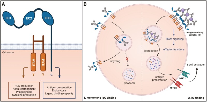
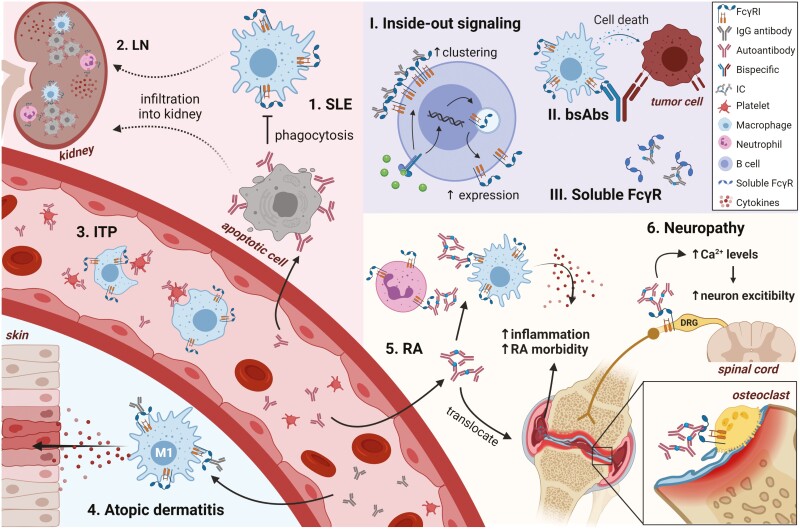
The Fc gamma receptor I (FcγRI or CD64) is the only human Fc receptor with a high affinity for monomeric IgG. It plays a crucial role in immunity, as it mediates cellular effector functions of antibodies including phagocytosis, antigen presentation, and cytokine production. FcγRI is constitutively saturated with monomeric IgG and this feeds the dogma that it has no role in immune responses. However, recent findings have implicated a role for FcγRI in various autoimmune disorders, neuropathies, and antibody therapy in tumor models. By a process known as 'inside-out' signaling, stimulation of myeloid cells with cytokines such as tumor necrosis factor alpha (TNF-α) and interferon-gamma (IFN-γ) enhances FcγRI binding to immune complexes (ICs), including antibody-opsonized pathogens or tumor cells. This review focuses on the current knowledge on interaction of FcγRI with IgG and ICs and the effect of inside-out signaling on FcγRI functioning. Additionally, this review will address potential clinical applications of targeting FcγRI, and the tools that can be used to overcome IC-mediated autoimmune diseases on the one hand, and to enhance antibody-based anti-cancer therapy on the other.

Keywords: CD64; autoimmunity; cancer; inside-out signaling; neuropathy; therapeutic antibodies.


Figures

Fig. 1.
Fig. 1.
Schematic representation of FcγRI and FcγRI functions. (A) Schematic representation of FcγRI with three extracellular domains; EC1, EC2, and EC3, the transmembrane part and intracellular tail (FcγRI-CY). The γ-chain and α-chain are responsible for different actions of the receptor. (B) FcγRI functions are regulated on multiple levels [1]. Interaction of FcγRI with monomeric IgG leads to rapid internalization and recycling of the receptor-IgG complex to the plasma membrane [2]. Crosslinking of FcγRI by ICs induces immunoreceptor tyrosine-based activation motif (ITAM) signaling via FcRγ and internalization and degradation of the antigen-receptor complex in the lysosome. The degraded peptides can be presented on MHC class I (MHC I) or MHC class II (MHC II), which leads to T cell activation.
Fig. 2.
Fig. 2.
Schematic representation of Fc-FcγRI interaction. (A) Extracellular domains EC1, EC2, and EC3 are depicted in cyan, purple, and blue, respectively. The Fc-tail of the antibody is depicted in grey. CH stands for constant heavy chain, CL for constant light chain, VL for variable light chain, and VH for variable heavy chain. Orange dashed boxes indicate binding sites 1 and 2. (B) Detailed depiction of binding site 1 (FcγRI and Fc-chain). The surface of EC2 is shown in purple and the surface of EC1 in cyan. The residues of the Fc-chain involved in the interaction of FcγRI are represented with lines (grey = Fc-chain, yellow = carbon, red = oxygen, and blue = nitrogen). The blue dashed line represents the hydrophobic pocket where Leu235 can bind. (C) Detailed depiction of binding site 2. The same color scheme as in (B) is used. Here the interaction surface lies relatively flat. For additional details see review by Kiyoshi et al. [17].
Fig. 3.
Fig. 3.
Role of FcγRI in autoimmune diseases and possible clinical applications for targeting FcγRI. Numbers depict autoimmune diseases and Roman numerals the possible intervention techniques [1]. In SLE, IgG autoantibodies are produced against self-antigens, which opsonize late apoptotic cells. Blocking FcγRI (not depicted) increases phagocytosis of apoptotic cells [2, 47]. ICs, formed by autoantibodies and self-antigens, reside in the kidney, causing the infiltration of FcγRI-expressing monocytes and macrophages, which release pro-inflammatory cytokines and chemokines, leading to inflammation and subsequently LN [3]. In ITP, IgG autoantibody-coated platelets get cleared by FcγRI-expressing macrophages and blocking FcγRI reduced clearance by 50% [20] (not depicted) [4]. In atopic dermatitis, FcγRI expression is increased on M1 macrophages, making them readily bind to the increased total and antigen-specific serum IgG4 [48]. FcγRI-targeted immunotoxins alter the polarization towards M2 phenotype [5, 49]. In RA, IgG autoantibody ICs can bind macrophages and neutrophils causing the release of pro-inflammatory cytokines which increase inflammation and RA morbidity. The IgG-ICs also translocate to the joints, causing further inflammation. They can also directly bind to osteoclasts. Capturing the IgG-ICs with recombinant soluble FcγRs (III) reduces cartilage degradation (not depicted) [6, 50, 51]. Activation of FcγRI on DRG neurons caused increased neuron excitability and subsequent pain. Blocking the receptor relieved pain in mice [52, 53]. (I) Cytokines cause inside-out signaling, leading to enhanced clustering of FcγRI in the membrane. Cytokines also regulate expression of FcγRI by inducing transfer of intracellular FcγRI to the plasma membrane as well as de novo expression. (II) bsAbs can be engineered to bind FcγRI-expressing target cells and a tumor-target, thereby inducing tumor-killing [54–56]. (III) Recombinant soluble FcγR can ‘capture’ circulating ICs. SLE = systemic lupus erythematous, LN = lupus nephritis, ITP = immune thrombocytopenic purpura, RA = rheumatoid arthritis, and bsAbs = bispecific antibodies.

References

    1. Nimmerjahn F, Ravetch JV. Fcγ receptors as regulators of immune responses. Nat Rev Immunol 2008;8(8):34–47. 10.1038/nri2206 - DOI - PubMed
    1. Takai T. Fc receptors and their role in immune regulation and autoimmunity. J Clin Immunol 2005;25(1):1–18. 10.1007/s10875-005-0353-8 - DOI - PubMed
    1. Groselj-Grenc M, Ihan A, Derganc M. Neutrophil and monocyte CD64 and CD163 expression in critically ill neonates and children with sepsis: comparison of fluorescence intensities and calculated indexes. Mediat Inflamm 2008;2008. 10.1155/2008/202646 - DOI - PMC - PubMed
    1. Bruhns P, Iannascoli B, England P et al. . Specificity and affinity of human Fcγ receptors and their polymorphic variants for human IgG subclasses. Blood 2009;113(16):3716–25. 10.1182/blood-2008-09-179754 - DOI - PubMed
    1. Barnes N, Gavin AL, Tan PS et al. . FcγRI-deficient mice show multiple alterations to inflammatory and immune responses. Immunity 2002;16(3):379–89. 10.1016/S1074-7613(02)00287-X - DOI - PubMed

LinkOut - more resources