

Knotted double j ureteral stent: a case report and literature review
- PMID: 36284889
- PMCID: PMC9557805
- DOI: 10.11604/pamj.2022.43.5.34538
Knotted double j ureteral stent: a case report and literature review
Abstract
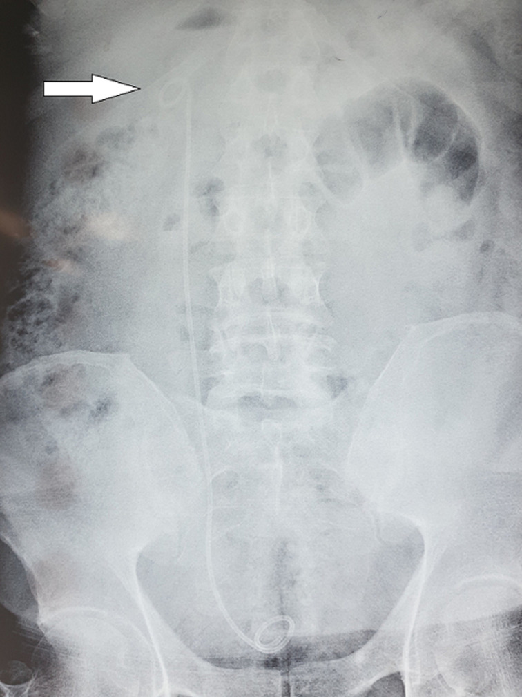
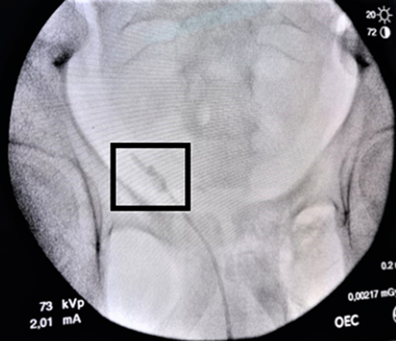
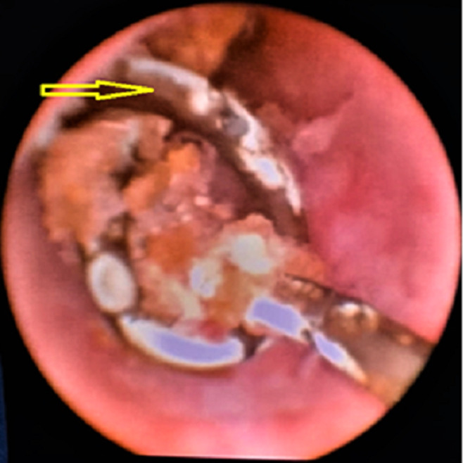
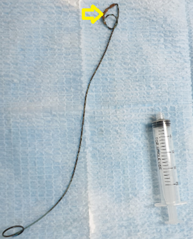
Many complications due to double j (DJ) stent placement have been reported. DJ stent knotting is a rare complication, with only a few cases reported in the literature. We presented a case of DJ stent knotting and reviewed the literature regarding this complication. We reported a 20-year-old man with a history of cystinuria and ureteral stone managed with retrograde ureteroscopy and holmium laser three months ago. The patient comes for DJ stent removal. Firstly, we tried to remove the DJ stent via the cystoscopic procedure, which failed. A fluoroscopic image revealed a knotted DJ stent lodged at the ureteropelvic junction and was removed via holmium laser ureteroscopic procedure without complications. In conclusion, when cystoscopic procedure with simple traction fails to remove DJ stents, multimodality urological procedures such as holmium laser should be tried, especially in patients with urolithiasis predisposing factors.
Keywords: Knot ureteral stent; case report; encrustation; urological complication.
Copyright: Omar Jendouzi et al.
Conflict of interest statement
The authors declare no competing interests.
Figures
References
-
- Zimskind PD, Fetter TR, Wilkerson JL. Clinical use of long-term indwelling silicone rubber ureteral splints inserted cystoscopically. J Urol. 1967 May;97(5):840–4. - PubMed
-
- Ahmed F, Al-wageeh S, Ghabisha S, Al-shami E, Al-naggar K, Obaid G, et al. A case report of forgotten double J stent with giant calculus formation from the renal pelvis to the bladder. JEMTAC. 2021;2021(3)
-
- Groeneveld AE. The role of ESWL in the treatment of large kidney stones. Singapore Med J. 1989 Jun;30(3):249–54. - PubMed
Publication types
MeSH terms
LinkOut - more resources
Full Text Sources
