

Emerging roles of centrosome cohesion
- PMID: 36285440
- PMCID: PMC9597181
- DOI: 10.1098/rsob.220229
Emerging roles of centrosome cohesion
Abstract
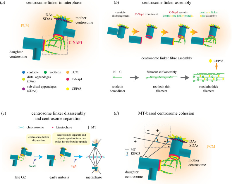
The centrosome, consisting of centrioles and the associated pericentriolar material, is the main microtubule-organizing centre (MTOC) in animal cells. During most of interphase, the two centrosomes of a cell are joined together by centrosome cohesion into one MTOC. The most dominant element of centrosome cohesion is the centrosome linker, an interdigitating, fibrous network formed by the protein C-Nap1 anchoring a number of coiled-coil proteins including rootletin to the proximal end of centrioles. Alternatively, centrosomes can be kept together by the action of the minus end directed kinesin motor protein KIFC3 that works on interdigitating microtubules organized by both centrosomes and probably by the actin network. Although cells connect the two interphase centrosomes by several mechanisms into one MTOC, the general importance of centrosome cohesion, particularly for an organism, is still largely unclear. In this article, we review the functions of the centrosome linker and discuss how centrosome cohesion defects can lead to diseases.
Keywords: centrosome cohesion; centrosome linker; centrosomes.
Conflict of interest statement
We declare we have no competing interests.
Figures
Similar articles
-
The centrosomal linker and microtubules provide dual levels of spatial coordination of centrosomes.PLoS Genet. 2015 May 22;11(5):e1005243. doi: 10.1371/journal.pgen.1005243. eCollection 2015 May. PLoS Genet. 2015. PMID: 26001056 Free PMC article.
-
The balance between KIFC3 and EG5 tetrameric kinesins controls the onset of mitotic spindle assembly.Nat Cell Biol. 2019 Sep;21(9):1138-1151. doi: 10.1038/s41556-019-0382-6. Epub 2019 Sep 2. Nat Cell Biol. 2019. PMID: 31481795
-
Centlein mediates an interaction between C-Nap1 and Cep68 to maintain centrosome cohesion.J Cell Sci. 2014 Apr 15;127(Pt 8):1631-9. doi: 10.1242/jcs.139451. Epub 2014 Feb 19. J Cell Sci. 2014. PMID: 24554434
-
Breaking the ties that bind: new advances in centrosome biology.J Cell Biol. 2012 Apr 2;197(1):11-8. doi: 10.1083/jcb.201108006. J Cell Biol. 2012. PMID: 22472437 Free PMC article. Review.
-
Separate to operate: control of centrosome positioning and separation.Philos Trans R Soc Lond B Biol Sci. 2014 Sep 5;369(1650):20130461. doi: 10.1098/rstb.2013.0461. Philos Trans R Soc Lond B Biol Sci. 2014. PMID: 25047615 Free PMC article. Review.
Cited by
-
The Dual Function of RhoGDI2 in Immunity and Cancer.Int J Mol Sci. 2023 Feb 16;24(4):4015. doi: 10.3390/ijms24044015. Int J Mol Sci. 2023. PMID: 36835422 Free PMC article. Review.
-
Protecting centrosomes from fracturing enables efficient cell navigation.Sci Adv. 2025 Apr 25;11(17):eadx4047. doi: 10.1126/sciadv.adx4047. Epub 2025 Apr 25. Sci Adv. 2025. PMID: 40279414 Free PMC article.
-
Loss of Primary Cilia Potentiates BRAF/MAPK Pathway Activation in Rhabdoid Colorectal Carcinoma: A Series of 21 Cases Showing Ciliary Rootlet CoiledCoil (CROCC) Alterations.Genes (Basel). 2023 Apr 27;14(5):984. doi: 10.3390/genes14050984. Genes (Basel). 2023. PMID: 37239344 Free PMC article.
-
Immunolocalization and 3D modeling of three unique proteins belonging to the costa of Tritrichomonas foetus.Parasitol Res. 2025 Mar 7;124(3):30. doi: 10.1007/s00436-025-08466-4. Parasitol Res. 2025. PMID: 40053153 Free PMC article.
-
A potential patient stratification biomarker for Parkinson´s disease based on LRRK2 kinase-mediated centrosomal alterations in peripheral blood-derived cells.NPJ Parkinsons Dis. 2024 Jan 8;10(1):12. doi: 10.1038/s41531-023-00624-8. NPJ Parkinsons Dis. 2024. PMID: 38191886 Free PMC article.
References
-
- Buchwalter R, Chen J, Zheng Y, Megraw T. 2016. Centrosome in cell division, development and disease. eLS. (10.1002/9780470015902.a0020872) - DOI
Publication types
MeSH terms
Substances
LinkOut - more resources
Full Text Sources
