. 2022 Dec;9(35):e2204513.
doi: 10.1002/advs.202204513. Epub 2022 Oct 26.

A Novel Protein Encoded by Exosomal CircATG4B Induces Oxaliplatin Resistance in Colorectal Cancer by Promoting Autophagy

Affiliations

A Novel Protein Encoded by Exosomal CircATG4B Induces Oxaliplatin Resistance in Colorectal Cancer by Promoting Autophagy

Zihao Pan et al. Adv Sci (Weinh). 2022 Dec.

Abstract

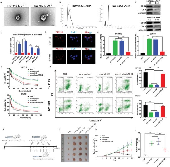
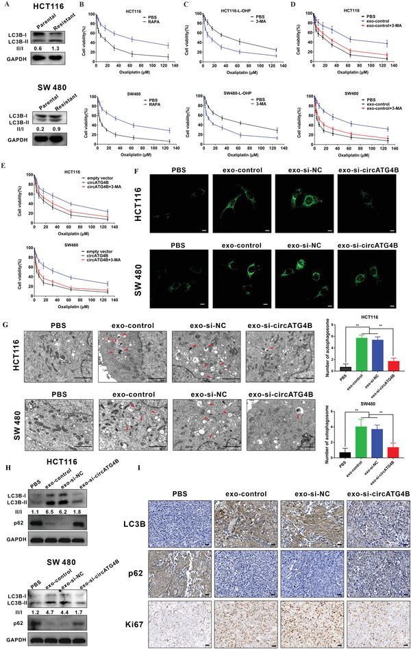
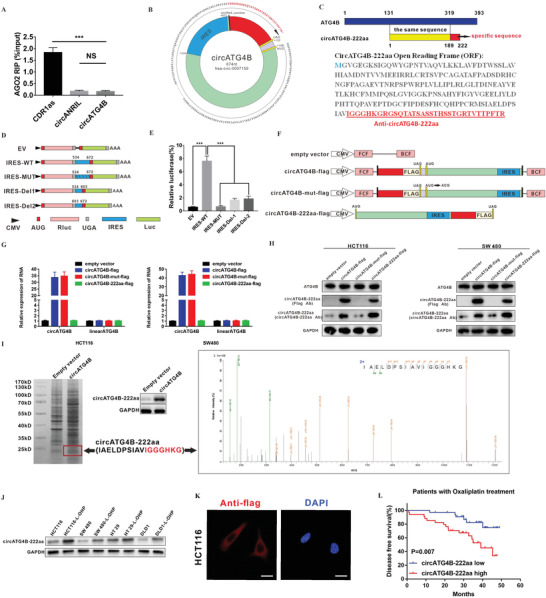
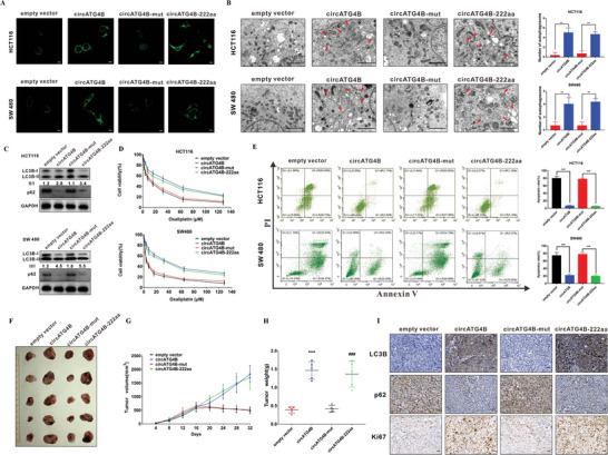
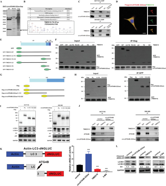
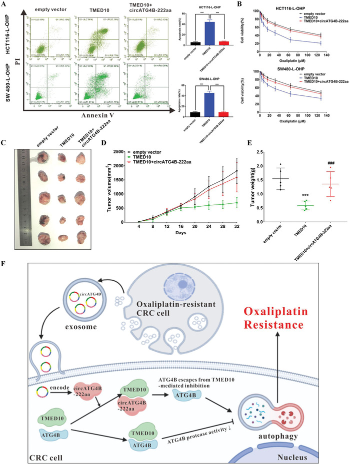
Oxaliplatin is commonly used in chemotherapeutic regimens for colorectal cancer (CRC) after surgical resection. However, acquired chemoresistance seriously affects the curative effect in CRC patients, and the mechanism is still unclear. Here, a circular RNA, circATG4B is identified, which plays an important role in oxaliplatin resistance in CRC. circATG4B expression is found to be increased in exosomes secreted by oxaliplatin-resistant CRC cells. In addition, the results suggest that circATG4B induces oxaliplatin resistance by promoting autophagy. Further in vivo and in vitro studies indicate that the effect of circATG4B is attributed to its potential to encode a novel protein, circATG4B-222aa. Next, circATG4B-222aa is found to function as a decoy to competitively interact with TMED10 and prevent TMED10 from binding to ATG4B, which leads to increased autophagy followed by induction of chemoresistance. Therefore, this study reveals that exosomal circATG4B participates in the decreased chemosensitivity of CRC cells, providing a new rationale for a potential therapeutic target for oxaliplatin resistance in CRC.

Keywords: autophagy; chemoresistance; circular RNA; colorectal cancer (CRC).


Conflict of interest statement

The authors declare no conflict of interest.

Figures

Figure 1
Figure 1
CircATG4B is overexpressed in oxaliplatin‐resistant CRC cells. A) Heatmap showing ATG4B gene‐derived circRNAs in CRC chemoresistant tissues compared with parental tissues analyzed by qRT‐PCR. B) The expression of circRNAs in HCT116 and HCT116‐L‐OHP (chemoresistant) cells. C) Divergent primer detected circular RNAs in cDNA but not gDNA. D) Six exons form circATG4B by back splicing from chromosomal region and Sanger sequencing of circATG4B showed the back‐splice junction (∇). E) Relative RNA level of circATG4B and ATG4B mRNA in different time points. F) Relative RNA level of circATG4B and ATG4B mRNA treated with RNase R. G) Fluorescence in situ hybridization assay was conducted to determine the subcellular localization of circATG4B in chemoresistant and parental CRC cells. Scale bars, 20 µm. H) The circATG4B expression of chemoresistant CRC cells was significantly higher than the parental CRC cells. I) Kaplan–Meier survival curves analysis of DFS in low‐circATG4B‐expression CRC patients with or without oxaliplatin therapy (n = 64). J) Kaplan–Meier survival curves analysis of DFS in high‐circATG4B‐expression CRC patients with or without oxaliplatin therapy (n = 64). K) Kaplan–Meier survival curves analysis of DFS in different circATG4B‐expression CRC patients with oxaliplatin therapy. The median value of circATG4B expression level was used as a cut‐off. *p < 0.05, **p < 0.01, ***p < 0.001.
Figure 2
Figure 2
CircATG4B transferred by an exosomal manner induces oxaliplatin resistance. A) Exosomes were isolated from the supernatant of the culture medium of HCT116‐L‐OHP and SW480‐L‐OHP cells, and the morphology and size were confirmed by transmission electron microscopy. Scale bars, 100 nm. B) NTA distribution of CRC cell‐derived exosomes. C) CRC cell‐derived exosomes were analyzed by Western blotting using anti‐CD54, anti‐CD9, and anti‐Annexin antibodies. Cellular lysates were used as positive loading controls. D) The circATG4B expression of the chemoresistant‐CRC exosome was significantly higher than the exosome of parental CRC cells. E) PKH26‐labeled CRC‐L‐OHP exosomes could fuse into CRC cells. Scale bars, 10 µm. F) qRT‐PCR quantification of circATG4B with the treatment of diverse CRC‐L‐OHP exosomes in CRC cells. G) CCK‐8 detection of cell viability by oxaliplatin in CRC cells treated with diverse CRC‐L‐OHP exosomes. H) Flow cytometry analysis of cell apoptosis by oxaliplatin in CRC cells treated with diverse CRC‐L‐OHP exosomes. I) A flow chart depicting the in vivo experimental design. J) The tumor of different groups treated with oxaliplatin. K) Tumor grow rates are showed in different groups. L) Tumor weights were monitored in different groups. *p < 0.05, **p < 0.01, ***p < 0.001.
Figure 3
Figure 3
Exosomal circATG4B promotes autophagy level. A) Western blotting showed that oxaliplatin‐resistant CRC cells induced the increased proportion of LC3B‐II/LC3B‐I. B,C) RAPA increased oxaliplatin resistance of CRC cells (B) while 3‐MA attenuated in chemoresistance of oxaliplatin‐resistant cells (C). D) 3‐MA reversed the chemoresistance induced by exosome‐control in sensitive cells. E) 3‐MA abolished circATG4B‐promoted oxaliplatin resistance in sensitive cells. F) Puncta‐like staining detection of CRC cells transfected with various treatments is shown. Scale bars, 10 µm. G) Transmission electron microscopy analysis of autophagy is shown. Arrows: autophagosomes/autolysosomes. Scale bars, 2 µm. H) LC3B‐II accumulation and p62 of CRC cells transfected with various treatments is shown. I) IHC analysis of Ki67, LC3B, and p62 in subcutaneous tumor. Scale bars, 20 µm.
Figure 4
Figure 4
Evaluation of the coding ability of circATG4B. A) RIP assay indicated that circATG4B was not associated with AGO2. B) The putative open reading frame (ORF) in circATG4B. C) Upper panel, circATG4B‐222aa shares the majority of its amino acids sequence with that of ATG4B with the exception of the unique amino acids. Lower panel, the sequences of putative ORF are shown. D) The putative IRES activity of circATG4B was tested. IRES sequences in circATG4B or its different truncation/mutation are cloned between the Rluc and Luc reporter genes with independent start (AUG) and stop (UGA) codons. E) The relative luciferase activity of Luc/Rluc in the above vectors was tested. F) Four vectors were constructed. Empty vector; circATG4B‐flag: flag‐tagged circATG4B sequence cloned into a CMV‐induced expression vector; circATG4B‐mut‐flag: flag‐tagged circATG4B sequence with a mutated start codon (ATG→ACG) cloned into a CMV‐induced expression vector; circATG4B‐222aa‐flag: flag‐tagged circATG4B‐222aa sequence cloned into a CMV‐induced expression vector. Forward circRNA frame (FCF) and backward circRNA frame (BCF) are sequences which could circularize the sequence of circRNA. G) Relative RNA expression of circATG4B and linearATG4B was detected by qRT‐PCR. H) Flag antibody and circATG4B‐222aa antibody were used to detect circATG4B‐222aa expression in CRC cells transfected with the above vectors. I) Left panel, total proteins from circATG4B or control plasmid‐transfected HEK‐293T cells were separated via SDS‐PAGE. CircATG4B‐222aa overexpression was confirmed by immunoblotting. Right panel, circATG4B‐222aa junction‐specific peptides were identified by LC/MS. J) CircATG4B‐222aa in chemoresistant and parental CRC cells was detected by immunoblotting. K) Flag‐tagged circATG4B was transfected into HCT116 cells. Immunofluorescence staining by anti‐flag was performed to show the circATG4B‐222aa. Scale bars, 20 µm. L) Semi‐quantitative analysis of circATG4B‐222aa expression level and CRC patient disease‐free survival (DFS) in the 68 patient cohort treated with Oxaliplatin. ***p < 0.001.
Figure 5
Figure 5
CircATG4B‐222aa but not circATG4B increases the autophagy and induces oxaliplatin in CRC cells in vitro and in vivo. A) Puncta‐like staining detection of HCT116 and SW480 transfected with four vectors. Scale bars, 10 µm. B) Transmission electron microscopy analysis of autophagy of HCT116 and SW480 transfected with four vectors. Scale bars, 2 µm. C) LC3B‐II/LC3B‐I proportion of HCT116 and SW480 transfected with four vectors. D) CCK‐8 detection of cell viability by oxaliplatin in HCT116 and SW480 transfected with four vectors. E) Flow cytometry analysis of cell apoptosis by oxaliplatin in HCT116 and SW480 transfected with four vectors. F) Nude mice xenografts were formed by HCT116 cells transfected with empty‐vector, circATG4B, circATG4B‐mut, and circATG4B‐222aa, respectively. Then, sixteen days after injection, oxaliplatin was injected intraperitoneally. G) Tumor volumes were monitored during the time course. H) Tumor weights were also measured at the end of this study. I) IHC analysis of LC3B, p62, and Ki67 in tumor nodules. Scale bars, 20 µm. **p < 0.01, ***p < 0.001; ***p < 0.001 empty vector versus circATG4B group, ### p < 0.001 circATG4B‐mut versus circATG4B‐222aa group (H).
Figure 6
Figure 6
CircATG4B‐222aa protects against the effect of ATG4B induced by TMED10‐mediated inhibition. A) Total proteins from flag‐circATG4B‐222aa plasmid‐transfected HEK‐293T cells were extracted. The proteins coimmunoprecipitated with antibody against flag were separated via SDS‐PAGE. B) Upper panel, list of the top five differentially expressed proteins identified by mass spectrometry. Lower panel, TMED10 was identified by LC/LC‐MS. C) The interaction of TMED10 and circATG4B‐222aa was detected by immunoprecipitation in CRC cells. D) Flag‐tagged circATG4B‐222aa was transfected into HCT116 cells and immunofluorescence was performed using anti‐flag and anti‐TMED10 antibody. Scale bars, 10 µm. E) Schematic of domain structure of TMED10 and GFP‐tagged TMED10 mutants. F) HEK‐293T cells were transfected with flag‐tagged circATG4B‐222aa and GFP‐tagged full‐length or TMED10 fragments, followed by IP using anti‐flag antibody. G) Schematic diagrams showed the wild‐type circATG4B‐222aa and its truncation mutants. H) HEK‐293T cells were transfected with GFP‐tagged TMED10 and flag‐tagged circATG4B‐222aa mutants, followed by IP with anti‐GFP antibody. I) Western Blotting assays showed the levels of autophagy in cells after transfection of wild‐type circATG4B‐222aa ORF or truncation mutants. J) After overexpression of circATG4B‐222aa, the binding capacity between TMED10 and ATG4B was monitored by Co‐IP. K) Left panel, schematic diagram of the quantification of ATG4B activity using an assay based on a luciferase‐release system. Right panel, HCT116 cells transfected with pEAK12‐Actin‐LC3‐dNGLUC were transfected with control, or circATG4B‐222aa overexpression or TMED10 overexpression, or treated with 3‐MA. The supernatants were collected, and the relative luciferase activity was measured. L) The collected supernatants were analyzed by Western blotting using an anti‐luciferase and anti‐LC3 antibody.
Figure 7
Figure 7
CircATG4B‐222aa reverses chemosensitivity induced by TMED10 in oxaliplatin‐resistant CRC cells. A) CircATG4B‐222aa reversed the increases in apoptosis induced by TMED10 in chemoresistant CRC cells. B) CircATG4B‐222aa reversed the increases in chemosensitivity induced by TMED10 in chemoresistant CRC cells. C) Nude mice xenografts were formed by HCT116‐L‐OHP cells transfected with empty‐vector, TMED10, and TMED10+circATG4B‐222aa, respectively. Then, sixteen days after injection, oxaliplatin was injected intraperitoneally. D) Tumor volumes were monitored during the time course. E) Tumor weights were also measured at the end of this study. F) CircATG4B‐222aa could induce oxaliplatin resistance by interacting with TMED10 and increasing autophagic activity in CRC cells. ***p < 0.001; ***p < 0.001 empty vector versus TMED10 group, ### p < 0.001 TMED10 versus TMED10+circATG4B‐222aa group (e).

References

    1. Sung H., Ferlay J., Siegel R. L., Laversanne M., Soerjomataram I., Jemal A., Bray F., CA Cancer J. Clin. 2021, 71, 209. - PubMed
    1. Jardim D. L., Rodrigues C. A., Novis Y. A. S., Rocha V. G., Hoff P. M., Ann. Oncol. 2012, 23, 1937. - PubMed
    1. Théry C., Zitvogel L., Amigorena S., Nat. Rev. Immunol. 2002, 2, 569. - PubMed
    1. van Niel G., Porto‐Carreiro I., Simoes S., Raposo G., J. Biochem. 2006, 140, 13. - PubMed
    1. Kulcheski F. R., Christoff A. P., Margis R., J. Biotechnol. 2016, 238, 42. - PubMed

Publication types

LinkOut - more resources